Atomic-Scale MXene Surface Analysis: STM and STS Mapping for Biomedical Material Characterization
This article provides a comprehensive guide to using Scanning Tunneling Microscopy (STM) and Spectroscopy (STS) for the nanoscale chemical and electronic mapping of MXene surfaces.
Atomic-Scale MXene Surface Analysis: STM and STS Mapping for Biomedical Material Characterization
Abstract
This article provides a comprehensive guide to using Scanning Tunneling Microscopy (STM) and Spectroscopy (STS) for the nanoscale chemical and electronic mapping of MXene surfaces. Targeted at researchers and material scientists, it covers foundational principles, practical methodologies for sample preparation and imaging, troubleshooting for common artifacts, and validation techniques against complementary methods like XPS and Raman spectroscopy. The focus is on extracting quantitative electronic structure data—work function, local density of states (LDOS), defect characterization—critical for tailoring MXenes in biomedical applications such as biosensing, drug delivery, and antimicrobial surfaces.
MXene Surfaces Demystified: Why Atomic-Scale STM/STS Mapping is Revolutionary
MXenes are a rapidly growing class of two-dimensional transition metal carbides, nitrides, and carbonitrides with the general formula Mn+1XnTx, where M is an early transition metal, X is carbon and/or nitrogen, and Tx represents surface termination groups. These terminations, primarily -O, -OH, and -F, are acquired during the synthesis process (typically by etching the "A" layer from a MAX phase using aqueous fluoride-containing solutions). The type, distribution, and ratio of these terminal groups fundamentally dictate MXene's electronic properties, chemical reactivity, surface energy, and catalytic/electrochemical performance. Within the context of a thesis focused on MXene surface chemical mapping with Scanning Tunneling Microscopy/Spectroscopy (STM/STS), precise characterization and control of these terminations is paramount for correlating local surface chemistry with electronic structure (e.g., local density of states).
Quantitative Data on MXene Terminal Groups
The composition of surface terminations varies significantly based on synthesis and post-processing conditions. The following table summarizes key quantitative data on the effects of these groups and their typical ranges.
Table 1: Influence and Characteristics of Primary MXene Terminal Groups
| Terminal Group | Typical Binding Energy (XPS, eV) | Effect on Work Function (eV) | Common Synthesis Route | Key Influence on Electronic Properties |
|---|---|---|---|---|
| -O | Ti 2p3/2 ~ 530.0-530.5 | Increases (~5.2 for Ti3C2O2) | Etching with LiF/HCl, thermal annealing, or oxidation in water. | Leads to semiconductor-like behavior; high catalytic activity for HER. |
| -OH | Ti 2p3/2 ~ 531.5-532.0 | Decreases (~4.6 for Ti3C2(OH)2) | Etching with HF or LiF/HCl, followed by storage in water. | Can induce metallic conductivity; enhances hydrophilicity and ion intercalation. |
| -F | Ti 2p3/2 ~ 684.5-685.0 (F 1s) | Intermediate (~4.9 for Ti3C2F2) | Direct product of HF-based etching processes. | Often reduces electronic conductivity; can block active sites. |
| Mixed Terminations | Multiple peaks in ranges above | Tunable (~4.6 - 5.2) | Controlled by post-etch washing, intercalation, or thermal treatment. | Properties are a weighted average, allowing for precise engineering. |
Table 2: Summary of Common MXene Synthesis Protocols and Resultant Terminal Group Profiles
| Synthesis Protocol | Etchant | Typical Duration | Post-Etch Treatment | Predominant Terminations (as-made) | Suitability for STM/STS |
|---|---|---|---|---|---|
| Direct HF Etching | 50% Aqueous HF | 24-48 hours | Washing with DI water to pH ~6 | High -F, some -OH, -O | Low: High F, poorly conductive, inhomogeneous. |
| In-situ HF (LiF/HCl) | LiF + 12M HCl | 24 hours at 35°C | Washing with DI water & centrifugation; delamination | Lower -F, higher -OH/-O | High: Larger flakes, more -O/-OH, better for imaging. |
| Molten Salt Etching | Fluoride salts (e.g., KF, LiF) in eutectic mixture | 1-2 hours at 550°C | Washing with dilute HCl/water | Primarily -O, -Cl | Moderate: Clean surfaces, but high temp may cause defects. |
Experimental Protocols
Protocol 1: Synthesis of Ti3C2TxMXene for STM/STS Studies via LiF/HCl Etching
Objective: To produce high-quality, few-layer Ti3C2Tx flakes with a controlled surface termination profile suitable for atomic-scale surface analysis.
Materials:
- Ti3AlC2 MAX phase powder (particle size < 40 µm)
- Lithium fluoride (LiF), powder
- Hydrochloric acid (HCl), 12 M (concentrated)
- Deionized (DI) water
- Argon gas supply
Procedure:
- Etchant Preparation: In a polypropylene vial, slowly add 4.0 g of LiF to 40 mL of 12 M HCl under constant stirring in a fume hood. Allow the mixture to stir until fully dissolved (~10 minutes). The solution temperature will increase.
- Etching: Gradually add 2.0 g of Ti3AlC2 powder to the etchant over 5 minutes to avoid violent reactions. Maintain the reaction at 35°C for 24 hours with continuous stirring (300 rpm).
- Washing: After etching, transfer the suspension to 50 mL conical tubes and centrifuge at 3500 RCF for 5 minutes. Decant the acidic supernatant.
- Neutralization: Resuspend the sediment in 40 mL of DI water. Centrifuge and decant. Repeat this washing step until the supernatant pH reaches approximately 6 (typically 8-10 washes). Critical: The final low-pH washes control the -OH/-F ratio.
- Delamination: After the final wash, add 40 mL of DI water to the sediment and manually shake for 5 minutes. Subject the bottle to argon gas bubbling for 1 hour under constant stirring to facilitate delamination into few-layer flakes.
- Isolation: Centrifuge the delaminated suspension at 1500 RCF for 30 minutes. Collect the dark green, colloidal supernatant containing few-layer MXene flakes.
- Sample Preparation for STM: Deposit a 10-20 µL droplet of the MXene colloidal solution onto a clean, annealed Au(111) on mica substrate. Allow it to adsorb for 2 minutes, then gently rinse with HPLC-grade isopropanol and dry under a stream of ultra-high purity Argon. Immediately transfer to the STM load lock.
Protocol 2: Thermal Annealing for Terminal Group Modification
Objective: To selectively remove -F and -OH groups and enrich the MXene surface with -O terminations, enabling the study of termination-dependent electronic structure.
Materials:
- As-synthesized Ti3C2Tx film or powder on substrate.
- Tube furnace with quartz tube.
- High-purity Argon gas.
Procedure:
- Loading: Place the MXene sample in a quartz boat inside a quartz tube furnace.
- Purging: Seal the tube and purge with Argon gas (200 sccm) for at least 30 minutes to remove oxygen and moisture.
- Annealing: Under a constant Argon flow (50 sccm), heat the furnace to the target temperature (commonly 350°C for partial dehydroxylation/defluorination, or 550°C for complete conversion to -O). Maintain the temperature for 2 hours.
- Cooling: Allow the furnace to cool naturally to room temperature under continuous Argon flow.
- Transfer: Transfer the annealed sample to the STM analysis chamber using an inert atmosphere transfer box to avoid re-adsorption of contaminants.
Visualizations
Title: MXene Synthesis and Termination Engineering Workflow
Title: STM/STS Correlation for Chemical Mapping
The Scientist's Toolkit: Research Reagent Solutions
Table 3: Essential Materials for MXene Surface Chemistry Studies
| Item | Function/Description | Example (Supplier) |
|---|---|---|
| Ti3AlC2 MAX Phase | The precursor material for synthesizing Ti3C2Tx. Purity and particle size affect etching efficiency. | Carbon Ukraine (>98% purity) |
| Lithium Fluoride (LiF) | Component of the mild "in-situ HF" etchant (with HCl). Provides fluoride ions while allowing Li+ intercalation. | Sigma-Aldrich, 99.99% trace metals basis |
| Hydrofluoric Acid (HF) / Hydrochloric Acid (HCl) | Standard etching solutions. HF is direct, aggressive; HCl is used with LiF for a gentler process. | Fisher Chemical, ACS grade |
| Argon Gas | Inert atmosphere for annealing, sample drying, and storage to prevent oxidation of MXenes. | Ultra High Purity (UHP, 99.999%) |
| Au(111)/Mica Substrate | Atomically flat, conductive substrate essential for high-resolution STM/STS of 2D materials. | Georg Albert PVD, 250 nm Au on Mica |
| Deionized Water (DI), >18 MΩ·cm | For washing and delaminating MXenes. Low ion content is critical to avoid unwanted surface species. | Millipore Milli-Q system |
| Anhydrous Isopropanol | For rinsing STM samples to remove salts and water without damaging the flakes or substrate. | Sigma-Aldrich, 99.5% |
| STM Tungsten Tips | Electrochemically etched tips for scanning tunneling microscopy. Must be sharp and stable. | Prepared in-lab from 0.25mm W wire |
Application Note: MXene Surface Characterization for Drug Delivery Vector Optimization
Thesis Context: This application note details the integration of Scanning Tunneling Microscopy/Spectroscopy (STM/STS) for the chemical mapping of MXene surfaces within a broader research program aimed at understanding and engineering these materials for targeted biomedical applications, such as drug delivery and biosensing.
Background: The functional efficacy of MXenes (e.g., Ti₃C₂Tₓ) in biomedicine is critically dependent on their surface termination groups (Tₓ = -O, -OH, -F). These groups dictate hydrophilicity, colloidal stability, drug-loading capacity, and biocompatibility. Nanoscale heterogeneity in these terminations directly impacts functional consistency. STM provides atomic-scale topographic mapping, while STS allows local electronic structure characterization, linking surface chemistry to electronic properties that influence biomolecular interactions.
Quantitative Data Summary: Table 1: Comparative Analysis of MXene Surface Terminations and Functional Properties
| Surface Termination | Relative Abundance (%) (XPS Data) | Hydrophilicity (Contact Angle) | Zeta Potential (mV) in PBS | Doxorubicin Loading Capacity (mg/g) |
|---|---|---|---|---|
| -O dominant | ~60 | <10° | -25 ± 3 | 120 ± 15 |
| -OH dominant | ~30 | ~5° | -35 ± 4 | 180 ± 20 |
| -F dominant | ~10 | ~15° | -15 ± 5 | 75 ± 10 |
Table 2: STM/STS Operational Parameters for MXene Analysis
| Parameter | Typical Setting (STM) | Typical Setting (STS) | Function |
|---|---|---|---|
| Bias Voltage (V) | 0.05 - 1.0 V | -2.0 to +2.0 V (Sweep) | Controls tunneling current/ probes LDOS |
| Tunneling Current (I) | 0.5 - 2.0 nA | Held Constant During Sweep | Determines tip-sample distance |
| Modulation Voltage (for dI/dV) | N/A | 10 - 50 mV (rms) | Enables conductance (dI/dV) mapping |
Protocol: STM/STS Mapping of MXene (Ti₃C₂Tₓ) Flakes for Surface Chemical Heterogeneity Analysis
Objective: To acquire correlated topographic and local density of states (LDOS) maps of MXene flakes to identify and characterize regions with varying surface terminations.
Materials & Reagent Solutions:
- MXene Sample: Aqueous colloidal dispersion of single/few-layer Ti₃C₂Tₓ flakes (≥0.5 mg/mL), synthesized via minimally intensive layer delamination (MILD) method.
- Substrate: Atomically flat, highly oriented pyrolytic graphite (HOPG) or Au(111) on mica.
- STM/STS System: Ultra-high vacuum (UHV) or inert atmosphere glovebox-compatible system with a low-temperature option (77K) for enhanced stability.
- Electrochemical Etching Unit: For preparation of sharp Pt/Ir or tungsten STM tips.
- Micro-syringe & Heated Stage: For precise substrate deposition and solvent evaporation.
Procedure:
Part A: Sample Preparation (Inert Atmosphere Recommended)
- Substrate Cleaving: Cleave HOPG using adhesive tape immediately before deposition to obtain a fresh, clean surface.
- MXene Deposition: Pipette 5 µL of the MXene dispersion onto the freshly cleaved HOPG substrate.
- Solvent Evaporation: Allow the droplet to dry under a gentle argon flow or on a heated stage at 40°C for 15 minutes to produce well-dispersed flakes.
Part B: STM/STS Measurement
- System Loading: Transfer the prepared sample into the STM chamber. If possible, perform a mild degas (≤100°C) under UHV to remove adsorbed water.
- Tip Approach: Approach the STM tip to the sample surface using coarse motors, then engage the automatic coarse approach until a tunneling current is detected.
- Topographic Imaging:
- Set the feedback loop to "Imaging" mode.
- Set parameters: Bias Voltage (Vbias) = 0.1 V, Tunneling Current (It) = 1.0 nA.
- Acquire a large-scale scan (e.g., 1 µm x 1 µm) to locate suitable isolated flakes.
- Select a region of interest (e.g., 50 nm x 50 nm) on a flake and acquire a high-resolution constant-current topographic image.
- Spectroscopic (STS) Point Mapping:
- Freeze the feedback loop at a specific X,Y location on the acquired image.
- Set STS parameters: Sweep Vbias from -1.0 V to +1.0 V, with a modulation voltage of 20 mV (rms, 531 Hz).
- Record the I-V curve and the simultaneously acquired differential conductance (dI/dV) signal.
- Repeat this measurement on a predefined grid (e.g., 32x32 points) over the previously scanned area.
- Data Analysis: Convert the grid of dI/dV spectra into spatial maps of LDOS at specific energies (e.g., at the Fermi level, or at energies corresponding to known termination-induced states). Correlate regions with distinct electronic signatures to topographic features.
Visualization of Key Concepts
Title: Nanoscale Analysis Links MXene Surface to Function
Title: STM/STS Protocol Workflow for MXene
The Scientist's Toolkit: Key Research Reagents & Materials
Table 3: Essential Materials for MXene Surface Analysis Experiments
| Item | Function/Description |
|---|---|
| Ti₃AlC₂ MAX Phase Powder | Precursor material for synthesizing Ti₃C₂Tₓ MXene via selective etching of the Al layer. |
| Lithium Fluoride (LiF) | Etchant component (in HCl). Source of F⁻ ions for etching and surface -F terminations. |
| Hydrochloric Acid (HCl, 9M) | Etching solution medium. Provides H⁺ for the etching process. |
| Deionized Water (O₂-free) | For washing and delaminating etched MXene. Must be degassed to prevent oxidation during processing. |
| HOPG Substrate | Provides an atomically flat, conductive, and inert surface for depositing MXene flakes for STM/STS analysis. |
| Pt/Ir (80/20) Wire | Material for fabricating sharp, stable STM tips via electrochemical etching. |
| Calcium Chloride (CaCl₂) | Desiccant for maintaining a dry environment in sample storage and transfer chambers to prevent MXene degradation. |
| Argon Gas (Ultra-high Purity) | Inert atmosphere for sample preparation, storage, and transfer to minimize surface oxidation prior to analysis. |
Introduction and Thesis Context Scanning Tunneling Microscopy (STM) and Spectroscopy (STS) are complementary techniques central to nanoscale surface science. Within the research on MXenes—a class of two-dimensional transition metal carbides/nitrides—their combined application is pivotal for correlating surface topography with local electronic structure. This is essential for mapping surface chemistry, such as identifying terminal functional groups (-O, -OH, -F) and their impact on MXene's properties for applications in energy storage, catalysis, and sensing. This document provides detailed application notes and protocols for employing STM/STS in this context.
Core Principles and Quantitative Comparison
Table 1: Core Principles and Operational Parameters of STM vs. STS
| Feature | Scanning Tunneling Microscopy (STM) | Scanning Tunneling Spectroscopy (STS) |
|---|---|---|
| Primary Output | Real-space topographic map of surface atoms/structures. | Local density of states (LDOS) as a function of energy at a specific point/area. |
| Core Physical Principle | Quantum tunneling of electrons between a sharp tip and a conductive sample. Tunneling current (I) is kept constant via feedback. | Measurement of the differential conductance (dI/dV) as a function of applied sample bias (V). |
| Key Operational Mode | Constant current mode: Tip height (z) is adjusted to maintain constant I. | Spectroscopy mode: Feedback loop is disabled at a specific location; I-V curves are acquired. |
| Information Type | Topographic: Atomic corrugation, step edges, defect locations, adsorbate positions. | Electronic: Band gap, surface potential, charge density waves, molecular orbital resonances. |
| Critical Parameter | Setpoint current (Iset) and bias voltage (Vbias). | Bias voltage sweep range and modulation voltage (for lock-in detection). |
| Spatial Resolution | Sub-Ångström vertical; Atomic lateral resolution under UHV and low temperature. | Lateral resolution dictated by tip sharpness and electronic delocalization; can be atomic. |
| MXene Application | Imaging basal plane termination, step edges, and defect sites. | Mapping heterogeneity in LDOS due to mixed surface terminations, identifying functional group signatures. |
Table 2: Representative Quantitative Data from MXene STM/STS Studies
| Material (MXene) | Key STM Finding (Topography) | Key STS Finding (Spectroscopy) | Experimental Conditions |
|---|---|---|---|
| Ti₃C₂Tₓ | Ordered atomic lattice with periodicity ~3.0 Å; presence of nanoscale pits. | Local band gap variation from 0.2 eV to 0.6 eV correlated with terminations. | Ultra-high vacuum (UHV), 77 K, electrochemically etched W tip. |
| Mo₂CTₓ | Triangular moiré patterns observed on terraces. | Peak in dI/dV at -0.5V below Fermi level attributed to -O termination sites. | UHV, 4.2 K, in-situ cleavage. |
| Nb₂CTₓ | Linear defects aligned along principal crystallographic directions. | Metallic character (finite LDOS at E_F) at defects vs. semiconducting on terraces. | UHV, 300 K (RT). |
Experimental Protocols
Protocol 1: MXene Sample Preparation for UHV-STM/STS Objective: To obtain an atomically clean, conductive MXene surface suitable for high-resolution STM/STS.
- Delamination: Starting from multilayer clay-like Ti₃AlC₂ MAX phase, etch Al layers using concentrated HF (or LiF+HCl mixture) to produce multilayer Ti₃C₂Tₓ.
- Intercalation & Separation: Intercalate with tetraalkylammonium hydroxide (e.g., TMAOH) and perform gentle sonication under Ar atmosphere. Centrifuge to obtain a colloidal suspension of few/single-layer flakes.
- Substrate Preparation: Clean a conductive substrate (highly oriented pyrolytic graphite - HOPG or Au(111) on mica) via exfoliation (HOPG) or Ar⁺ sputtering/annealing cycles (Au).
- Drop-casting: Under an inert glovebox atmosphere, deposit a dilute MXene suspension onto the substrate. Allow to dry.
- UHV Transfer: Load the sample into a UHV load-lock, pump to high vacuum (<10⁻⁸ mbar), and outgas at 120-150°C for several hours to desorb water and solvents before transferring to the STM stage.
Protocol 2: Combined STM Imaging and Point STS on MXenes Objective: To acquire topographic data and subsequent local electronic spectra on a region of interest.
- Tip Preparation: Electrochemically etch a tungsten (W) wire in 2M NaOH to a sharp point. Clean tip in UHV via electron bombardment or field emission.
- Coarse Approach: Use a coarse approach motor (inertial slider or stepper motor) to bring the tip within tunneling range (~1 mm to <1 µm).
- STM Imaging:
a. Set feedback parameters:
V_bias = +100 mV(sample),I_set = 50 pA. b. Engage the feedback loop. Scan area (e.g., 50 nm x 50 nm) in constant current mode. c. Flatten the image line-by-line to correct for tilt and bow. - Point Spectroscopy:
a. Select a specific site on the acquired image (e.g., on a terrace, near a pit).
b. Disable the feedback loop (
Feedback Off). Set the tip at the desired z-position. c. Sweep the sample biasVacross a predefined range (e.g., -1.0 V to +1.0 V). d. At each voltage step, measure the tunneling currentI. Average over multiple cycles to reduce noise. d. (Alternative: Lock-in Detection) Apply a small AC modulation (e.g., 10 mV, 1 kHz) to the bias. Use a lock-in amplifier to measure the differential conductancedI/dVdirectly, which is proportional to the LDOS. - Data Processing: For I-V curves, numerically differentiate to obtain
dI/dV. Normalize spectra by dividing(dI/dV)by(I/V)to minimize topographic artifacts.
Visualizations
Title: MXene Sample Prep Workflow for UHV STM/STS
Title: STS Measurement Protocol Logic Flow
The Scientist's Toolkit: Essential Research Reagent Solutions
Table 3: Key Materials and Reagents for MXene STM/STS Research
| Item | Function & Relevance |
|---|---|
| Hydrofluoric Acid (HF), 48-49% | Primary etchant to selectively remove the 'A' layer (Al) from the MAX phase to produce multilayer MXene. EXTREME CAUTION required. |
| Lithium Fluoride (LiF) + Hydrochloric Acid (HCl) | Alternative, milder etchant system (in-situ generation of HF). Often yields MXenes with larger flake size and different termination ratios. |
| Tetraalkylammonium Hydroxide (e.g., TMAOH) | Organic base used as an intercalant to swell MXene layers and facilitate delamination into single sheets via sonication. |
| Argon (Ar) Gas | Inert atmosphere for all post-etching steps (washing, delamination, storage) to prevent oxidation of MXenes, especially Ti₃C₂Tₓ. |
| Highly Oriented Pyrolytic Graphite (HOPG) | Atomically flat, conductive, and easily cleanable substrate for drop-casting MXene flakes for STM. |
| Tungsten (W) Wire, 0.25mm diameter | Standard material for fabricating sharp STM tips via electrochemical etching. |
| Sodium Hydroxide (NaOH) pellets, 2M solution | Electrolyte for electrochemical etching of W wire to produce sharp STM tips. |
| Deionized Water (18.2 MΩ·cm) | Used for washing MXene sediments to neutral pH post-etching and for preparing solutions. Residual ions degrade STM performance. |
This application note details the protocols for Scanning Tunneling Spectroscopy (STS) to measure key electronic properties of MXene surfaces. Within the broader thesis on MXene surface chemical mapping with STM/STS, these measurements are fundamental for correlating surface terminations (e.g., -O, -OH, -F) with electronic structure, which is critical for applications in catalysis, energy storage, and sensing relevant to researchers and drug development professionals investigating nanomaterial interfaces.
Work Function (Φ) Measurement via STS
Theoretical Basis
The work function is extracted from the onset of field emission resonances (FER) in dI/dV spectra or from the shift in the tunneling barrier height with tip-sample distance (I-z spectroscopy).
Experimental Protocol
Materials & Setup:
- Ultra-high vacuum (UHV) STM/STS system (base pressure <1×10⁻¹⁰ mbar).
- Electrically grounded, atomically clean MXene sample (e.g., Ti₃C₂Tₓ flake on Au(111) substrate).
- Chemically etched W tip, cleaned in-situ by electron bombardment.
Procedure:
- Sample Preparation: MXene flakes are transferred in an argon glovebox and introduced into the UHV system via a load-lock, followed by overnight degassing at ~150°C.
- Tip Conditioning: Perform controlled crashes and voltage pulses on a clean metal surface (Au) to achieve a stable tip.
- I-z Spectroscopy: a. Set the STM to a specific location on the MXene terrace (Vbias = 0.1 V, Iset = 100 pA). b. Disable feedback loop. c. Ramp the tip towards the sample over a predefined distance (Δz, e.g., 1 nm) while recording the tunneling current (I). d. Fit the I-z data to the relationship: I ∝ V_bias * exp(-2κz), where κ = √(2mΦ)/ħ. e. Extract the apparent local work function Φ from the decay constant κ.
Key Considerations
- Measurements must be performed on flat terraces to avoid geometric artifacts.
- The work function is highly sensitive to surface adsorbates; cleanliness is paramount.
Band Gap Determination via STS
Theoretical Basis
The band gap (E_g) of a semiconductor, like many MXenes, is determined from the region of zero conductance in a spatially averaged dI/dV spectrum, representing the energy difference between the conduction band minimum (CBM) and valence band maximum (VBM).
Experimental Protocol
Materials & Setup:
- UHV STM/STS system with lock-in amplifier (modulation frequency ~1 kHz, modulation amplitude 10-30 mV_rms).
- Stable, spectroscopic-grade STM tip.
Procedure:
- Stabilize Tip-Sample Junction: At the point of interest, set feedback parameters (Vbias, Iset) to ensure stable tunneling (e.g., Vset = 1.0 V, Iset = 200 pA).
- Disable Feedback: Turn off the feedback loop to maintain constant tip-sample distance.
- Spectral Acquisition: a. Ramp the sample bias voltage through a range spanning the suspected gap (e.g., -2.0 V to +2.0 V). b. Simultaneously, use the lock-in amplifier to measure the differential conductance (dI/dV) directly. c. Average multiple spectra (≥50) from the same point to improve signal-to-noise.
- Data Analysis: a. Plot dI/dV vs. Vbias. b. Identify the energy range where *dI/dV* is flat and near zero. c. Define the VBM and CBM as the onset energies where the conductance increases significantly from the noise floor. Eg = CBM - VBM.
Key Considerations
- Surface states or defect-induced in-gap states can obscure the intrinsic band gap.
- Thermal broadening limits resolution; cryogenic temperatures (e.g., 4.2 K) are preferred for precise measurement.
Local Density of States (LDOS) Mapping
Theoretical Basis
At low temperatures and small modulation voltages, the normalized differential conductance approximates the LDOS: (dI/dV)/(I/V) ∝ ρs(r, E), where ρs is the sample LDOS at energy E=eV_bias and position r.
Experimental Protocol
Materials & Setup:
- Low-temperature STM/STS system (4.2 K or 77 K).
- Vibration isolation system.
Procedure:
- Topographic Imaging: Acquire a high-resolution STM image of the MXene surface area.
- Grid Definition: Define a matrix of points (e.g., 64x64) over the image.
- Spectral Mapping: a. At each grid point, pause the scan and disable the feedback. b. Acquire a dI/dV spectrum using the lock-in technique as described in Section 2. c. Store the full spectrum or the conductance value at a specific bias of interest. d. Re-enable feedback and move to the next point.
- Data Reconstruction: Create 2D spatial maps of the LDOS by plotting the dI/dV intensity at a given energy for all (x,y) positions.
Key Considerations
- This is a time-intensive measurement; stability over hours is required.
- Normalization (dI/dV)/(I/V) is crucial for correcting the exponential decay of the tunneling probability with energy.
Table 1: Typical STS-measured Electronic Properties of Select MXenes
| MXene Formula | Surface Termination (Tₓ) | Work Function (eV) | Band Gap (eV) | Measurement Conditions | Reference Year |
|---|---|---|---|---|---|
| Ti₃C₂ | -O, -OH | 4.2 - 4.8 | 0.05 - 0.2 (metallic/semimetallic) | UHV, 77 K | 2023 |
| Mo₂TiC₂ | -O | 4.5 - 5.0 | ~0.2 | UHV, RT | 2022 |
| Ti₂C | -F | 3.8 - 4.3 | ~0.8 (semiconducting) | UHV, 4.2 K | 2023 |
| Nb₂C | -O | 4.0 - 4.5 | ~0.4 | UHV, 77 K | 2024 |
Table 2: Key Parameters for Core STS Protocols
| Protocol | Key Measured Signal | Critical Lock-in Parameters | Typical Bias Range | Primary Output |
|---|---|---|---|---|
| Work Function (I-z) | Tunneling Current (I) | N/A | Fixed at low bias (0.1-0.2 V) | Barrier height κ, Work Function Φ |
| Band Gap (Point STS) | dI/dV | fmod=873 Hz, Vmod=20 mV | Wide range (±2-3 V) | dI/dV spectrum, E_g onset |
| LDOS Mapping | dI/dV at each (x,y,E) | fmod=1.1 kHz, Vmod=10 mV | User-defined (single energy or range) | 2D spatial map of ρ_s(r, E) |
Visualized Workflows
Title: STS Measurement Workflow for MXene Analysis
Title: From Tunneling Current to Sample LDOS
The Scientist's Toolkit: Key Research Reagent Solutions & Materials
Table 3: Essential Materials for MXene STM/STS Research
| Item Name | Function/Brief Explanation | Typical Specification/Supplier Note |
|---|---|---|
| MAX Phase Precursor | Starting material for MXene synthesis (e.g., Ti₃AlC₂). Determines the MXene composition. | Purity >98%, 200 mesh powder (e.g., Forsman Scientific). |
| Etching Agent (LiF/HCl) | Selective etching solution to remove the 'A' layer (e.g., Al) from the MAX phase to produce multilayered MXene. | "Minimally Intensive Layer Delamination" (MILD) method standard. |
| Intercalant (DMSO, TMAOH) | Organic solvent used to intercalate between MXene layers and aid in delamination to produce single/few-layer flakes. | Anhydrous grade, stored over molecular sieves. |
| Anodic Stripping Foil | Substrate for MXene flake deposition (e.g., Au(111) on mica, HOPG). Provides an atomically flat, conductive, inert surface. | Epitaxial Au on mica, freshly annealed in UHV by Ar⁺ sputtering and heating. |
| UHV Transfer Case | Sealed, argon-filled vessel for transferring air-sensitive MXene samples from glovebox to UHV load-lock without air exposure. | Custom or commercial (e.g., Kurt J. Lesker Company). |
| Electrochemical Etching Solution | For preparation of sharp, reproducible tungsten STM tips (e.g., 2M KOH or NaOH solution). | Analytical grade, used with Pt or carbon counter electrode. |
| Lock-in Amplifier | Enables sensitive detection of the small differential conductance (dI/dV) signal by measuring response at a specific modulation frequency. | Required for STS (e.g., Stanford Research Systems SR830). |
| Cryogenic Liquids (LHe, LN₂) | For cooling STM stages to 4.2 K or 77 K, reducing thermal noise and broadening for high-resolution STS and LDOS mapping. | Essential for studying subtle electronic features and superconducting gaps. |
Within the broader thesis on "MXene Surface Chemical Mapping with STM/STS for Catalytic and Biosensing Applications," a fundamental and persistent challenge is the preparation of atomically clean, environmentally stable, and electronically homogeneous MXene surfaces. This is a prerequisite for obtaining reliable scanning tunneling microscopy (STM) topographic data and spatially resolved scanning tunneling spectroscopy (STS) electronic maps. This document outlines the standardized protocols and material considerations necessary to overcome these challenges, enabling reproducible high-resolution imaging crucial for researchers and drug development professionals investigating MXene-biomolecule interactions or catalytic active sites.
Table 1: Primary Challenges in MXene STM/STS Preparation
| Challenge | Impact on STM/STS | Quantitative Manifestation |
|---|---|---|
| Surface Oxidation & Degradation | Introduces non-conductive oxides (e.g., TiO₂), disrupting tunneling current and masking intrinsic electronic structure. | Increased surface roughness (RMS > 5 nm vs. <1 nm for fresh), loss of atomic features within hours in ambient air. |
| Residual Water & Intercalants | Alters local density of states (LDOS), causes tip instability, and prevents true surface imaging. | Unstable tunneling current (>10% fluctuation), irreproducible I-V curves, presence of "bubble-like" artifacts in STM. |
| Flake Inhomogeneity & Terminations | Leads to spatially variable STS spectra, complicating chemical mapping. | Work function variations of 0.3-0.8 eV across a single flake dependent on -O, -OH, -F termination ratio. |
| Substrate Interaction | Poor adhesion or charge transfer can electronically couple or decouple the MXene from the substrate. | Apparent height variations >50% of true flake thickness, shifting of Fermi level in STS by hundreds of meV. |
Table 2: Comparative Efficacy of Common MXene Transfer Solvents
| Solvent | Primary Function | Boiling Point (°C) | Residual Film (AFM measured) | Recommended for STM? |
|---|---|---|---|---|
| Deionized Water | Standard transfer medium | 100 | High (several nm) | No - Promotes oxidation. |
| Ethanol | Displaces water, faster drying | 78 | Moderate (~1-2 nm) | Conditional - Use spectroscopic grade. |
| Isopropanol (IPA) | Low surface tension, displaces water | 82 | Low (<1 nm) | Yes - Preferred. |
| Toluene | Non-polar, water-free transfer | 111 | Very Low (negligible) | Yes - For inert-atmosphere glovebox use. |
Detailed Experimental Protocols
Protocol 3.1: Inert-Environment MXene Flake Preparation for STM
Objective: To deposit isolated, clean MXene (Ti₃C₂Tₓ) flakes onto an atomically flat substrate with minimal atmospheric exposure.
Materials:
- MXene suspension (single-layer, ~1 mg/mL in degassed, deionized water), stored in glovebox antechamber.
- Atomically flat substrate: Highly Ordered Pyrolytic Graphite (HOPG) or Au(111) on mica.
- Anoxic transfer chamber (glovebox) with O₂ & H₂O < 1 ppm.
- Centrifuge, ultrasonic bath (inside glovebox).
- Spectroscopic grade Isopropanol (IPA), anhydrous.
- Spin coater or drop-cast setup within glovebox.
Procedure:
- Substrate Preparation: Inside the glovebox, cleave HOPG using adhesive tape immediately before use to ensure a pristine, uncontaminated surface.
- Suspension Dilution & Dispersion: Dilute the MXene stock suspension 1:100 in degassed IPA. Sonicate for 5 minutes at low power (50W) to break up any aggregates.
- Deposition: Using a micropipette, deposit 20 µL of the diluted suspension onto the freshly cleaved HOPG surface.
- Drying: Allow the solvent to evaporate fully under a gentle argon flow for 10 minutes. For more uniform coverage, spin-coat at 2000 rpm for 60 seconds.
- Immediate Transfer: Place the prepared sample into a vacuum-sealed, inert-atmosphere transfer module. This module must be directly connected to the STM load-lock to prevent any air exposure.
Protocol 3.2:In-situPlasma Cleaning for MXene Surfaces in UHV-STM
Objective: To remove adsorbed hydrocarbons and thin oxide layers from MXene surfaces immediately prior to UHV-STM imaging.
Materials:
- UHV-STM system with integrated argon ion sputter gun and sample heating stage.
- High-purity (99.9999%) Argon gas.
Procedure:
- Sample Introduction: Transfer the glovebox-prepared sample (from Protocol 3.1) into the UHV chamber via the inert transfer module. Base pressure should be < 5×10⁻¹⁰ mbar.
- Gentle Ar⁺ Sputtering:
- Backfill the chamber with Ar to a pressure of 5×10⁻⁶ mbar.
- With the sample at room temperature, expose it to a low-energy (300 eV) Ar⁺ beam for 30-60 seconds. Use a low ion current density (~1 µA/cm²).
- Rotate the sample during sputtering for uniform cleaning.
- Post-Cleaning Anneal: Optionally, anneal the sample at 150-200°C for 15 minutes to heal defects and promote surface ordering. Caution: Higher temperatures may induce de-functionalization or phase transformation.
- STM Verification: Cool the sample to the desired imaging temperature (often 77 K or 4.2 K for highest stability) and approach with an STM tip. Confirm surface cleanliness by atomic-resolution imaging on the HOPG substrate surrounding the MXene flake.
Visualizations
STM Sample Prep & UHV Cleaning Workflow
Instability Causes & Controlled Env Solutions
The Scientist's Toolkit: Essential Research Reagents & Materials
Table 3: Essential Materials for Reliable MXene STM
| Item | Specification / Brand Example | Critical Function |
|---|---|---|
| MXene Suspension | Single-layer Ti₃C₂Tₓ, concentration ≤ 1 mg/mL, stored under argon. | The core material. Single-layer flakes are essential for substrate coupling and minimizing artifacts. |
| HOPG Substrate | ZYA or ZYB grade, 10x10 mm. | Provides an atomically flat, conductive, and inert staging surface for MXene flakes. Easily cleaved for renewal. |
| Anhydrous Isopropanol | Spectroscopic grade, 99.9+%, packaged under inert gas (e.g., Sigma-Aldrich). | Low-surface-tension solvent for final MXene deposition, minimizing residue and water content. |
| Argon Gas | Ultra-high purity (99.9999%), with additional purifier for glovebox and UHV use. | Creates and maintains an inert atmosphere throughout preparation and transfer, suppressing oxidation. |
| UHV-Compatible Transfer Module | Portable, sealable container with electrical feedthroughs for heating (if needed). | Physically bridges the glovebox and UHV-STM, preventing atmospheric exposure of the prepared sample. |
| Electrochemically Etched STM Tips | Tungsten or PtIr alloy, cleaned in-situ via electron bombardment or field emission. | The probing tool. Must be atomically sharp and clean to resolve MXene atomic structure and acquire precise STS. |
Step-by-Step Protocol: STM/STS of MXenes from Sample Prep to Data Acquisition
This application note details protocols for preparing high-quality MXene (primarily Ti₃C₂Tₓ) samples for Scanning Tunneling Microscopy (STM) and Spectroscopy (STS) studies. Within the broader thesis on MXene surface chemical mapping with STM/STS, consistent and atomically clean sample preparation is paramount. The electronic structure and surface chemistry probed by STS are exquisitely sensitive to flake thickness, substrate interaction, and adsorbates. These protocols aim to produce reproducible samples with large, clean terraces suitable for atomic-scale imaging and local density of states measurements.
Research Reagent Solutions & Essential Materials
The following table lists key materials required for optimal MXene sample preparation for STM/STS studies.
| Item | Function in Protocol |
|---|---|
| Ti₃C₂Tₓ MXene Colloidal Solution | The material of interest. Typically etched and delaminated, stored in aqueous colloidal suspension at ~1-5 mg/mL under argon. |
| Highly Ordered Pyrolytic Graphite (HOPG) | Atomically flat, conductive substrate. Ideal for initial STM characterization due to ease of cleaving and inert surface. |
| Au(111) on Mica Substrate | Atomically flat, conductive gold film. Provides a different work function and interaction for MXene flakes compared to HOPG. |
| Isopropanol (IPA), HPLC Grade | Used for substrate cleaning and as a solvent for MXene deposition in certain methods to improve spreading. |
| Deionized Water (Degassed) | Primary solvent for drop-casting. Degassing minimizes bubble formation under flakes during drying. |
| Argon Gas | Inert atmosphere for glovebox or bag use to prevent MXene oxidation during drying and storage. |
| Anodic Aluminum Oxide (AAO) Filter | Used for vacuum-assisted filtration to create uniform films (alternative method). |
| Programmable Hotplate | Provides controlled, uniform temperature for thermal annealing or controlled drying. |
| Micropipettes & Tips | For precise, small-volume deposition of MXene dispersion. |
Substrate Preparation Protocols
HOPG Substrate Preparation
Objective: Achieve a pristine, atomically clean graphite surface.
- Cleaving: Using adhesive tape, perform a "peel-off" cleave on the HOPG surface immediately before use.
- Inspection: Visually inspect for a smooth, reflective surface. Avoid previously cleaved areas.
- Mounting: Immediately mount the cleaved HOPG onto the STM sample disk using conductive tape (e.g., copper tape). Transfer to the deposition environment.
Au(111)/Mica Substrate Preparation
Objective: Obtain a clean, reconstructed Au(111) surface.
- Annealing: Place the Au/mica substrate on a hotplate inside a argon-filled glovebox.
- Thermal Treatment: Anneal at 350°C for 5-10 minutes to remove organic contaminants and promote surface reconstruction.
- Cooling: Allow the substrate to cool to room temperature under argon before deposition.
MXene Flake Deposition & Drying Protocols
Three primary methods are compared for STM suitability.
Protocol A: Drop-Casting with Controlled Drying (Recommended for STM)
Methodology:
- Dilution: Dilute the as-received MXene colloidal solution (e.g., 1 mg/mL) with degassed DI water at a 1:3 ratio.
- Deposition: Using a micropipette, deposit a 5-10 µL droplet onto the center of the prepared substrate (HOPG or Au).
- Environment: Immediately place the sample inside an argon-filled glove bag or glovebox.
- Drying: Allow the droplet to dry slowly under a static argon atmosphere for 2-4 hours. Do not apply heat.
Protocol B: Spin-Coating
Methodology:
- Dilution: Dilute MXene solution with a 1:1 mixture of degassed DI water and IPA to ~0.1 mg/mL.
- Dispense: Load 50-100 µL onto the substrate mounted on the spin coater.
- Coating: Spin at 3000 rpm for 30 seconds.
- Drying: The sample will be dry immediately post-spin. Store under argon.
Protocol C: Vacuum-Assisted Filtration & Transfer
Methodology:
- Filtration: Filter 5-10 mL of diluted MXene dispersion (0.05 mg/mL) through an AAO filter under mild vacuum.
- Drying: Keep the filter cake under vacuum for 5 minutes.
- Transfer: Press the target substrate (HOPG/Au) onto the filter cake manually. Gently peel the substrate away, transferring the film.
Quantitative Comparison of Deposition Methods
The table below summarizes key outcomes from recent studies relevant to STM/STS research.
| Parameter | Drop-Casting (Controlled) | Spin-Coating | Vacuum Filtration & Transfer |
|---|---|---|---|
| Typical Flake Size | 1-5 µm | 0.2-1 µm | Can form large continuous films |
| Flake Thickness (Layers) | Predominantly 1-3 layers | 1-10 layers (broader distribution) | Often multi-layer (>10 layers) |
| Sample Uniformity | Low (coffee-ring effect possible) | High across central area | Very high |
| Surface Cleanliness | High (slow drying reduces adsorbate trapping) | Moderate (rapid drying) | Moderate (polymer filter contact) |
| Suitability for STM | Excellent for finding isolated, clean flakes | Good for statistical surveys | Poor (too thick for STS) |
| Primary Reference | ACS Nano 2023, 17, 811 | Langmuir 2022, 38, 10216 | Nat. Protoc. 2021, 16, 548 |
Post-Deposition Treatment
Thermal Annealing (Optional but Recommended for STS):
- After drying, place the sample on a hotplate inside an argon glovebox.
- Heat to 120°C for 30 minutes to desorb residual water and volatile organics.
- Cool to room temperature before transferring to the STM load lock. Minimize air exposure (< 1 minute).
Workflow and Pathway Diagrams
Diagram Title: MXene Sample Prep Workflow for STM
Diagram Title: Thesis-Driven Logic for Prep Protocol
This application note details critical scanning tunneling microscopy (STM) protocols for the nanoscale characterization of MXenes, a class of two-dimensional transition metal carbides, nitrides, and carbonitrides. The procedures are framed within the broader thesis objective of achieving high-fidelity surface chemical mapping of MXenes using STM and scanning tunneling spectroscopy (STS). Success in this endeavor hinges on the precise optimization of tunneling parameters to accommodate the unique electronic structure, surface terminations, and ambient stability challenges presented by MXene materials.
Fundamental Tunneling Conditions for MXenes
Key Parameter Definitions
- Bias Voltage (V_bias): The voltage applied between the STM tip and the sample. It determines the energy window of the electronic states being probed. For MXenes, the polarity can dramatically affect imaging stability and contrast due to their asymmetric density of states.
- Tunnel Current (I_set): The target current flowing through the quantum mechanical tunnel junction. It is the primary feedback parameter for maintaining a constant tip-sample distance.
- Gap Resistance (R = Vbias / Iset): The effective resistance of the tunnel junction, defining the "hardness" of contact. This is the most critical combined parameter for initial setup.
MXene-Specific Considerations
MXenes (e.g., Ti₃C₂Tₓ, Mo₂CTₓ, where Tₓ represents surface terminations like -O, -OH, -F) present distinct challenges:
- Surface Reactivity: Terminations can be mobile or reactive under the electric field of the tip.
- Mixed Electronic Character: Can exhibit metallic, semiconductor-like, or localized state behavior depending on composition and termination.
- Surface Contamination: Prone to adsorbates in ambient or poor vacuum conditions, requiring careful preparation and handling.
Quantitative Parameter Tables
Table 1: Recommended Starting Parameters for Common MXenes in Ultra-High Vacuum (UHV)
| MXene Formula | Typical Termination (Tₓ) | Suggested Bias Voltage (V_bias) | Suggested Setpoint Current (I_set) | Approx. Gap Resistance | Primary Imaging Goal |
|---|---|---|---|---|---|
| Ti₃C₂Tₓ | -O, -OH, -F | +0.05 V to +0.2 V (Sample Positive) | 50 - 200 pA | 0.25 GΩ - 4 GΩ | Atomic structure of termination |
| Mo₂CTₓ | -O, -OH | -0.1 V to -0.3 V (Sample Negative) | 20 - 100 pA | 1 GΩ - 15 GΩ | Defect and domain imaging |
| V₂CTₓ | -O, -F | +0.15 V to +0.5 V | 100 - 500 pA | 0.3 GΩ - 5 GΩ | Electronic heterogeneity mapping |
| Ti₂CTₓ | -O, -OH | ±0.05 V to ±0.15 V | 10 - 50 pA | 1 GΩ - 15 GΩ | High-res termination order |
Table 2: Feedback Loop Optimization Parameters
| Parameter | Typical Value Range | Effect on Imaging | MXene-Specific Adjustment |
|---|---|---|---|
| Proportional Gain (P) | 0.1 - 5.0 | Speed of response to topographic change. | Use lower values (0.1-0.5) for atomically flat terraces to prevent oscillation; higher (2-4) for rough, non-uniform surfaces. |
| Integral Gain (I) | 0.1 - 10.0 Hz | Corrects long-term drift from setpoint. | Increase (5-10 Hz) for stable, conductive regions; decrease drastically (0.1-1 Hz) near edges or adsorbates to avoid tip crashes. |
| Scan Speed | 50 - 800 Hz (line freq.) | Balances speed vs. signal-to-noise. | Slower speeds (50-200 Hz) for initial characterization and STS; faster (400-800 Hz) for stable, clean surfaces once optimized. |
| Setpoint Tolerance | 1% - 5% | Allows current deviation before feedback reacts. | Set higher (5%) on heterogeneous surfaces to prevent "hopping"; set tight (1-2%) for atomic resolution on uniform terraces. |
Detailed Experimental Protocols
Protocol: Establishing Stable Tunneling on a Fresh MXene Flake
Objective: To safely engage the tip and establish a stable tunnel junction without damaging the delicate MXene surface or the tip.
Preparation:
- Mount MXene sample (preferably exfoliated or deposited on an atomically flat conductive substrate like Au(111) or HOPG in an inert environment).
- Transfer quickly to UHV system (P < 1x10⁻⁹ mbar).
- Outgas sample at 120-150°C for 4-12 hours to remove adsorbates.
- Prepare a clean PtIr or W tip via field emission and/or gentle indentation into a clean metal surface.
Coarse Approach:
- With bias voltage set to 0.1 V and current setpoint to 1 pA (R = 100 GΩ), initiate automated coarse approach.
- This high-resistance, low-current condition minimizes the risk of a tip crash upon contact.
Initial Engagement and Conditioning:
- Once the tip is engaged (feedback on), immediately pause scanning.
- Gradually increase the setpoint current to 10 pA.
- Apply a series of short-duration (1 ms) bias pulses (±3-5 V) to condition the tip apex. Monitor for sudden changes in I_set, indicating tip restructuring.
Parameter Optimization for Imaging:
- Set the scan area to a small region (e.g., 50 nm x 50 nm).
- Begin with parameters from Table 1 corresponding to your MXene.
- Adjust V_bias first. If the image is noisy or unstable, incrementally increase the bias magnitude by 0.02 V steps. If the surface appears distorted or termination features are mobile, decrease the bias.
- Adjust Iset second. To increase corrugation contrast, decrease Iset. To improve signal-to-noise on a stable terrace, increase I_set.
- Fine-tune feedback gains (P and I from Table 2) until the line trace is smooth without oscillations or overshoot.
Protocol: Feedback Optimization for High-Resolution Chemical Mapping
Objective: To differentiate surface terminations (-O vs. -OH vs. -F) via subtle topographic and electronic contrasts.
- Locate a Stable Terrace: Using Protocol 4.1, find a large, atomically flat region.
- Switch to Slow-Scan Mode: Reduce scan speed to 100 Hz or lower. Set a low pass filter on the feedback loop if available.
- Minimize Lateral Forces: Reduce the Integral Gain (I) to 0.5-2.0 Hz to allow the tip to "float" gently over variable regions without aggressive correction.
- Optimize for Contrast: On a known termination (e.g., -O region from prior literature), adjust V_bias to the value that yields the clearest atomic lattice image. This is the optimal bias for that electronic state.
- Perform Reference Spectroscopy: Acquire a point spectroscopy (I-V) curve on this reference region. Note the shape of the curve near Fermi level.
- Map Termination Regions: Scan a larger area. Change in atomic contrast or apparent height suggests different termination.
- Verify with STS: Move the tip to a suspected different termination region (e.g., darker spot). Acquire a new I-V curve. Compare with the reference. A shift in the onset of states or a change in curve shape confirms chemical difference.
- Iterate: Use the V_bias that maximizes the spectroscopic difference between terminations for subsequent constant-current imaging. This often yields the best chemical map.
Visualizations
The Scientist's Toolkit: Essential Research Reagents & Materials
Table 3: Key Materials for MXene STM/STS Research
| Item | Function in MXene STM Research | Example/Notes |
|---|---|---|
| Single-Layer MXene Flakes | The primary sample. Must be as large and defect-free as possible for reliable imaging. | Ti₃C₂Tₓ produced by MILD etching/DMSO intercalation, deposited via spin-coating or drop-casting. |
| Atomically Flat Substrates | Provides a conductive, stable base for MXene deposition and a reliable imaging reference. | Highly Ordered Pyrolytic Graphite (HOPG), Au(111) on mica, or epitaxial graphene on SiC. |
| Pt₀.₈Ir₀.₂ or W STM Tips | The scanning probe. PtIr is robust and less oxide-prone. W is sharp but requires in-situ etching. | Chemically etched (W) or mechanically cut (PtIr) tips, often cleaned in UHV via field emission. |
| Inert Atmosphere Glovebox | Prevents MXene oxidation and degradation during sample preparation and mounting. | H₂O and O₂ levels < 0.1 ppm. Used for substrate cleavage and MXene deposition. |
| UHV System with STM | Provides contamination-free environment and necessary instrumentation for atomic-scale imaging. | Base pressure < 5x10⁻¹¹ mbar. Equipped with in-situ sample heating and tip treatment capabilities. |
| Electrochemical Etching Setup | For preparing sharp, clean tungsten tips. | 2M NaOH solution, Pt or carbon ring counter electrode, DC power supply. |
| Low-Noise Current Amplifier | Measures the minute tunneling current (pA to nA range) with high fidelity. | Typically integrated into the STM electronics. Bandwidth and gain are critical specifications. |
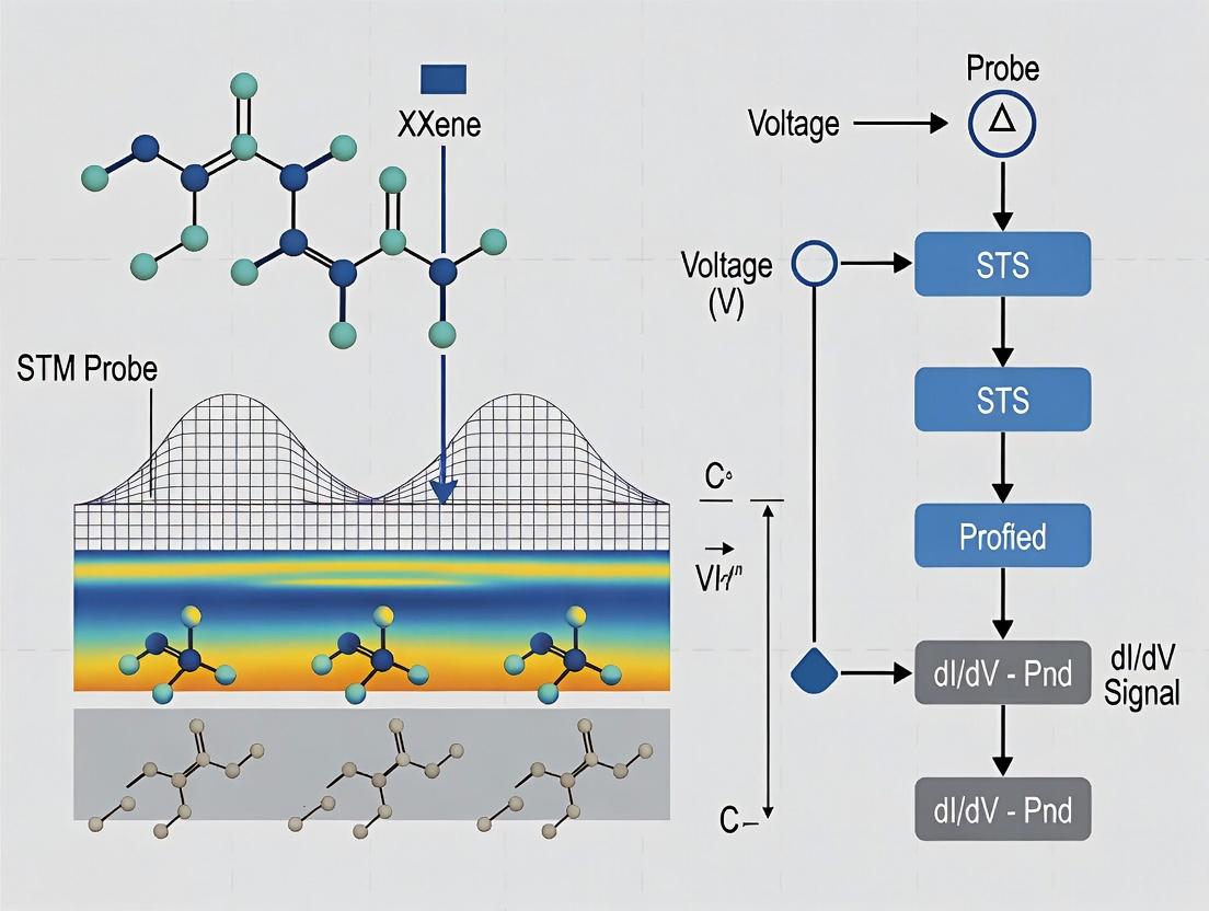
Within the broader thesis on "MXene surface chemical mapping with STM/STS research," Scanning Tunneling Spectroscopy (STS) is the critical technique for probing the local electronic density of states (LDOS) of MXene surfaces at the atomic scale. This application note details the protocols for performing point and grid spectroscopy (I-V and dI/dV), enabling the correlation of topographic features with electronic properties to map chemical heterogeneity, functional group distribution, and defect states on MXene sheets (e.g., Ti₃C₂Tₓ).
Fundamental Principles
STS measures the tunneling current (I) as a function of the sample bias voltage (V) at a fixed tip-sample separation. The first derivative (dI/dV) is approximately proportional to the LDOS of the sample. Two primary modes are used:
- Point Spectroscopy: I-V curves are acquired at discrete, user-selected points.
- Grid Spectroscopy (or Current Imaging Tunneling Spectroscopy - CITS): A full I-V curve is acquired at every pixel of a defined topographic scan grid, generating a 3D data cube (x, y, V).
Key Research Reagent Solutions & Materials
Table 1: Essential Materials and Reagents for STM/STS on MXenes
| Item | Function/Description |
|---|---|
| MXene Sample (e.g., Ti₃C₂Tₓ) | Primary substrate. Must be atomically flat, clean, and deposited on a conductive substrate (Au(111), HOPG). Tₓ denotes surface terminations (-O, -OH, -F). |
| Conductive Substrate (HOPG, Au(111) on mica) | Provides a flat, clean, and electrically conducting support for MXene flakes. |
| Electrochemically Etched STM Tip (Pt/Ir or W wire) | Scanning probe. Must be sharp and stable. Pt/Ir (80/20) is common for stability; W tips require in-situ cleaning (electron bombardment/field emission). |
| Inert Atmosphere Glovebox (Ar/N₂) | For sample preparation and loading to prevent oxidation of air-sensitive MXenes. |
| Ultrasonic Dispersion Solvent (Ethanol, IPA) | For dispersing MXene flakes onto the substrate. |
| Vibration Isolation System | Active or passive air/table system to achieve sub-angstrom stability. |
| Ultra-High Vacuum (UHV) System | Ideal: Provides pristine, contamination-free surfaces for fundamental studies. |
| Lock-in Amplifier (for dI/dV) | Essential for modulating the bias voltage and measuring the differential conductance (dI/dV) with high signal-to-noise ratio. |
Experimental Protocols
Protocol 4.1: Sample Preparation (MXene on HOPG)
- MXene Dispersion: In an Ar-glovebox, dilute few-layer MXene flakes (≈0.1 mg/mL) in degassed, anhydrous isopropanol.
- Substrate Preparation: Cleave HOPG substrate using adhesive tape to expose a fresh, atomically flat surface.
- Deposition: Drop-cast 5-10 µL of the MXene dispersion onto the HOPG surface.
- Drying: Allow the solvent to evaporate completely under an inert atmosphere.
- Transfer: Quickly transfer the sample to the STM sample holder. For UHV studies, transfer via a load-lock system.
Protocol 4.2: STM Tip Preparation (Pt/Ir)
- Mechanical Cutting: Use sharp, clean wire cutters to cut a 0.25mm Pt/Ir (80/20) wire at an angle.
- Electrochemical Etching (Optional): Immerse tip in a solution of CaCl₂/H₂O or NaCl/H₂O. Apply AC voltage (5-10 VAC) until the lower part drops off. Rinse with pure water and ethanol.
- In-situ Cleaning: Once in the STM, perform controlled voltage pulses (2-5 V) and gentle crashes into the substrate to stabilize the tip apex.
Protocol 4.3: Point Spectroscopy (I-V & dI/dV)
- Stable Imaging: Obtain a stable, atomically resolved constant-current topographic image of the MXene surface. Set your tunneling parameters (e.g., Vbias = 0.5 V, Iset = 100 pA).
- Point Selection: Select specific points of interest (e.g., on a basal plane, near a defect, or at an edge).
- Feedback Disable: Position the tip and disable the feedback loop to maintain a fixed tip-sample separation.
- Voltage Ramp & Data Acquisition:
- For I-V: Ramp the bias voltage over a predefined range (e.g., -1.5 V to +1.5 V) with a set number of points (512). Measure the tunneling current at each voltage step.
- For dI/dV: Superimpose a small AC modulation on the bias ramp (V_mod = 5-20 mV, f = 400-900 Hz). Use a lock-in amplifier to measure the in-phase component of the current response, which is dI/dV.
- Feedback Enable: Re-engage the feedback loop after the sweep.
- Repeat: Perform steps 2-5 for multiple points to build statistics.
Protocol 4.4: Grid Spectroscopy (CITS)
- Define Scan Area: Set the scan size (e.g., 10 nm x 10 nm) and pixel resolution (e.g., 128x128).
- Define Spectroscopy Parameters: Set the bias voltage sweep range and points (e.g., -1V to +1V, 100 points).
- Acquisition:
- At the first pixel, perform Protocol 4.3 steps 3-4.
- Move to the next pixel and repeat. The STM software automates this process, acquiring a full I-V/dI/dV curve at every pixel.
- Data Structure: The result is a 3D data cube: I(x, y, V) or dI/dV(x, y, V).
Data Presentation & Analysis
Table 2: Representative Quantitative STS Data from MXene (Ti₃C₂O₂)
| Measurement Type | Location on MXene | Bias Voltage at Peak (V) | Corresponding Feature in LDOS | Interpretation |
|---|---|---|---|---|
| dI/dV Point Spectra | Basal Plane (Terminated) | -0.8, +0.5 | Valence band edge, Conduction band edge | Band gap estimation (~1.3 eV). |
| dI/dV Point Spectra | Near -OH defect | +0.2 | In-gap state | Defect-induced state from functional group. |
| I-V Grid (CITS) | At metal adatom | -0.5 | Sharp resonance | Localized charge from adsorbate. |
| Averaged I-V | Over 5nm x 5nm area | N/A | Fitted barrier height Φ | Work function variation (Φ ≈ 4.2 eV). |
Analysis Steps:
- dI/dV Normalization: Normalize dI/dV curves by (I/V) to reduce the exponential tunneling background.
- Grid Data Extraction: From CITS data, extract constant-bias maps (dI/dV at a specific V) to visualize spatial distribution of specific electronic states.
- Band Gap Determination: Identify the bias range where dI/dV ~ 0 in point spectra on insulating areas.
Visualization Diagrams
Diagram 1: STS Experimental Workflow Decision Tree
Diagram 2: STS Setup for MXene Surface Electronic Mapping
Application Notes
This document details the application of scanning tunneling spectroscopy (STS) grid techniques for spatially resolved mapping of chemical and electronic heterogeneities on MXene surfaces. Framed within a broader thesis on MXene surface characterization, these methods are critical for researchers in nanoelectronics, catalysis, and energy storage, where surface termination and local work function directly influence performance metrics like conductivity, catalytic activity, and biocompatibility.
Core Principles: Differential conductance (dI/dV) spectra, acquired via grid-based STS at each pixel of a topographic scan, contain signatures of the local density of states (LDOS). Variations in spectral features—such as the energy position of peaks, dips, or the onset of conductance—correlate with local chemical identity (via elemental-specific electronic states) and work function (via shifts in the Fermi level alignment or vacuum level). Statistical clustering of spectral shapes allows for the deconvolution of mixed surface terminations common to MXenes (e.g., -O, -OH, -F).
Current Research Context: Recent advancements (2023-2024) in automated, high-speed grid STS have enabled the acquisition of dense spectroscopic grids (>10^4 spectra) on air-sensitive MXenes transferred under inert conditions. Coupled with machine learning (ML) analysis, particularly unsupervised clustering (e.g., k-means, hierarchical clustering) and principal component analysis (PCA), this allows for the generation of quantitative "chemical maps" where each cluster is assigned to a specific surface termination or defect state. Concurrently, the local work function (Φ) is extracted by fitting the logarithmic derivative of the I-V curve or by measuring the onset of field emission resonances in higher bias spectra.
Key Applications:
- Drug Development & Biocompatibility: Mapping -OH/-F termination ratios on Ti₃C₂ MXenes correlates with protein adsorption sites and cytotoxic responses.
- Catalysis: Identifying metallic vs. semiconducting domains on Mo-based MXenes to optimize hydrogen evolution reaction (HER) active sites.
- Nanoelectronics: Correlating work function variations with Schottky barrier heights at MXene/metal or MXene/semiconductor interfaces.
Experimental Protocols
Protocol 1: Inert Transfer and Sample Preparation for MXene STS
Objective: To prepare a pristine, atomically clean MXene surface for ultra-high vacuum (UHV) STM/STS analysis. Materials: See "Research Reagent Solutions" table. Procedure:
- MXene Synthesis: Synthesize Ti₃C₂Tₓ MXene via minimally intensive layer delamination (MILD) method from Ti₃AlC₂ MAX phase using LiF/HCl etchant.
- Film Deposition: Filter the colloidal solution through an anodic aluminum oxide (AAO) membrane to form a freestanding film. Dry under vacuum at 60°C for 12 hours.
- Inert Transfer: a. Load the MXene film into a glovebox-integrated vacuum transfer suitcase (Ar atmosphere, H₂O & O₂ < 0.1 ppm). b. Attach the suitcase to the load-lock of the UHV-STM system. c. Execute a staged pump-down procedure over 6 hours to UHV (<5×10⁻¹⁰ mbar). d. Transfer the sample onto the STM sample plate using a magnetic transfer rod without breaking vacuum.
- In-situ Cleaning (Optional): For studies requiring removal of adventitious carbon, perform gentle annealing at 250-300°C for 15-30 minutes via direct current heating. Higher temperatures may alter surface termination.
Protocol 2: Acquisition of a Spectroscopic Grid (dI/dV Grid)
Objective: To collect a spatially resolved array of I-V or dI/dV spectra. Materials: UHV-STM with lock-in amplifier and grid spectroscopy capability, PtIr or W tip. Procedure:
- Tip Preparation: Prepare a clean STM tip by electrochemical etching and subsequent in-situ cleaning via electron bombardment or field emission on a clean metal substrate (Au(111) or Cu(111)).
- Topography: Engage the tip on the MXene surface. Acquire a constant-current topographic image (e.g., 256×256 pixels) at parameters: Vbias = 0.5 V, Iset = 50 pA.
- Grid Definition: Overlay a spectroscopic grid on the topographic image. A typical grid for heterogeneity mapping is 64×64 spectra over the same area.
- Spectrum Acquisition Parameters:
- Setpoint stabilization: At each grid point, stabilize the tip at Vset = 0.5 V, Iset = 200 pA.
- Bias sweep range: -2.0 V to +2.0 V.
- Sweep points: 200 per spectrum.
- Lock-in modulation: For dI/dV, use a sinusoidal modulation V_mod = 10-20 mV (rms) at frequency f = 731 Hz.
- Acquire both I-V and synchronous dI/dV(V) signals.
- Acquisition: Execute automated grid acquisition. Total time is ~2-3 hours for a 64×64 grid.
Protocol 3: Data Processing and Map Generation
Objective: To convert raw spectroscopic grids into chemical and work function maps. Materials: Analysis software (e.g., WSxM, MATLAB, Python with scikit-learn). Procedure:
- Pre-processing: Flatten each dI/dV spectrum by subtracting a polynomial background. Normalize spectra to the conductance at a high bias (e.g., at +1.5 V) to account for tip-sample distance variations.
- Dimensionality Reduction & Clustering: a. Reshape the 3D data cube (x, y, energy) into a 2D matrix (pixel index, energy). b. Apply PCA to reduce noise and identify primary spectral components. c. Perform k-means clustering (k=3-5) on the first 5-10 principal component scores. d. Assign each pixel to a cluster label. This label map is the chemical probability map.
- Work Function Extraction: a. For each I-V curve, calculate the logarithmic derivative: L(V) = (d(ln I)/d(ln V)) ≈ (V/I)(dI/dV). b. Fit the linear region of L(V) near zero bias. The x-intercept of this fit yields the local barrier height ϕ, related to the average work function: ϕ ≈ (Φtip + Φsample)/2. c. If the tip work function (Φtip) is calibrated on a known metal (e.g., Au(111), Φ = 5.31 eV), solve for the local sample work function Φsample(x,y). d. Plot Φ_sample(x,y) as a work function map.
- Correlation: Overlay the work function map with the chemical map to identify structure-property relationships (e.g., -O terminations correspond to higher work function regions).
Data Tables
Table 1: Quantitative STS Signatures for Common Ti₃C₂Tₓ Terminations
| Surface Termination (-Tₓ) | Characteristic dI/dV Peak Positions (vs. Fermi Level) | Extracted Work Function Range (eV) | Assigned Electronic State |
|---|---|---|---|
| -O | Sharp peak at -0.8 eV; low conductivity near E_F | 5.2 - 5.6 | Titanium 3d- Oxygen 2p |
| -OH | Broad feature at -1.5 eV; moderate conductivity at E_F | 4.6 - 4.9 | Ti 3d - OH hybridized |
| -F | Featureless gap-like spectrum up to ±1.5 V | 4.3 - 4.7 | Insulating, large band gap |
| Bare Ti (defect) | High, constant density of states at E_F (metallic) | 4.0 - 4.3 | Ti 3d |
Table 2: Typical Parameters for STS Grid Acquisition on MXenes
| Parameter | Value / Range | Purpose / Rationale |
|---|---|---|
| Grid Size (pixels) | 32x32 to 128x128 | Balances spatial resolution and acquisition time. |
| Stabilization Setpoint (I) | 100 - 500 pA | Ensures stable tip-sample distance before sweep. |
| Bias Sweep Range | ±1.5 V to ±3.0 V | Captures relevant empty & filled states. |
| Modulation Voltage (V_mod) | 10 - 30 mV (rms) | Optimizes dI/dV signal-to-noise without broadening. |
| Lock-in Frequency (f) | 300 Hz - 1 kHz | Avoids 1/f noise and main interference (50/60 Hz). |
| Points per Spectrum | 100 - 256 | Determines energy resolution of spectral features. |
Diagrams
STS Grid Data Processing Workflow
The Scientist's Toolkit
Table 3: Key Research Reagent Solutions & Materials
| Item / Reagent | Function / Role in Experiment |
|---|---|
| Ti₃AlC₂ MAX Phase Precursor | Starting material for MXene synthesis. |
| Lithium Fluoride (LiF) / Hydrochloric Acid (HCl) Etchant | Selective etching of Al layer from MAX to produce multilayer MXene. |
| Anodic Aluminum Oxide (AAO) Membrane | Substrate for vacuum-assisted filtration to create freestanding MXene films. |
| Argon-filled Glovebox (H₂O/O₂ < 0.1 ppm) | Environment for handling air-sensitive MXene films prior to UHV transfer. |
| UHV-Compatible Transfer Suitcase | Enables vacuum-sealed sample transfer from glovebox to STM without air exposure. |
| Pt₀.₈Ir₀.₂ or Etched W STM Tip | Conducting probe for tunneling current and spectroscopy. |
| Lock-in Amplifier | Extracts the small dI/dV signal by detecting the current response at the modulation frequency. |
| UHV-STM System (Base Pressure < 1e-10 mbar) | Provides vibration-isolated, atomically clean environment for stable spectroscopy. |
| MATLAB or Python (scikit-learn, NumPy) | Software for multivariate analysis, clustering, and map generation from STS grids. |
This document provides application notes and protocols for interpreting Scanning Tunneling Spectroscopy (STS) data within the broader context of a thesis on MXene surface chemical mapping with STM/STS. The focus is on identifying terminal functional groups (e.g., -O, -OH, -F) and analyzing defects on MXene surfaces, which are critical for applications in catalysis, energy storage, and sensors.
Fundamentals of STS on MXenes
STS measures the differential conductance (dI/dV) as a function of sample bias, providing a local density of states (LDOS). For MXenes (e.g., Ti₃C₂Tₓ), the position and shape of spectral features are directly influenced by surface terminations (Tₓ) and defect states.
Key Spectral Signatures & Quantitative Data
The table below summarizes characteristic STS spectral features associated with common MXene terminations and defects.
Table 1: STS Spectral Signatures for MXene Surface Features
| Surface Feature | Typical Bias Range (V) | Spectral Signature (dI/dV peak) | Proposed Assignment | Notes on Line Shape |
|---|---|---|---|---|
| Hydroxyl Group (-OH) | -0.8 to -0.4 | Prominent peak in occupied states | Surface state near valence band maximum | Broad, asymmetric |
| Oxygen Group (-O) | -1.2 to -0.8 | Sharp peak in occupied states | Hybridized Ti-d / O-p state | Narrow, intense |
| Fluorine Group (-F) | +1.5 to +2.5 | Peak in unoccupied states | σ* resonance | Broad |
| Metal Vacancy Defect | -0.5 to +0.5 | Peak at/near Fermi level (E_F) | Localized defect state | Very sharp, high intensity |
| Carbon Vacancy Defect | +0.8 to +1.5 | Peak in unoccupied states | Ti-d orbital localized state | Moderate width |
| Oxidized Region | -1.5 & +1.5 | Multiple peaks, both biases | Band gap states from TiO₂ formation | Complex, multiple features |
| Pristine MXene Region | ~ -0.3, ~ +0.4 | Characteristic double peak | Density of states minima (pseudogap) | Defines intrinsic electronic structure |
Experimental Protocols
Protocol 4.1: Sample Preparation for MXene STM/STS
Objective: Prepare an atomically clean, termination-rich MXene surface for reliable STS mapping.
- Synthesis & Delamination: Synthesize Ti₃C₂Tₓ MXene via Min etching of Ti₃AlC₂ MAX phase, followed by delamination using LiCl or TMAOH.
- Substrate Preparation: Flame-anneal a Au(111) or highly ordered pyrolytic graphite (HOPG) substrate in argon atmosphere.
- Film Deposition: Drop-cast a dilute MXene suspension (0.1 mg/mL in deionized water) onto the substrate. Alternatively, use electrophoretic deposition for better control.
- Drying & Transfer: Dry the sample under a high-purity argon flow for >12 hours. Transfer to the STM load-lock immediately to minimize atmospheric contamination.
- In-Situ Cleaning (Optional): If chamber permits, perform mild annealing (<200°C) under ultra-high vacuum (UHV, <1×10⁻⁹ mbar) to remove physisorbed water.
Protocol 4.2: Acquisition of STS Point Spectra & Maps
Objective: Collect spatially resolved dI/dV spectra to correlate electronic structure with surface features.
- STM Setup: Load sample into UHV-STM system. Use a PtIr or etched W tip. Condition the tip by applying high voltage pulses and scanning over clean metal surfaces until stable feedback is achieved.
- Topography Imaging: Image the MXene flake at constant current mode (typical parameters: Vbias = 0.5 V, Iset = 50 pA). Identify regions of interest (terraces, edges, apparent defects).
- STS Parameter Setting: Halt the scan at a desired pixel (X,Y). Set the feedback loop off with a delay time (typically 1-5 ms).
- Sweep Bias: Ramp the sample bias voltage over a predetermined range (e.g., -2.5 V to +2.5 V) while measuring the tunneling current (I). The lock-in amplifier technique is standard: superimpose a small AC modulation (V_mod = 10-30 mV, f = 731 Hz) on the DC bias and measure the dI/dV signal directly.
- Data Point Collection: Record the (V_bias, dI/dV) pair at each step. Average over multiple sweeps (n≥50) per point to enhance signal-to-noise ratio.
- Spectral Mapping: Define a grid (e.g., 64x64 points) over the area of interest. Automatically execute Protocol 4.2 steps 3-5 at each grid point.
- Calibration: Record a reference spectrum on a clean metal substrate (e.g., Au(111)) to confirm tip integrity (should show the well-known surface state).
Protocol 4.3: Data Processing & Peak Assignment
Objective: Transform raw STS data into normalized LDOS for quantitative comparison and assignment.
- Averaging & Smoothing: Average all repeated sweeps for a single point. Apply a Savitzky-Golay filter to reduce high-frequency noise without distorting peak shapes.
- Normalization: Normalize each dI/dV spectrum by dividing by the (I/V) value at a high bias (e.g., at V = 2.0 V) to account for variations in tip-sample distance. This yields a quantity proportional to LDOS.
- Baseline Subtraction: Fit a polynomial to the background of the normalized spectrum and subtract it to flatten the baseline.
- Peak Identification: Use a second-derivative (d²I/dV²) method or a peak-finding algorithm (e.g., find_peaks in Python's SciPy) to locate local maxima/minima in the normalized dI/dV curve.
- Spatial Correlation: Overlay the position of identified peaks (e.g., -OH peak at -0.6V) onto the STM topography map to create chemical maps.
Visualization of Analysis Workflow
Diagram 1: STS Data Acquisition & Processing Workflow
Title: STS Analysis Workflow from Sample to Insight
Diagram 2: Spectral Signature Assignment Logic
Title: Logic Tree for Assigning STS Peaks
The Scientist's Toolkit: Key Research Reagent Solutions
Table 2: Essential Materials & Reagents for MXene STM/STS Studies
| Item | Function/Benefit | Critical Specification/Note |
|---|---|---|
| Ti₃AlC₂ MAX Phase Powder | Precursor for MXene synthesis. | Purity >98%, particle size <40 µm. |
| Lithium Fluoride (LiF) | Etching agent in Min etchant. | Anhydrous, 99.99% purity to control F-terminations. |
| Hydrochloric Acid (HCl) | Provides acidic environment for etching. | Concentrated, trace metal grade to avoid impurities. |
| Tetramethylammonium Hydroxide (TMAOH) | Delaminating intercalant. | 25% solution in water, for large flake production. |
| Deionized (DI) Water | Washing and suspension medium. | Resistivity >18.2 MΩ·cm (Milli-Q grade). |
| Argon Gas | Inert atmosphere for sample storage/transfer. | Ultra-high purity (99.999%) with oxygen getter. |
| Au(111) or HOPG Substrate | Atomically flat substrate for STM. | Freshly cleaved immediately before MXene deposition. |
| PtIr or Tungsten STM Tip | Probe for tunneling current. | Etched and in-situ cleaned for stable spectroscopy. |
| Lock-in Amplifier | Enables direct dI/dV measurement. | Essential for detecting small AC signals superimposed on DC bias. |
Solving Common STM/STS Challenges: Artifacts, Contamination, and Data Interpretation Pitfalls
Within the broader thesis on MXene surface chemical mapping using Scanning Tunneling Microscopy and Spectroscopy (STM/STS), achieving atomic-scale resolution and reliable spectroscopic data is paramount. MXenes, a class of two-dimensional transition metal carbides/nitrides, present unique surfaces terminated with functional groups (-O, -OH, -F) that dictate their electronic properties and chemical reactivity. Accurate STM/STS mapping of these heterogeneous surfaces is critical for applications in energy storage, catalysis, and sensing. However, this fidelity is routinely compromised by prevalent imaging artifacts: tip contamination, multiple tips, and thermal/mechanical drift. This application note details the identification and mitigation of these artifacts, providing protocols to ensure robust data acquisition for MXene surface analysis.
Artifact Identification & Quantitative Impact
Table 1: Common STM Artifacts in MXene Research
| Artifact | Key Identifying Features in STM/STS | Typical Impact on MXene Surface Data | Common Causes |
|---|---|---|---|
| Tip Contamination | Blurred or "ghost" images, sudden instability in tunneling current (It), loss of atomic resolution, step edges appearing smeared. In STS, noisy or non-reproducible I-V/dI/dV spectra. | Inaccurate topographic height measurement of functional groups. Misassignment of electronic states from noisy STS. | MXene flake debris/adherents, adsorbates from ambient transfer (hydrocarbons, water), residual surface contaminants. |
| Multiple Tips | Appearance of "double" or overlapping features, periodic duplication of surface defects at fixed spacing, inconsistent corrugation amplitudes. | False identification of surface periodicities or defect densities. Incorrect lateral measurement of MXene terraces and terminations. | Damaged or poorly etched tip apex, tip crashing into surface, picking up multiple asperities. |
| Drift (Thermal/Mechanical) | Continuous, time-dependent distortion of image features (stretching, compression, rotation). Non-closure of scan lines. In STS maps, spatial misregistration between spectroscopy points and topographic features. | Prevents reliable correlation of STS spectra with specific surface sites (e.g., -O vs. -OH regions). Distorts real-space dimensions of MXene domains. | Temperature fluctuations (>0.1°C), mechanical relaxation of scanner tube, acoustic or floor vibrations. |
Experimental Protocols for Artifact Mitigation
Protocol 3.1: In-situ Tip Conditioning and Cleaning for MXene Surfaces
Objective: To restore a single, atomically sharp, and clean tip apex for reliable MXene imaging. Materials: STM with tip conditioning capabilities (voltage pulse, heating), electrochemically etched W or PtIr tip, atomically flat reference sample (Au(111), HOPG). Procedure:
- Initial Approach: Engage the tip on a clean area of the MXene sample under standard imaging conditions (e.g., Vbias = 0.5 V, It = 100 pA).
- Diagnosis: If imaging is unstable or blurred, retract the tip by 1 µm.
- Voltage Pulsing: Apply a series of short-duration (1-10 ms) voltage pulses (Vpulse = +3V to +10V) between tip and sample. For MXenes, start at lower voltages to avoid surface modification.
- Field Emission/Heating: If pulsing is insufficient, use the field emission protocol by positioning the tip 1 µm from the surface and applying +150V for 10-30 seconds. Alternatively, use a tip heater if available.
- Recalibration: Re-approach on the reference sample (Au(111)) to confirm atomic resolution. Image the reference lattice to verify tip symmetry and sharpness.
- Return to MXene: Once a stable tip is confirmed on the reference, re-approach the MXene sample for imaging.
Protocol 3.2: Distinguishing True MXene Features from Multiple-Tip Artifacts
Objective: To confirm that observed surface structures are intrinsic to the MXene sample and not an artifact of the tip. Materials: STM, MXene sample (e.g., Ti3C2Tx), reference atomic lattice. Procedure:
- Variable-Scan Imaging: Acquire images of the same MXene region at different scan sizes (e.g., 50 nm x 50 nm, then 20 nm x 20 nm). A true multiple-tip artifact will maintain a constant offset between duplicated features regardless of scan size or angle.
- Scan Angle Rotation: Rotate the scan direction by 90°. If the duplicated features rotate with the scan, they are likely real. If their orientation relative to the scan frame changes, it suggests multiple tips.
- Spectroscopic Verification: Perform point STS on a suspected duplicated feature and its "original." True surface features (like a metal adatom or functional group cluster) will have distinct electronic signatures, while a multiple-tip artifact will show identical spectra.
- Tip Reformation: If multiple tips are confirmed, perform tip conditioning (Protocol 3.1).
Protocol 3.3: Drift Compensation for Long-Duration STS Mapping on MXenes
Objective: To spatially lock STS measurement points to specific surface chemistries over extended periods. Materials: STM with software-based drift compensation, MXene sample with identifiable fiducial markers (e.g., inherent step edges, defects). Procedure:
- Initial Topography: Obtain a high-resolution STM image of the MXene area of interest. Identify stable, sharp topographic landmarks (LMs) at the image corners.
- Set Measurement Grid: Define the matrix of (x,y) points for STS (dI/dV spectroscopy) based on this initial image.
- Implement Linear Drift Correction: a. Periodically (e.g., every 5-10 spectra), interrupt spectroscopy to quickly re-image the landmarks. b. Calculate the lateral drift vector (Δx, Δy) by measuring the displacement of the LMs. c. Apply a linear correction to all subsequent spectroscopic coordinates in the measurement queue.
- Post-Processing Correlation: After data acquisition, use the recorded drift vectors to reconstruct the true physical location of each STS spectrum relative to the final topographic image.
Visualization of Workflows
Title: STM Artifact Diagnosis & Mitigation Decision Tree
Title: Drift-Compensated STS Mapping Workflow
The Scientist's Toolkit: Key Research Reagent Solutions
Table 2: Essential Materials for Artifact-Free MXene STM/STS
| Item | Function & Relevance to MXene Studies |
|---|---|
| Electrochemically Etched Tungsten (W) Tips | Standard STM tip material. Requires in-situ cleaning. Good for most MXenes, but hard material may displace surface groups if crashed. |
| Platinum-Iridium (Pt90Ir10) Tips | More resistant to oxidation, mechanically robust. Less prone to forming multiple asperities. Ideal for initial survey scans of MXenes. |
| Atomically Flat Reference Samples (Au(111), HOPG) | Critical for tip quality verification post-conditioning. Provides a known lattice constant to calibrate scanner and confirm atomic resolution. |
| In-situ Tip Heater/Flash Unit | Applies high temperature to the tip shank to desorb contaminants. More controlled than high-voltage pulsing for delicate MXene surfaces. |
| Vibration Isolation System | Passive air tables or active isolation platforms minimize mechanical drift and noise, essential for stable imaging of atomic-scale MXene corrugation. |
| Ultra-High Vacuum (UHV) Preparation Chamber | Allows for in-situ MXene cleavage, heating (to ~200°C), or argon sputtering/annealing to remove surface adsorbates before STM analysis. |
| Low-Temperature STM (LT-STM) | Operates at 4.5K or 77K. Dramatically reduces thermal drift and suppresses vibrational noise, enabling definitive spectroscopy of MXene electronic states. |
1. Introduction & Thesis Context Within a broader thesis on atomic-scale surface chemical mapping of MXenes using Scanning Tunneling Microscopy and Spectroscopy (STM/STS), the paramount challenge is the rapid environmental degradation of the MXene surface. Oxidation and adsorbate contamination under ambient conditions introduce spurious electronic states, alter local density of states (LDOS), and render the surface morphologically unstable under the STM tip. These artifacts severely compromise the fidelity of chemical and electronic property mapping. These protocols outline a holistic strategy for fabricating, transferring, and maintaining pristine MXene surfaces for reproducible, high-resolution STM/STS research.
2. Quantitative Data Summary: Degradation Factors & Mitigation Efficacy
Table 1: Primary Degradation Pathways for MXenes (Ti₃C₂Tₓ) and Corresponding Mitigation Strategies
| Degradation Factor | Key Environmental Trigger | Observed Effect on STM/STS | Mitigation Protocol |
|---|---|---|---|
| Oxidation | H₂O, O₂ (Ambient) | Loss of metallic conductivity, introduction of oxide-based band gap in STS, surface roughening. | Synthesis/Storage in Ar glovebox (<0.1 ppm O₂/H₂O). |
| Intercalation | H₂O, cations | Layer expansion, fluctuation in tunnel current, altered stacking order. | Use of aprotic solvents (e.g., PC), electrochemical control. |
| Adsorbate Contamination | Organics, CO₂, H₂O | Non-periodic features in STM, masking intrinsic surface terminations. | In-situ cleaving, UHV annealing (<300°C). |
| Tip-Induced Degradation | High bias voltage, current | Drift, sudden changes in topography, irreversible surface modification. | Use of low bias (<100 mV) for imaging, current-limited spectroscopy. |
Table 2: Comparative Stability of MXene Surfaces Under Different Environmental Controls
| Surface Preparation Method | Approximate Time to Observable Degradation (STM) | Key STS Artifact Observed | Recommended for Chemical Mapping? |
|---|---|---|---|
| Ambient-exposed flake | Minutes | Disappearing atomic features, high noise. | No. |
| Glovebox-loaded, transferred via sealed vessel | 1-2 hours | Gradual broadening of LDOS peaks. | Limited, for initial surveys. |
| In-situ cleaved in UHV | >8 hours | Stable dI/dV spectra over repeated scans. | Yes, optimal. |
| Cryogenic temperatures (80K) in UHV | >24 hours | Suppressed adsorbate mobility, pristine spectra. | Yes, for highest resolution. |
3. Detailed Experimental Protocols
Protocol 3.1: Synthesis and Storage of STM-Grade MXene (Ti₃C₂Tₓ) Objective: To produce few-layer MXene flakes with minimal initial oxidation for surface science studies. Materials: Ti₃AlC₂ MAX phase powder, Lithium Fluoride (LiF) powder, 9 M Hydrochloric Acid (HCl), N₂-sparged Deionized Water, Dimethyl Sulfoxide (DMSO), Argon glovebox (O₂/H₂O < 0.1 ppm). Steps:
- Etching: Slowly add 1 g LiF to 20 mL of 9 M HCl under stirring in a polypropylene vial. Once dissolved, gradually add 1 g Ti₃AlC₂ powder. Etch at 35°C for 24 hours under continuous stirring.
- Washing: Transfer the slurry to glovebox immediately. Wash the sediment 5-7 times with N₂-sparged DI water via centrifugation (3500 rpm, 5 min) until supernatant pH > 6.
- Delamination: Resuspend the sediment in 50 mL DMSO. Shake for 6 hours.
- Final Wash & Storage: Centrifuge the DMSO mixture. Resuspend the pellet in anhydrous, degassed propylene carbonate (PC) inside the glovebox. Store the colloidal solution in a sealed, Teflon-lined vial. Do not use aqueous suspensions.
Protocol 3.2: In-Situ Preparation of Pristine MXene Surface in UHV-STM Objective: To prepare an atomically clean, stable MXene surface for chemical mapping. Materials: Au(111) or HOPG substrate, UHV system (base pressure < 5×10⁻¹¹ mbar), fast-entry load-lock, in-situ cleaver or annealer, home-built droplet applicator. Steps:
- Substrate Preparation: Clean Au(111) substrate via repeated Ar⁺ sputter (1 keV, 15 min) and annealing (450°C, 30 min) cycles in UHV. Confirm cleanliness with STM.
- Transfer & Deposition (Glovebox): Apply a 2 µL droplet of the MXene/PC suspension onto the substrate inside an Ar glovebox. Let it dry for 60 seconds before rinsing gently with 100 µL pure PC to remove excess salt. Dry completely.
- Sealed Transfer: Mount the sample on a compatible STM holder. Seal the holder in an Ar-filled, transparent bag or a dedicated transfer vessel.
- In-Situ Treatment (UHV): Quickly transfer the holder via load-lock into the UHV-STM preparation chamber. Anneal the sample at 200-250°C for 15 minutes to desorb residual PC and water. Avoid temperatures >300°C to prevent decomposition.
- Immediate Transfer: Move the sample to the pre-cooled STM stage (preferably 80K) without breaking vacuum. Begin scanning within 1 hour of annealing.
Protocol 3.3: Stable STM/STS Acquisition on MXenes Objective: To acquire spatially-correlated topography and LDOS maps without tip-induced degradation. STM Parameters: Set-point current: 10-50 pA. Imaging bias: < |100| mV. Use a chemically etched W tip, cleaned by electron bombardment and characterized on a clean metal surface. STS Acquisition:
- At each pixel in a grid, stop the feedback loop.
- Perform a current-voltage (I-V) sweep from -500 mV to +500 mV with a step size of 2-5 mV.
- Use lock-in detection (modulation frequency ~731 Hz, amplitude 5-10 mV rms) to acquire simultaneous dI/dV spectra.
- Restart feedback and move to the next pixel. Limit grid size (e.g., 64x64) to minimize total acquisition time.
- Store raw I-V and dI/dV data for each pixel.
4. Visualizations
Diagram 1: MXene Stability Control Workflow for STM.
Diagram 2: In-situ MXene Sample Prep Protocol & Key Parameters.
5. The Scientist's Toolkit: Key Research Reagent Solutions
Table 3: Essential Materials for MXene STM Surface Preparation
| Item | Function & Rationale |
|---|---|
| Anhydrous Propylene Carbonate (PC) | Aprotic, polar solvent for final MXene suspension. Prevents re-intercalation of water and cations, which cause swelling and degradation. |
| Argon Glovebox | Controlled inert atmosphere (O₂/H₂O < 0.1 ppm) for all post-etching steps: washing, storage, and substrate deposition. |
| Sealed Sample Transfer Vessel | Maintains inert atmosphere during physical transport from glovebox to UHV load-lock, preventing ambient exposure. |
| UHV-Compatible Annealer | For in-situ thermal treatment to desorb solvents and light adsorbates without causing MXene decomposition. |
| Cryogenic STM Stage (80K or lower) | Dramatically reduces surface diffusion of adsorbates and oxidation kinetics, extending stable measurement window to >24 hours. |
| Chemically Etched Tungsten Tips | Standard STM tips, cleaned in UHV, provide stable tunneling on MXenes. PtIr tips can be used but may be softer. |
| Au(111) on Mica Substrate | Provides an atomically flat, inert, and conductive surface for depositing MXene flakes. Easy to clean via sputter/anneal. |
Within a broader thesis on MXene surface chemical mapping using Scanning Tunneling Microscopy and Spectroscopy (STM/STS), optimizing the signal-to-noise ratio (SNR) is paramount. The chemical heterogeneity and rich surface termination landscape of MXenes (e.g., Ti₃C₂Tₓ) require high-fidelity differential conductance (dI/dV) spectra to correlate electronic structure with local chemistry. This application note details the critical interplay of lock-in amplifier parameters, spatial grid density, and spectral averaging to achieve the SNR necessary for reliable chemical mapping.
Core Principles & Parameter Optimization
Lock-in Amplifier Fundamentals for STS
In STS, a small, high-frequency AC voltage modulation (V_mod) is superimposed on the DC bias. The lock-in amplifier detects the corresponding AC component of the tunneling current at the modulation frequency, providing a direct measure of dI/dV. Key settings define the SNR and spatial/energy resolution.
Table 1: Lock-in Amplifier Parameters and Optimization Guidelines
| Parameter | Symbol | Typical Range for MXene STS | Effect on SNR & Resolution | Optimization Tip for MXenes |
|---|---|---|---|---|
| Modulation Frequency | f_mod | 0.5 - 3 kHz | Higher f reduces 1/f noise but risks cable capacitance effects. | Use 1-2 kHz to balance noise rejection and system bandwidth. Avoid harmonics of mains (50/60 Hz). |
| Modulation Amplitude | V_mod | 5 - 30 mV RMS | Larger amplitude increases signal but degrades energy resolution (ΔE ~ 2.5V_mod). | Start with 20 mV for survey spectra. Reduce to 10 mV or lower for resolving sharp features (e.g., van Hove singularities). |
| Time Constant | τ | 3 - 100 ms | Longer τ reduces noise bandwidth, increasing SNR but slowing acquisition. | Set τ ≥ 1/(π * f_mod) to integrate fully over one modulation cycle. Use 10-30 ms for point spectroscopy. |
| Roll-off Slope | n | 6 - 24 dB/oct | Steeper slopes provide better noise rejection. | Use 24 dB/oct for maximum noise suppression in typical lab environments. |
| Sensitivity (Current) | I_sens | 10 nA - 1 μA | Must be set to accommodate the AC current without overload. | Auto-range initially, then fix to a range where signal is >10% of full scale. |
Spatial Grid Density and Spectral Averaging
The choice between a dense spectroscopic grid for high-resolution mapping and the necessity of averaging per point is a fundamental trade-off.
Table 2: Grid Density vs. Averaging Strategy Trade-off
| Mapping Strategy | Grid Density (points/nm²) | Spectra per Point (N_avg) | Effective SNR Gain* | Best Use Case for MXene Research |
|---|---|---|---|---|
| High-Resolution Chemical Map | High (e.g., 64x64 over 50nm² = ~1.6 /nm²) | Low (1-5) | √N_avg | Identifying termination-dependent electronic heterogeneity at grain boundaries. |
| High-Fidelity Point Spectroscopy | Single point or coarse grid | High (50-200) | √N_avg | Precisely measuring local density of states (LDOS) on pristine terraces vs. defect sites. |
| Balanced Mapping | Moderate (32x32 over 50nm² = ~0.4 /nm²) | Moderate (10-20) | √N_avg | Correlating medium-scale topographic features with electronic structure. |
*SNR improves with the square root of the number of averaged spectra (N_avg).
Experimental Protocols
Protocol 1: Systematic Lock-in Parameter Calibration for MXene STS
Objective: To establish optimized lock-in settings for a specific MXene sample (e.g., Ti₃C₂Tₓ) and STM system. Materials: Freshly prepared MXene sample (transferred in glovebox), STM with lock-in capability, low-noise current preamplifier. Procedure:
- Initial Setup: Load sample into STM under inert atmosphere. Approach with standard W or PtIr tip. Set DC bias (V_bias) to -0.5 V, set-point current (I_set) to 100 pA.
- Frequency Selection: With V_mod = 20 mV, τ = 10 ms, test f_mod at 413 Hz, 831 Hz, 1.23 kHz, and 2.57 kHz. Acquire single dI/dV spectrum at a clean terrace region from -1V to +1V.
- Noise Assessment: Calculate RMS noise in a flat region of the spectrum (e.g., from 0.6 to 0.8 V). Select the f_mod yielding the lowest noise, typically the highest feasible frequency.
- Amplitude Optimization: At chosen f_mod, test V_mod at 5, 10, 20, and 30 mV. Acquire spectra on a known sharp feature (e.g., a step edge or defect). Determine the amplitude where the feature's full width at half maximum (FWHM) begins to broaden significantly (ΔE > 50 meV). Select the largest amplitude before significant broadening.
- Time Constant Setting: Set τ to be 3-5 times the period of the modulation frequency (e.g., for 1 kHz, period = 1 ms, so τ = 3-5 ms). Increase if noise persists, but note the increased acquisition time per point.
- Documentation: Record final parameters for all subsequent experiments on this sample system.
Protocol 2: Grid-Based STS Mapping with Adaptive Averaging
Objective: To acquire a spatially resolved dI/dV map balancing resolution, SNR, and acquisition time. Materials: As in Protocol 1. Procedure:
- Topographic Imaging: Acquire a constant-current STM image of the target area (e.g., 50 nm x 50 nm). Select a region of interest (ROI) containing features of chemical interest (terraces, terminations, defects).
- Pilot Spectroscopy: At 3-5 representative points within the ROI (terrace, edge, defect), acquire a high-quality reference spectrum using optimized lock-in settings and N_avg = 50.
- Noise Floor Determination: Calculate the RMS noise level (σ_signal) from a flat region of these pilot spectra.
- Define SNR Target & Averaging: For the map, define a minimum acceptable SNR (e.g., 10:1 for prominent features). Estimate the single-sweep signal amplitude (A_signal) from the pilot data. Calculate required averages: Navg ≥ (SNRtarget * σsignal / Asignal)².
- Set Grid Density: Based on the feature size and available time (Total Time ∝ Npoints * Navg * τ), choose the maximum grid density (e.g., 64x64, 128x128). If N_avg from step 4 is too high, reduce grid density to maintain feasible total acquisition time.
- Mapping Execution: Program the STM to perform point spectroscopy at each pixel in the grid. At each point, the tip stabilizes at (V_bias, I_set), the feedback is interrupted, and dI/dV is measured N_avg times (or via a single slow sweep with long τ). The average value is stored as the pixel intensity for each bias value.
- Data Cube Formation: The result is a 3D data cube: I(x, y, V). Generate constant-bias dI/dV maps or extract spectra from specific (x,y) locations.
Diagrams
Title: Lock-in Parameter Optimization Workflow
Title: SNR Optimization Trade-off Strategies
The Scientist's Toolkit
Table 3: Essential Research Reagent Solutions & Materials for MXene STS
| Item | Function in MXene STS Research |
|---|---|
| MXene Single/Few-Layer Flakes (Ti₃C₂Tₓ, Mo₂CTₓ) | The core material under study. Surface terminations (Tₓ = -O, -OH, -F, -Cl) dictate local electronic structure probed by STS. |
| Anhydrous, Oxygen-Free Solvents (e.g., Degassed Ethanol, Toluene) | Used for spin-coating or drop-casting MXene dispersions onto atomically flat substrates (Au(111), HOPG) under inert atmosphere to prevent oxidation. |
| Highly Oriented Pyrolytic Graphite (HOPG) or Au(111) on Mica Substrate | Provides an atomically flat, chemically inert, and conductive surface for depositing MXene flakes for STM/STS. |
| Electrochemically Etched Tungsten (W) or PtIr Tips | The scanning probe. W tips are often sharp and stable but may oxidize. PtIr tips are more chemically inert. Both require in-situ cleaning (e.g., electron bombardment, gentle collisions) for stable spectroscopy. |
| Ultra-High Purity Argon/Nitrogen Glovebox (H₂O, O₂ < 0.1 ppm) | Essential environment for sample preparation, transfer, and loading into the STM to prevent surface degradation of air-sensitive MXenes. |
| Lock-in Amplifier with Internal Oscillator & Low-Pass Filters | The core electronic component for performing STS. Extracts the small dI/dV signal from the noisy tunneling current using frequency-domain detection. |
| Low-Noise Current Preamplifier (Gain: 10^8 - 10^9 V/A) | Converts the minute tunneling current (pA-nA) into a measurable voltage before processing by the lock-in amplifier. Its noise floor limits ultimate SNR. |
| Vibration Isolation System (e.g., Pneumatic Table, Spring Suspension) | Critically dampens mechanical vibrations from the environment to achieve atomic-scale spatial resolution and stable tunneling junction. |
Within a broader thesis on MXene surface chemical mapping with Scanning Tunneling Microscopy and Spectroscopy (STM/STS), the integrity of the scanning probe tip is paramount. The atomic-scale electronic and topographic data of novel materials like MXenes are critically dependent on a well-defined tip apex. This protocol details the essential preparatory step of verifying and calibrating the STM tip state on a standard Au(111) surface prior to investigating complex, often reactive, MXene surfaces. A known tip state ensures that subsequent spectroscopic features (e.g., local density of states maps on MXenes) are intrinsic to the sample and not artifacts of tip asymmetry or contamination.
The Scientist's Toolkit: Research Reagent Solutions & Essential Materials
| Item | Function in Tip Calibration/Functionalization |
|---|---|
| Single-crystal Au(111) substrate | Standard calibration surface providing a known, atomically flat terrace structure with a characteristic herringbone reconstruction and surface state for electronic verification. |
| Electrochemical etching setup (Pt ring, KOH/NaOH) | For initial preparation of sharp tungsten (W) or platinum-iridium (PtIr) STM tips via controlled electrochemical dissolution. |
| UHV Chamber (Base pressure <1e-10 mbar) | Essential environment for maintaining atomically clean Au(111) and tip surfaces, preventing contamination during calibration. |
| In-situ tip treatment facilities (e.g., e-beam heater, ion sputter gun) | For cleaning and annealing the metal tip to remove oxides and adsorbed contaminants prior to calibration. |
| STM/STS Control Electronics | High-stability current amplifier and voltage sources for precise I-V and dI/dV spectroscopy during tip state verification. |
| Molecular beam epitaxy (MBE) source (optional) | For depositing controlled amounts of gold or other metals onto a contaminated tip to reform the apex via "tip coating." |
Experimental Protocol: Tip State Verification on Au(111)
Protocol 1: Au(111) Substrate Preparation
- Cleaning: Insert a single-crystal Au(111) substrate into the UHV chamber.
- Sputtering: Clean the surface with several cycles of Ar⁺ ion sputtering (Ar⁺ energy: 1-2 keV, sample current: 1-2 µA, duration: 15-30 minutes per cycle) at room temperature.
- Annealing: After each sputtering cycle, anneal the substrate at 450-500°C for 15-30 minutes to restore crystallinity and the characteristic herringbone reconstruction.
- Verification: Confirm surface quality with LEED (Sharp (1x1) pattern) and a preliminary large-scale STM scan to confirm large, atomically flat terraces separated by monatomic steps (height ~2.5 Å).
Protocol 2: STM Tip Preparation and Initial Approach
- Fabrication: Prepare a polycrystalline tungsten tip via electrochemical etching in 2M NaOH using a drop-off method. For PtIr tips, mechanically cut with fine wire cutters.
- In-situ Cleaning: Prior to use, clean the W tip in UHV via repeated cycles of high-temperature resistive flashing (by direct current heating) or electron bombardment heating until a stable field emission current is achieved. For PtIr, mild ion sputtering may be used.
- Approach: Using the optical microscope and coarse approach mechanism, bring the tip to within ~1 mm of the Au(111) surface. Use the automated coarse approach to engage until a tunneling current is detected (typical setpoint: 1 nA, bias: 0.5 V).
Protocol 3: Tip State Calibration and Verification
- Topographic Calibration:
- Acquire a constant-current topographic image over a 200 nm x 200 nm area (setpoint: 0.1 nA, sample bias: -0.5 V).
- Requirement: Verify the presence of the 22 x √3 herringbone reconstruction. Measure the step height between terraces.
- Acceptance Criterion: Step height must be 2.5 ± 0.2 Å. The reconstruction must be clearly resolved, indicating a stable, single-apex tip.
- Electronic (Spectroscopic) Calibration:
- On a clean terrace, acquire scanning tunneling spectroscopy (STS) point spectra.
- Method: Disable the feedback loop (z-fixed), sweep the sample bias from -1.0 V to +1.0 V, and record the I-V curve. Perform numerical differentiation or use a lock-in amplifier (modulation: 10-20 mV rms, frequency: ~1 kHz) to obtain the dI/dV spectrum.
- Acceptance Criterion: The dI/dV spectrum must clearly show the onset of the Au(111) surface state at approximately -0.5 eV relative to the Fermi level (see quantitative table).
- Tip Functionalization Check (Optional):
- If a specific tip functionalization (e.g., with a CO molecule for high-resolution imaging) is required for the subsequent MXene study, it should be performed at this stage. The functionalized tip must then be re-verified on Au(111) to confirm its modified but stable electronic response before transferring to the MXene sample.
Table 1: Key Topographic and Electronic Benchmarks for Tip Verification on Au(111)
| Parameter | Expected Value | Tolerance | Measurement Technique | Indication of Good Tip |
|---|---|---|---|---|
| Atomic Step Height | 2.5 Å | ± 0.2 Å | STM Topography (line profile) | Single, metallic apex. |
| Herringbone Periodicity | ~6.3 nm (along reconstruction) | ± 0.5 nm | STM Topography (FFT analysis) | Stable imaging without double tips. |
| Surface State Onset (dI/dV) | -0.48 eV to -0.52 eV | ± 0.03 eV | STS point spectroscopy | Clean, unperturbed electronic structure of tip. |
| Surface State Peak | ~ -0.02 eV (relative to EF) | ± 0.05 eV | STS point spectroscopy | Well-defined local density of states at tip apex. |
| Tunneling Current Noise (RMS) | < 0.5 pA | -- | I-t measurement at fixed bias/gap | Mechanically and electronically stable junction. |
Title: STM Tip Calibration and Verification Workflow on Au(111)
Title: Impact of Tip Calibration on MXene STM/STS Data Quality
This Application Note provides essential protocols for validating scanning tunneling microscopy/spectroscopy (STM/STS) data, specifically within a broader thesis investigating MXene surface chemistry and electronic structure. MXenes, a class of 2D transition metal carbides/nitrides, exhibit tunable surface terminations (-O, -OH, -F) that critically influence their electronic properties, such as local density of states (LDOS) and work function. A core challenge in high-resolution STM/STS mapping is the unambiguous differentiation of genuine electronic features (e.g., defect states, termination-dependent band edges, quantum confinement patterns) from ubiquitous measurement artifacts (e.g., tip changes, thermal drift, feedback loop oscillations, capacitive coupling). This document details systematic approaches for this validation, ensuring reliability in correlating MXene surface chemistry with electronic function for downstream applications, including in biosensing or catalytic platforms relevant to drug development.
Table 1: Key Distinguishing Characteristics
| Feature / Artifact | Typical Manifestation in STM/STS | Diagnostic Validation Test | Expected for MXene Surfaces |
|---|---|---|---|
| Genuine Surface State | Sharp peak in dI/dV spectra at fixed bias across spatial locations. | Spectrum reproducibility over multiple scans; correlation with atomic structure in STM topo. | Termination-dependent bandgap states near Fermi level (EF). |
| Tip Change Artifact | Sudden, global change in image resolution or spectral shape. | Acquire reference spectrum on known feature (e.g., Au(111)) before/after suspect measurement. | N/A - indicates tip instability. |
| Thermal Drift | Linear distortion or stretching of topographic images over time. | Perform bidirectional fast-scan analysis; measure feature displacement vs. time. | Can obscure true arrangement of surface terminations. |
| Feedback Loop Oscillation | Periodic corrugations or waves in topography, especially on flat terraces. | Adjust PI gains; increase time constant; compare images at different scan speeds. | Misinterpreted as moiré patterns or charge density waves. |
| Capacitive Coupling | Bias-dependent vertical offset in dI/dV spectra, mimicking a true LDOS shift. | Acquire I-V curves with different setpoint currents; use lock-in detection with proper shielding. | Can distort measurement of MXene work function or band bending. |
| Surface Contamination | Unstructured "blobs" or high tunneling barrier regions. | Perform in-situ cleaning (annealing, sputter-anneal cycle); check with Auger or XPS. | Hydrocarbon adsorbates on MXene can mimic functional groups. |
| Genuine Edge State | Enhanced conductance at step edges or termination boundaries. | Verify spatial localization to physical edge across multiple bias voltages. | Predicted for certain MXene nanoribbons or termination boundaries. |
Experimental Protocols for Data Validation
Protocol 3.1: Reproducible STS on MXene Flakes
Objective: To acquire statistically valid dI/dV spectra representing genuine LDOS. Materials: Freshly prepared/sputtered MXene (e.g., Ti3C2Tx) on conductive substrate, UHV STM/STS system. Procedure:
- Tip Preparation: Briefly indent tip into Au substrate or apply voltage pulses to achieve stable tunneling on Au(111). Acquire reference dI/dV spectrum on Au to confirm Shockley surface state presence.
- Locate Flake: Image large area to identify atomically flat terraces of MXene.
- Grid Spectroscopy: On a selected terrace, define a grid (e.g., 10x10 points). At each point, pause scanning, disable feedback loop, and acquire a dI/dV point spectrum using lock-in detection (typical parameters: Vmod = 10-20 mVrms, f = 400-900 Hz).
- Intra-grid Validation: Compute the standard deviation of the spectral intensity at each bias across all points. Genuine features show low spatial variance at their characteristic bias.
- Inter-scan Validation: Move to a new, non-overlapping terrace and repeat the grid. Compare average spectra from both grids. Genuine features persist.
Protocol 3.2: Drift Compensation and Topography Validation
Objective: To correct for thermal drift enabling accurate correlation of chemistry and electronic maps. Procedure:
- Fast Bidirectional Imaging: Acquire the same topographic line both left-to-right and right-to-left. The separation between features in the two directions quantifies the drift rate.
- Marker Point Tracking: Select a stable, isolated topographic protrusion as a marker. After each spectroscopic grid, return to this point and record its displacement.
- Software Correction: Use drift-corrected spatial coordinates for all spectroscopic data points. Discard data from scans where drift exceeds 10% of the scan size.
Protocol 3.3: Tip Artifact Diagnosis and Mitigation
Objective: To confirm spectroscopic features originate from the sample, not the tip. Procedure:
- Pre- and Post-Series Reference: Before and after a series of measurements on MXene, acquire dI/dV spectra on a known reference sample (e.g., Au(111)) under identical conditions.
- Symmetry Test: Acquire dI/dV spectra at both positive and negative sample bias on a featureless MXene terrace. Genuine LDOS features are not necessarily symmetric, but many tip-induced artifacts are.
- Setpoint Dependence Test: Acquire spectra on the same spot with different tunneling currents (e.g., 50 pA, 100 pA, 200 pA). Genuine LDOS features scale with tip-sample distance in a predictable way; abrupt changes suggest artifact.
Visualization of Workflows
Diagram 1: STM/STS Validation Decision Tree
Title: STM/STS Data Validation Decision Tree
Diagram 2: MXene Termination Mapping Workflow
Title: MXene Termination Mapping & Validation Workflow
The Scientist's Toolkit: Research Reagent Solutions
Table 2: Essential Materials for MXene STM/STS Validation
| Item | Function & Relevance to Validation |
|---|---|
| Ultra-High Vacuum (UHV) System (< 5x10⁻¹⁰ mbar) | Provides pristine surface stability, prevents oxidation and adsorbate contamination of MXene surfaces, which are critical for reproducible spectroscopy. |
| Electrochemically Etched Tungsten Tips | Standard STM probes. Must be in-situ cleaned (e.g., electron beam heating) to remove oxides for stable tunneling and to minimize tip artifact generation. |
| Single-Crystal Gold (Au(111)) Substrate | Essential reference sample. Its known surface state provides a critical benchmark for tip condition and spectroscopic calibration before/after MXene measurement. |
| Lock-in Amplifier | Enables sensitive dI/dV (conductance) detection by measuring the differential response to a small AC voltage modulation, separating signal from noise. |
| In-situ Argon Sputter Gun | For cleaning MXene surfaces by gentle removal of surface adsorbates and oxides, revealing the intrinsic termination chemistry. |
| Resistive Heating Sample Stage | Allows for in-situ thermal processing of MXenes to induce controlled changes in surface termination (e.g., dehydroxylation) for comparative studies. |
| Low-Temperature STM/STS System (4K/77K) | Drastically reduces thermal drift and broadens spectral resolution, making genuine electronic features (e.g., sharp defect states) more discernible from noise. |
| Multichannel Data Acquisition System | Simultaneously records topography, current, and dI/dV signals, enabling precise spatial correlation essential for validating feature localization. |
Benchmarking STM/STS Insights: Correlative Microscopy and Validation for Biomedical Relevance
This application note details a methodology for the cross-validated analysis of MXene surfaces, integrating Scanning Tunneling Spectroscopy (STS) electronic state measurements with X-ray Photoelectron Spectroscopy (XPS) chemical composition mapping. The protocol is designed to establish definitive correlations between localized density of states (LDOS) and surface terminal groups (e.g., -O, -OH, -F), a critical requirement for advancing MXene-based applications in catalysis and energy storage.
The functional properties of MXenes (Mn+1XnTx) are governed by their surface termination (Tx). STM/STS provides atomic-scale electronic structure (e.g., defect states, band edges), while XPS offers quantitative chemical composition. This cross-validation framework is essential for a thesis aiming to construct predictive models of MXene surface reactivity and functionality by directly linking electronic signatures from STS with chemical identities from XPS.
Key Research Reagent Solutions & Materials
| Item | Function/Description |
|---|---|
| Single-layer Ti3C2Tx MXene Flake | Primary specimen. Tx represents mixed surface terminations (-O, -OH, -F). |
| Highly Oriented Pyrolytic Graphite (HOPG) | Atomically flat substrate for MXene deposition for STM/STS. |
| Ultra-High Vacuum (UHV) System (<1×10⁻¹⁰ mbar) | Essential environment for clean surface analysis for both XPS and STS. |
| Monochromatic Al Kα X-ray Source | XPS excitation source (1486.6 eV) for high-resolution spectra. |
| Electron Energy Analyzer | Measures kinetic energy of photoelectrons for XPS. |
| Scanning Tunneling Microscope (STM) Tip (Pt/Ir) | For topographical imaging and local spectroscopic probing. |
| Lock-in Amplifier | Used in STS for sensitive dI/dV (conductance) measurement, proportional to LDOS. |
Experimental Protocols
Protocol 1: Correlative Sample Preparation & Transfer
Objective: Prepare a pristine, contamination-free MXene sample for sequential analysis.
- MXene Deposition: Drop-cast a dilute colloidal suspension of single-layer Ti3C2Tx onto an HOPG substrate. Dry under argon flow.
- UHV Load-Lock Transfer: Introduce the sample into a UHV system equipped with interconnected XPS and STM chambers.
- In-situ Annealing: Gently anneal the sample at 150-200°C in UHV for 1 hour to remove physisorbed contaminants without altering intrinsic surface terminations.
- Transfer: Use a magnetic linear translator or trolley to shuttle the sample between the XPS analysis chamber and the STM chamber without breaking vacuum.
Protocol 2: XPS Surface Chemical Mapping Protocol
Objective: Quantify chemical composition and map distribution of surface terminations.
- Survey Spectrum: Acquire a wide scan (0-1200 eV) to identify all elements present.
- High-Resolution Regional Scans: Acquire high-resolution spectra for:
- Ti 2p region: Deconvolute to identify Ti-C, Ti-X, Ti-O bonds.
- C 1s region: Deconvolute for C-Ti, C-C, C-O.
- O 1s region: Deconvolute for O in Ti-O, H2O/OH.
- F 1s region: Quantify F-terminal groups.
- Data Analysis: Fit peaks using appropriate software (e.g., CasaXPS). Use Shirley background and Gaussian-Lorentzian line shapes. Calculate atomic percentages from peak areas corrected with sensitivity factors.
Protocol 3: STS Electronic States Measurement Protocol
Objective: Acquire local electronic density of states (LDOS) spectra at pre-defined locations.
- STM Imaging: In constant-current mode, image the MXene flake at atomic resolution. Identify regions of interest (ROIs): basal plane, edges, defects.
- STS Point Spectroscopy:
- Position the tip over the ROI.
- Disable feedback loop.
- Apply a bias voltage modulation (typical ∆V = 10-20 mV, f = 2-5 kHz) superimposed on the ramped sample bias (V).
- Measure the differential conductance (dI/dV) using the lock-in amplifier. The signal is proportional to LDOS.
- Ramp bias from -1.5 V to +1.5 V through 0V.
- dI/dV Mapping: Acquire a full spectrum at each pixel of a defined area to create a spatial map of electronic states at specific energies.
Protocol 4: Direct Correlation & Cross-Validation Analysis
Objective: Overlay XPS chemical data with STS electronic data.
- Coordinate Registration: Use identifiable topographic features (e.g., step edges, distinctive defects) visible in both optical microscopy (pre-UHV) and STM to locate the same flake and general region.
- Grid-based Correlation: For a specific ROI (e.g., 500 nm x 500 nm), create an analytical grid.
- Data Alignment: Perform ex-situ correlation by aligning the spatially resolved XPS elemental/chemical maps (from a parallel experiment or large-area averaging per grid sector) with the STS dI/dV maps at characteristic energies (e.g., -0.8 eV for a defect state, +0.5 eV for a conduction band onset).
Data Presentation & Correlation
Table 1: Quantitative XPS Analysis of Ti3C2Tx Surface Composition
| Binding Energy Region | Peak Assignment | Binding Energy (eV) | Atomic % | Chemical State Inference |
|---|---|---|---|---|
| Ti 2p₃/₂ | Ti-C | 455.0 | 18.2 | Ti in carbide core |
| Ti-X | 455.9 | 32.5 | Ti bonded to mixed -O/-OH | |
| Ti-O | 458.8 | 9.3 | Oxidation (e.g., TiO₂) | |
| O 1s | Ti-O-Mxene | 530.4 | 25.1 | Lattice oxygen in termination |
| OH/H₂O | 532.1 | 10.8 | Hydroxyl/adsorbed water | |
| F 1s | Ti-F | 685.0 | 4.1 | Fluorine termination |
Table 2: STS Spectral Features vs. XPS-Assigned Chemistry
| STS ROI Description | Characteristic dI/dV Peak (eV) | LDOS Feature | Correlated XPS Measurement (from grid sector) | Proposed Assignment |
|---|---|---|---|---|
| Basal Plane, Area 1 | -0.5, +0.7 | Band gap ~1.2 eV | O%: High; F%: Low | Region rich in -O/-OH termination |
| Basal Plane, Area 2 | Fermi Level Pinning | High zero-bias conductance | F%: Elevated; O%: Lower | Region with higher -F termination density |
| Edge Site | -0.8, +0.3 | In-gap states | C% (C-O): High; O%: High | Defective edge with oxidized carbon |
Workflow & Relationship Diagrams
Diagram Title: Cross-Validation Workflow from Sample to Thesis Model
Diagram Title: Data Correlation Logic Between XPS and STS
This protocol details the integration of Atomic Force Microscopy (AFM) modes—topography, nanomechanics, and conductive mapping—to provide a comprehensive surface characterization platform. Within the broader thesis on "MXene Surface Chemical Mapping with STM/STS Research," these AFM techniques serve as a critical correlative microscopy suite. They bridge the gap between ultra-high-resolution electronic structure data from Scanning Tunneling Microscopy/Spectroscopy (STM/STS) and the nanoscale distribution of surface chemistry, functional groups, and mechanical properties that define MXene performance in applications ranging from energy storage to biomedical sensors.
Key Experimental Protocols
Protocol 2.1: Correlative AFM-STM Workflow for MXene Flakes
Objective: To spatially correlate surface topography, modulus, adhesion, and local conductivity on a single-layer MXene flake prior to targeted STS interrogation. Materials: Ti₃C₂Tₓ MXene monolayer on degenerately doped Si/SiO₂ substrate, conductive AFM probe (PtIr-coated), STM tip (etched W). Procedure:
- Sample Preparation: Spin-coat a dilute MXene dispersion (0.1 mg/mL in deionized water) onto a clean Si/SiO₂ substrate. Dry under Argon flow.
- AFM Topography & Mechanics:
- Mount sample on AFM stage. Engage contact mode with a standard silicon probe (k ~ 0.4 N/m).
- Acquire a 5 µm x 5 µm topography image in air (RH < 30%) to locate isolated flakes.
- Switch to PeakForce Quantitative Nanomechanical Mapping (PF-QNM) mode.
- Calibrate probe sensitivity and spring constant on a clean silica region.
- On a selected flake, acquire a 1 µm x 1 µm map with a PeakForce frequency of 1 kHz, recording topography, DMT modulus, and adhesion simultaneously.
- Conductive AFM (c-AFM) Mapping:
- Retract the non-conductive probe. Load a conductive, PtIr-coated probe (k ~ 2.8 N/m).
- Navigate to the same flake using optical microscope coordinates.
- Engage in contact mode with a applied DC bias of 10 mV to the sample.
- Acquire a 1 µm x 1 µm map of the current channel simultaneously with topography.
- Data Correlation & STM Targeting:
- Overlay modulus, adhesion, and current maps onto topography using analysis software.
- Identify regions of interest (e.g., high conductivity vs. low modulus spots).
- Transfer the sample to the STM system under inert atmosphere. Use AFM-generated coordinates to navigate the STM tip to the pre-characterized ROI for atomic-resolution imaging and point spectroscopy.
Protocol 2.2: Torsional Harmonic Kelvin Probe Force Microscopy (TH-KPFM) for Surface Potential
Objective: To map the surface contact potential difference (CPD) of MXenes with high spatial resolution, correlating with functional group (-O, -OH, -F) distribution. Materials: MXene film, conductive doped-diamond probe for KPFM (k ~ 5 N/m, f₀ ~ 75 kHz). Procedure:
- Mount the sample in the AFM and locate a region with topographic variation.
- Perform a single pass in dual-pass (interleave) mode.
- First Pass (Topography): Use tapping mode to acquire the topographic line.
- Second Pass (CPD): Lift the tip 5 nm above the topographic trace. Use a nulling technique where an AC voltage (ω, 2V) and a DC bias are applied to the tip. The feedback loop adjusts the DC bias to nullify the electrostatic force at ω.
- Record the DC bias value as the CPD at each pixel. The CPD map reveals variations in work function/local surface charge.
Data Presentation
Table 1: Representative Quantitative Data from Multi-Modal AFM on Ti₃C₂Tₓ MXene
| Property Measured | AFM Mode | Typical Value (Single Flake) | Spatial Resolution | Key Correlation to Surface Chemistry |
|---|---|---|---|---|
| RMS Roughness | Tapping Mode | 0.2 - 0.5 nm | < 5 nm | Indicates surface uniformity and defect density. |
| DMT Elastic Modulus | PF-QNM | 80 - 250 GPa* | < 10 nm | Softer regions may correlate with higher -OH/-F termination density or intercalated water. |
| Adhesion Force | PF-QNM | 5 - 25 nN | < 10 nm | Higher adhesion suggests hydrophilic -OH groups or capillary forces. |
| Local Current (10 mV) | c-AFM | 10 pA - 1 nA | < 15 nm | Directly maps conductive pathways; lower current at oxidized/defective sites. |
| Contact Potential (CPD) | TH-KPFM | -0.1 to -0.4 V (vs. tip) | < 20 nm | Lower (more negative) CPD indicates higher local work function, often linked to -O terminations. |
Note: Modulus varies significantly with hydration, intercalation, and termination.
Visualized Workflows and Relationships
Title: Correlative AFM Workflow for MXene Characterization
Title: From AFM Properties to Chemical Inference
The Scientist's Toolkit: Research Reagent Solutions & Essential Materials
Table 2: Key Reagents and Materials for AFM-based MXene Surface Mapping
| Item Name / Category | Function & Rationale | Example Product / Specification |
|---|---|---|
| MXene Dispersant | Provides stable, monolayer-rich suspensions for uniform film deposition. Aqueous, oxygen-free media prevents oxidation. | Degassed, deionized water or anhydrous propylene carbonate. |
| Conductive AFM Probes | Enables c-AFM and KPFM. Metal coating (PtIr, Cr/Pt) ensures stable electrical contact and defined tip work function. | Bruker SCM-PIT-V2 (PtIr-coated, k ~ 2.8 N/m). |
| Nanomechanical AFM Probes | Calibrated probes with well-defined spring constant and sharp tip for quantitative modulus/adhesion mapping. | Bruker RTESPA-150 (k ~ 5 N/m, tip radius < 8 nm). |
| Inert Environment Kit | Prevents MXene oxidation during preparation and measurement, crucial for reproducible electronic property mapping. | Glovebox (Ar atmosphere, O₂/H₂O < 0.1 ppm) or sealed fluid cell. |
| Reference Sample for Calibration | Essential for validating nanomechanical and electrical measurements. Provides known modulus and work function. | Polystyrene (E ~ 2-3 GPa), Gold on mica (clean CPD standard). |
| Vibration Isolation System | Critical for high-resolution mapping, especially for STM correlation. Minimizes acoustic/floor noise. | Active or passive isolation platform with resonant frequency < 1 Hz. |
1. Introduction & Thesis Context Within the broader thesis focusing on MXene surface chemical mapping using Scanning Tunneling Microscopy/Spectroscopy (STM/STS), confirming the exact surface termination and local structural modifications is paramount. STM/STS provides exceptional topographical and electronic density of states information but offers limited direct chemical and bond-specific data. This application note details the synergistic use of Raman Spectroscopy and Scanning Transmission Electron Microscopy-Electron Energy Loss Spectroscopy (STEM-EELS) as complementary techniques to provide the structural and chemical confirmation needed to interpret STM/STS maps of MXenes (e.g., Ti₃C₂Tₓ) accurately.
2. Core Principles and Complementary Information
Table 1: Complementary Data from Raman, STEM-EELS, and STM/STS
| Technique | Probe Type | Information Gained | Spatial Resolution | Key Role in MXene Thesis Context |
|---|---|---|---|---|
| Raman Spectroscopy | Photon (Laser) | Molecular bond vibrations, functional groups (O-H, C-F), defect density, layer stacking order. | ~1 µm (Confocal: ~300 nm) | Confirms surface terminations (Tₓ) and identifies oxidation products (e.g., TiO₂) on regions of interest identified by STM. |
| STEM-EELS | High-energy Electron | Elemental composition (core-loss), bonding/valence state (low-loss), local atomic structure. | ≤1 Å (imaging), ~1 nm (spectroscopy) | Directly images atomic layers, confirms MXene formula (Ti/C ratio), maps element-specific surface terminations at atomic scale. |
| STM/STS | Tunneling Electron | Surface topography at atomic scale, local electronic density of states (LDOS). | ~1 Å (topo), ~1 nm (spectra) | Thesis Core: Maps surface electronic heterogeneity, identifies defect sites, and measures work function; requires chemical input from Raman/EELS. |
3. Detailed Experimental Protocols
Protocol 3.1: Confocal Raman Spectroscopy for MXene Surface Termination Analysis Objective: To obtain a vibrational fingerprint of the MXene flake being studied via STM/STS, confirming the presence of specific surface functional groups. Materials: MXene flake on SiO₂/Si or conducting substrate, Confocal Raman microscope with 532 nm laser. Procedure:
- Sample Transfer: Locate the specific MXene flake previously analyzed by STM using optical microscopy coordinates or marker alignment.
- Laser Calibration: Calibrate the spectrometer using a silicon reference peak at 520.7 cm⁻¹.
- Acquisition Parameters: Set laser power to <1 mW at sample to avoid laser-induced heating/oxidation. Use a 600 lines/mm grating. Accumulate 3-10 spectra with 10-30s integration time.
- Spectral Collection: Focus laser to a diffraction-limited spot (~300 nm) on the flake. Collect spectra in the range 100-2000 cm⁻¹.
- Mapping (Optional): Perform a 2D raster scan over a 10x10 µm area with 500 nm step size to create spatial maps of key peak intensities (e.g., ~200 cm⁻¹ for Ti-C vibrations, ~725 cm⁻¹ for out-of-plane carbon vibrations).
- Data Analysis: Fit peaks with Lorentzian functions. Identify key MXene peaks and signatures of surface groups (broad band ~3000-3600 cm⁻¹ for O-H). Correlate spectral changes with STM-identified surface features.
Protocol 3.2: STEM-EELS for Atomic-Scale Structural and Chemical Confirmation Objective: To provide direct atomic-resolution imaging and elemental/valence state analysis of the MXene structure, confirming surface termination atoms. Materials: Suspended MXene flake on TEM grid (e.g., lacey carbon), Probe-corrected STEM equipped with high-resolution EELS spectrometer. Procedure:
- Sample Preparation: Prepare a dilute suspension of MXene flakes in ethanol. Drop-cast onto a TEM grid. Use a plasma cleaner for 30s to reduce contamination.
- STEM Alignment: Align the microscope in STEM mode with a sub-Ångstrom probe size. Set accelerating voltage to 80 or 100 keV to minimize knock-on damage.
- High-Angle Annular Dark-Field (HAADF) Imaging: Acquire atomic-resolution images of MXene edges and basal planes. Confirm monolayer/few-layer structure and uniform atomic lattice.
- EELS Acquisition: a. Core-Loss: Position probe on region of interest. Acquire Ti-L₂₃ edge (455-470 eV) and C-K edge (280-320 eV). For surface termination, acquire F-K edge (685-690 eV) and O-K edge (530-540 eV). b. Low-Loss: Acquire spectrum in the 0-50 eV range to analyze plasmon peaks and interband transitions.
- Spectral Processing: Perform dark current subtraction, deconvolution (for plural scattering), and power-law background removal for core-loss edges.
- Quantification: Use Hartree-Slater cross-sections to estimate approximate Ti:C ratio from integrated edge intensities. Compare fine structures of C-K edge (π, σ peaks) to known references.
4. Visualization of the Complementary Workflow
Title: Integrated Workflow for MXene Surface Analysis
5. The Scientist's Toolkit: Research Reagent Solutions & Essential Materials
Table 2: Key Reagents and Materials for MXene Characterization
| Item | Function/Description | Example/Supplier |
|---|---|---|
| Ti₃AlC₂ MAX Phase Powder | Precursor for synthesizing Ti₃C₂Tₓ MXene via selective etching of Al. | Typically sourced from academic labs (e.g., Drexel University) or companies like Carbon Ukraine. |
| Lithium Fluoride (LiF) / Hydrochloric Acid (HCl) Etchant | The standard "Minimal Intensive Layer Delamination" (MILD) etchants. LiF provides F⁻ ions, HCl controls pH for selective Al removal. | Sigma-Aldrich (LiF, 99.98%; HCl, 37%). |
| Dimethyl Sulfoxide (DMSO) | Intercalation solvent used after etching to weaken interlayer bonds for delamination into single/few-layer flakes. | Anhydrous, ≥99.9%, Sigma-Aldrich. |
| Deionized Water (Degassed) | Used for washing and final dispersion of etched MXene. Degassing minimizes oxidation during processing. | 18.2 MΩ·cm resistivity. |
| SiO₂/Si Substrates (90 nm Oxide) | Standard substrate for optical identification and Raman analysis of 2D materials. | University Wafer or similar. |
| Holey Carbon/Cu TEM Grids | Support film for STEM-EELS sample preparation, allowing analysis of suspended, clean material regions. | Ted Pella, Inc. (e.g., Lacey Carbon, 200 mesh). |
| Calibration Reference for Raman | Silicon wafer with known peak at 520.7 cm⁻¹ for precise Raman shift calibration. | Any single-crystal Si chip. |
| Conductive STM Substrates | Atomically flat surfaces for depositing MXenes for STM (e.g., Au(111) on mica, HOPG). | SPI Supplies or George F. |
This work is presented within the broader thesis context of advancing MXene surface chemical mapping using Scanning Tunneling Microscopy (STM) and Scanning Tunneling Spectroscopy (STS). The precise spatial distribution of surface functional groups (-O, -OH, -F) on Ti3C2Tx MXenes is a critical, yet poorly quantified, determinant of their drug loading capacity and efficiency. This case study establishes a protocol to correlate nanoscale functional group mapping with macroscopic drug loading performance, enabling predictive design of MXene-based drug delivery systems.
Key Experimental Protocols
Protocol 2.1: Preparation of Monolayer Ti3C2Tx on HOPG Substrate
Objective: To create an atomically flat, conductive substrate suitable for high-resolution STM/STS analysis. Materials: Ti3AlC2 MAX phase powder, Concentrated Hydrofluoric Acid (HF, 49%), Deionized Water (DI H2O), 1-Butanol, Highly Ordered Pyrolytic Graphite (HOPG, 10mm x 10mm). Procedure:
- Etching & Delamination: Slowly add 1g of Ti3AlC2 powder to 20 mL of HF (49%) under constant stirring at 35°C for 24 hours. Wash the resulting multilayer Ti3C2Tx sediment with DI water via centrifugation (3500 rpm, 5 min) until pH >6.
- Intercalation & Sonication: Resuspend the washed sediment in 50 mL of 1-butanol. Sonicate the mixture in an ice bath using a probe sonicator (400W, 30% amplitude) for 1 hour under Ar flow.
- Langmuir-Schaefer Deposition: Filter the supernatant containing monolayer flakes onto an anodized aluminum oxide (AAO) membrane. Transfer the filter cake to the surface of DI water in a Langmuir-Blodgett trough. Compress the film to a surface pressure of 25 mN/m.
- Substrate Transfer: Dip a freshly cleaved HOPG substrate horizontally (Schaefer method) through the compressed film. Dry the substrate under vacuum overnight.
Protocol 2.2: STM/STS Mapping of Functional Groups
Objective: To acquire simultaneous topographical and electronic maps correlating with functional group identity. Equipment: Ultra-High Vacuum (UHV) STM/STS system (<1×10⁻¹⁰ Torr), PtIr tip. Procedure:
- STM Imaging: Load the prepared sample into the UHV chamber. Outgas at 150°C for 12 hours. Acquire constant-current topographic images at setpoints of 0.5 V bias and 50 pA current at room temperature.
- STS Point Spectroscopy: At predefined grid points (256 x 256) over a 100 nm x 100 nm area, halt the tip, open the feedback loop, and acquire I-V curves. Parameters: Bias range -2.0 V to +2.0 V, step size 0.01 V.
- dI/dV Mapping: Convert I-V curves to dI/dV (differential conductance) vs. V spectra via lock-in amplification (modulation voltage 20 mV rms, frequency 531 Hz). The value of dI/dV at specific biases is proportional to the Local Density of States (LDOS).
Protocol 2.3: Functional Group Assignment via STS Fingerprinting
Objective: To assign specific functional groups based on spectroscopic signatures. Analysis:
- Reference STS Peak Catalog: Use density functional theory (DFT)-calculated LDOS spectra for pristine Ti3C2 and Ti3C2 terminated with -O, -OH, and -F as reference.
- Peak Matching: Identify experimental peaks in the acquired dI/dV spectra and assign functional groups based on established energy level positions:
- -O groups: Prominent peak near -0.8 eV below Fermi level.
- -OH groups: Characteristic peak near +0.5 eV above Fermi level.
- -F groups: Broad feature centered near +1.2 eV.
- Pixel Assignment: Generate a functional group map where each pixel (STM location) is assigned a primary terminal based on the highest correlation to reference spectra.
Protocol 2.4: Drug Loading and Quantification
Objective: To measure the loading efficiency (LE) of a model drug (Doxorubicin, DOX) on mapped MXene samples. Materials: Doxorubicin hydrochloride (DOX), Phosphate Buffered Saline (PBS, pH 7.4), MXene-coated HOPG substrates. Procedure:
- Incubation: Immerse the characterized MXene/HOPG substrate in 5 mL of DOX solution (50 µg/mL in PBS) for 24 hours in the dark at 4°C.
- Washing: Gently rinse the substrate with PBS to remove unbound drug and blot dry.
- Desorption & Quantification: Place the substrate in 3 mL of acidic PBS (pH 5.0) for 12 hours to desorb bound DOX. Measure the absorbance of the desorption solution at 480 nm using a UV-Vis spectrophotometer.
- Calculation: Determine the amount of loaded DOX using a standard calibration curve. Calculate Loading Efficiency (LE) as: LE (%) = (Mass of drug loaded / Initial mass of drug offered) x 100.
Data Presentation
Table 1: Correlation Between Functional Group Areal Density and DOX Loading Efficiency
| Sample ID | Areal Density -O (%) | Areal Density -OH (%) | Areal Density -F (%) | Measured LE (%) | Predicted LE (%) (from Model) |
|---|---|---|---|---|---|
| MXene-A | 45.2 | 38.1 | 16.7 | 78.3 | 77.1 |
| MXene-B | 62.8 | 22.4 | 14.8 | 85.6 | 84.9 |
| MXene-C | 28.5 | 55.3 | 16.2 | 72.1 | 71.8 |
| MXene-D | 20.1 | 18.9 | 61.0 | 41.5 | 42.2 |
Table 2: Characteristic STS Peaks for Ti3C2Tx Surface Terminations
| Functional Group | DFT-Predicted Peak (eV rel. to EF) | Experimental Peak Range (eV rel. to EF) | Assigned Electronic Feature |
|---|---|---|---|
| Terminal -O | -0.85 | -0.75 to -0.90 | Ti-d / O-p hybridized state |
| Terminal -OH | +0.55 | +0.45 to +0.60 | O-p state from hydroxyl |
| Terminal -F | +1.25 | +1.10 to +1.35 | Ti-d / F-p hybridized state |
| Bare Ti site | +0.10 | +0.05 to +0.15 | Undercoordinated Ti state |
Visualizations
Title: MXene Drug Loading Prediction Workflow
Title: STS Spectral Fingerprints for Surface Groups
The Scientist's Toolkit: Research Reagent Solutions
Table 3: Essential Materials for MXene Surface Mapping & Drug Loading Studies
| Item | Function/Benefit |
|---|---|
| Ti3AlC2 MAX Phase Powder (≤40 µm) | High-purity precursor for synthesizing Ti3C2Tx MXenes. Particle size influences etching kinetics. |
| Hydrofluoric Acid (HF, 49%, ACS Grade) | Selective etchant to remove Al layers from MAX phase, creating multilayer MXene with -F, -O, -OH terminations. |
| 1-Butanol (Anhydrous, 99.8%) | Intercalation solvent that aids delamination and reduces restacking of MXene sheets during monolayer preparation. |
| Highly Ordered Pyrolytic Graphite (HOPG) | Atomically flat, conductive substrate critical for high-resolution STM imaging of deposited MXene monolayers. |
| Platinum-Iridium STM Tip (Pt0.8Ir0.2) | Robust, chemically inert tip for stable tunneling and spectroscopy in UHV conditions. |
| Doxorubicin Hydrochloride (DOX) | Model chemotherapeutic drug with strong fluorescent/absorbance properties for facile loading quantification. |
| Anodized Aluminum Oxide (AAO) Filter (0.2 µm pore) | For filtering and transferring MXene monolayers via the Langmuir-Schaefer method. |
| Phosphate Buffered Saline (PBS, pH 7.4 & 5.0) | Physiological buffer for drug loading (pH 7.4) and simulated acidic endosomal drug release (pH 5.0). |
Within the broader thesis on "Advanced MXene Surface Chemical Mapping via Scanning Tunneling Microscopy/Spectroscopy (STM/STS)," establishing the statistical reliability of spectroscopic data is paramount. MXenes, such as Ti₃C₂Tₓ, exhibit heterogeneous surface termination (Tₓ = -O, -OH, -F) which locally modulates electronic properties. Reliable chemical mapping via STS requires robust protocols to ensure that measured variations in local density of states (LDOS) are intrinsic to surface chemistry and not artifacts of measurement instability, tip variability, or environmental noise. This application note details standardized protocols for acquiring, processing, and statistically validating STS data across multiple MXene samples and regions to ensure reproducible and statistically significant conclusions.
Research Reagent Solutions & Essential Materials
| Item | Function in MXene STM/STS Research |
|---|---|
| High-Quality MXene Flakes (e.g., Ti₃C₂Tₓ, Mo₂CTₓ) | The core sample. Must be synthesized and delaminated to produce large, clean, atomically flat terraces suitable for STM. Termination (Tₓ) controls surface chemistry. |
| Atomically Flat Substrate (HOPG, Au(111) on mica) | Provides a conductive, inert, and flat mounting surface for MXene flakes. Critical for achieving stable tunneling conditions. |
| Electrochemical Etching Solution (e.g., 2M NaOH for W tips) | For preparing sharp, clean Scanning Tunneling Microscope tips. Tip quality directly impacts resolution and spectroscopic reliability. |
| Inert Environment Glovebox (Ar atmosphere, O₂ & H₂O < 1 ppm) | For sample storage and transfer. Prevents oxidation and contamination of air-sensitive MXene surfaces prior to measurement. |
| Ultra-High Vacuum (UHV) STM System (p < 1×10⁻¹⁰ mbar) | Provides a pristine, vibration-damped environment for atomic-scale imaging and spectroscopy, minimizing surface adsorbates. |
| Lock-in Amplifier | Used in conjunction with the STM controller to perform differential conductance (dI/dV) measurements, the standard mode for STS, with high signal-to-noise ratio. |
Experimental Protocols for Reproducible STS on MXenes
Protocol 3.1: Sample Preparation & Transfer
- MXene Deposition: Under inert atmosphere (glovebox), deposit a dilute suspension of delaminated MXene flakes (≈0.1 mg/mL in deaerated ethanol) onto a freshly cleaved HOPG substrate.
- Drying: Allow the solvent to evaporate completely within the glovebox antechamber.
- UHV Transfer: Load the dried sample into a UHV-compatible transfer module without air exposure. Evacuate and transfer into the main UHV-STM chamber.
- In-situ Annealing: Anneal the sample at 150-200°C for 1-2 hours in UHV to desorb residual water and solvents. Caution: Higher temperatures may alter surface terminations.
Protocol 3.2: STS Grid Acquisition for Statistical Sampling
- Tip Preparation: In UHV, prepare a clean PtIr or W tip by controlled indentation into a clean metal surface (e.g., Au(111)) or by electron beam heating.
- Imaging: Acquire a stable, atomic-resolution STM topograph of a MXene terrace (e.g., 50 nm x 50 nm, 512x512 pixels). Set tunneling parameters (e.g., Vbias = 0.1 V, It = 50 pA).
- Grid Definition: Overlay a virtual grid (e.g., 10x10 points) on the topographic image, ensuring points are placed on visually distinct regions (e.g., atop presumed termination sites, near defects, on flat terraces).
- Spectroscopic Acquisition: At each grid point, pause scanning. Disable feedback loop. Acquire a current-voltage (I-V) curve over a set bias range (e.g., -1.5 V to +1.5 V, 201 points). Use lock-in detection (modulation voltage V_mod = 10-20 mV, f = 873 Hz) to simultaneously record the dI/dV signal. Re-engage feedback and move to the next point.
- Replication: Repeat this grid acquisition on at least three distinct terraces per sample and across a minimum of three independently prepared samples from the same synthesis batch.
Data Processing & Statistical Analysis Protocol
Protocol 4.1: Standardized STS Curve Processing
- Averaging: For each defined "site type" (e.g., "Terminal Site A," "Terminal Site B," "Grain Boundary"), average all dI/dV spectra from that specific site across all grids and samples.
- Normalization: Normalize each averaged dI/dV curve by dividing by the spatially averaged conductance (dI/dV) at a high bias where LDOS is presumed featureless (e.g., at V_bias = 1.0 V). This minimizes topographic/electronic coupling artifacts.
- Background Subtraction: Optionally, subtract a polynomial or Shirley-type background to flatten the spectrum if analyzing sharp features (e.g., defect states) on a sloping density of states.
Protocol 4.2: Statistical Comparison & Significance Testing
- Feature Identification: For each processed spectrum type, identify key electronic features: apparent band edges (ECBM, EVBM), peak positions (P1, P2), and mid-gap states.
- Data Compilation: Compile the measured values (e.g., P1 energy) for each instance of a site type into a population. Use the following statistical measures:
- Central Tendency & Spread: Mean (μ) and Standard Deviation (σ).
- Precision Indicator: Calculate the Standard Error of the Mean (SEM = σ/√n), where n is the number of measurements.
- Comparison Test: Perform an unpaired two-sample t-test (or non-parametric Mann-Whitney U test if data is not normally distributed) to compare the distributions of a feature (e.g., P1 energy) between two different site types (e.g., Site A vs. Site B). The null hypothesis (H₀) is that the means of the two populations are equal.
Table 1: Statistical Analysis of STS-Derived Peak Positions (P1) for Two Putative Termination Sites on Ti₃C₂Oₓ
| Site Type | Sample Count (n) | Mean Peak Energy, μ (mV) | Std. Dev., σ (mV) | Std. Error, SEM (mV) | 95% Confidence Interval (mV) | |
|---|---|---|---|---|---|---|
| Site A | 127 | -345 | 28 | 2.5 | [-350, -340] | |
| Site B | 118 | -210 | 35 | 3.2 | [-216, -204] | |
| Comparison (A vs. B) | p-value | < 0.001 | t-statistic | 32.1 | Conclusion | Distinct Populations |
Table 2: Reproducibility Metrics for Apparent Band Gap Across Multiple Samples
| MXene Batch | Sample ID | Measured Band Gap (eV) | Number of Grids | Inter-Grid Std. Dev. (eV) |
|---|---|---|---|---|
| Synthesis #1 | A1 | 0.45 | 5 | 0.03 |
| A2 | 0.43 | 5 | 0.04 | |
| A3 | 0.46 | 5 | 0.05 | |
| Synthesis #2 | B1 | 0.44 | 4 | 0.06 |
| B2 | 0.42 | 4 | 0.04 | |
| Pooled Data | Mean ± SEM | 0.44 ± 0.01 | - | - |
Visualized Workflows & Relationships
Experimental & Analysis Workflow for Reliable STS
Logical Framework: From Challenge to Reliable Outcome
Conclusion
STM and STS provide an unparalleled, direct window into the nanoscale electronic and chemical landscape of MXene surfaces, bridging the gap between bulk synthesis and atomic-scale functionality. Mastering the methodologies and troubleshooting outlined enables researchers to reliably map terminal group distributions, quantify work function variations, and characterize defects—all critical parameters for designing next-generation biomedical MXenes. Future directions involve in-situ STM/STS under controlled liquid or electrochemical environments to study bio-interfacial interactions in real-time, and the integration of machine learning for automated analysis of vast STS datasets. This atomic-level understanding will accelerate the rational design of MXenes with optimized properties for targeted drug delivery, sensitive diagnostic platforms, and advanced antimicrobial coatings, solidifying their role in translational clinical research.