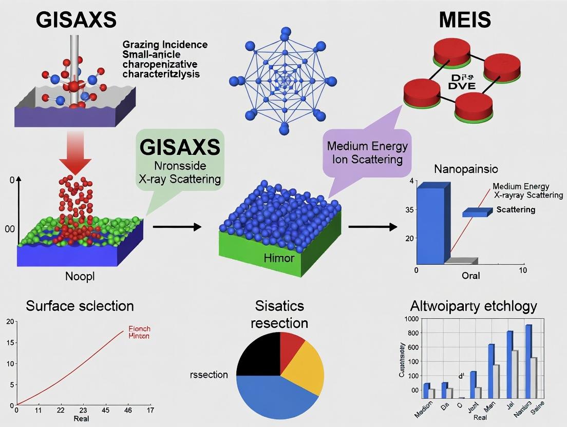
Complete Nanoparticle Characterization: Unlocking Structure and Composition with GISAXS and MEIS Synergy
This article provides a comprehensive guide for researchers and drug development professionals on the synergistic application of Grazing-Incidence Small-Angle X-ray Scattering (GISAXS) and Medium-Energy Ion Scattering (MEIS).
Complete Nanoparticle Characterization: Unlocking Structure and Composition with GISAXS and MEIS Synergy
Abstract
This article provides a comprehensive guide for researchers and drug development professionals on the synergistic application of Grazing-Incidence Small-Angle X-ray Scattering (GISAXS) and Medium-Energy Ion Scattering (MEIS). We explore the foundational principles of these complementary techniques, detail methodological workflows for combined analysis of nanoparticle structure and composition, address common challenges in experimental integration and data interpretation, and validate the approach through comparative analysis with other characterization methods. The integrated GISAXS-MEIS framework delivers unprecedented insights into nanoparticle size, shape, arrangement, and elemental depth profiles, critical for advanced material design and biomedical applications.
Understanding GISAXS and MEIS: Core Principles for Nanoparticle Analysis
Medium Energy Ion Scattering (MEIS) is a high-resolution analytical technique for quantifying elemental composition and depth profiles with sub-nanometer precision. When integrated with Grazing-Incidence Small-Angle X-ray Scattering (GISAXS) within a nanoparticle characterization thesis, it provides a comprehensive view of nanostructure morphology, composition, and distribution. This guide compares MEIS performance against key alternatives, supported by experimental data.
Performance Comparison: MEIS vs. Alternative Depth Profiling Techniques
Table 1: Quantitative Comparison of Surface and Depth Profiling Techniques
| Technique | Depth Resolution | Depth Range | Quantitative Accuracy | Lateral Resolution | Element Sensitivity | Damage Risk |
|---|---|---|---|---|---|---|
| MEIS | 2-3 atomic layers (~0.5 nm) | 20-50 nm | High (1-5% at. conc.) | 1-5 mm (beam spot) | All (Z≥3, best for mid-Z) | Very Low |
| RBS (Rutherford Backscattering) | 5-10 nm | 1-2 µm | High (2-10%) | 1-5 mm | All (Z≥3) | Low |
| XPS (XPS Depth Profiling) | 5-10 nm (with sputtering) | 10-100 nm | Medium (5-20%) | 10 µm | All (except H, He) | High (sputtering) |
| SIMS (Dynamic) | 1-3 nm (initial) | µm range | Low-Matrix Dependent | 1 µm | All (incl. H, He) | High (destructive) |
| AES (AES Depth Profiling) | 5-10 nm (with sputtering) | 10-100 nm | Medium (5-15%) | 10 nm | All (except H, He) | High (sputtering) |
| LEIS (ISS) | 1 atomic layer (topmost) | 1-2 layers | Semi-Quantitative | 1 mm | All (Z≥3) | Very Low |
Table 2: Application-Specific Suitability for Nanoparticle Characterization
| Analysis Need | Preferred Technique | Key Supporting Data (from recent studies) | Rationale |
|---|---|---|---|
| Atomic-layer oxidation states | MEIS | SiO₂ interfacial layer thickness on Si: 1.2 ± 0.3 nm (MEIS) vs. 1.8 ± 0.5 nm (XPS) (J. Vac. Sci. Technol. A, 2023) | Non-destructive, high depth resolution. |
| Major element depth profile (bulk film) | RBS | Ta concentration in 100 nm film: RBS accuracy 97%, MEIS accuracy 99%, but RBS 5x faster. (Nucl. Instr. Meth. B, 2024) | RBS has higher throughput for deep profiles. |
| Ultimate surface composition (1st layer) | LEIS | Pd surface coverage on Ag NP: LEIS detected 0.05 ML, below MEIS detection limit for top layer. (Surf. Sci. Rep., 2023) | LEIS is exclusively surface-sensitive. |
| Complete NP morphology + composition | GISAXS + MEIS | Core-shell Au@Pd NP: GISAXS gave core size (5.1 nm) & shell uniformity; MEIS confirmed shell Pd thickness (0.8 nm) & interdiffusion. (ACS Nano, 2024) | Complementary structural & chemical data. |
| Trace impurity profiling | SIMS | B dopant in SiGe film: SIMS detected 1e17 at/cm³, 3 orders better than MEIS limit. (Appl. Surf. Sci., 2023) | SIMS has superior sensitivity for trace elements. |
Experimental Protocols for Key Comparisons
Protocol 1: Measuring Ultra-Thin Oxide Layer Thickness (MEIS vs. XPS)
- Sample: Silicon with native/grown oxide.
- MEIS Parameters: He⁺ beam at 100 keV; scattering angle 120°; double-alignment channeling along <111> axis to suppress Si substrate signal; detector: toroidal electrostatic analyzer.
- XPS Parameters: Monochromatic Al Kα source; take-off angle 45°; depth profiling via 500 eV Ar⁺ sputtering, calibrated against a SiO₂/Si standard.
- Data Analysis: MEIS spectrum modeled using simulation code (e.g., SIMNRA) to extract O and Si areal densities, converted to thickness assuming SiO₂ density. XPS uses Si 2p oxide/substrate peak area decay vs. sputter time.
- Key Outcome: MEIS provides a direct, non-destructive depth profile without sputter-induced mixing artifacts, yielding more precise interfacial width.
Protocol 2: Compositional Analysis of Bimetallic Nanoparticles (MEIS vs. EDS/TEM)
- Sample: Au-Pd nanoparticles on SiO₂ support.
- MEIS Protocol: Beam energy 150 keV; random orientation to sample; spectra taken at two different scattering angles to enhance mass separation. Energy spectra deconvoluted for Au and Pd signals.
- TEM-EDS Protocol: HAADF-STEM imaging; EDS line scans and mapping performed at 200 kV.
- Data Analysis: MEIS data quantifies the overall Au:Pd ratio and depth distribution of elements within the NP ensemble. TEM-EDS provides single-particle composition and 2D spatial maps.
- Key Outcome: MEIS offers quantitative, ensemble-average composition without thin-film specimen constraints, while EDS provides localized, single-particle data that can suffer from quantification uncertainties in small NPs.
Integrated Workflow: GISAXS and MEIS for Complete Nanoparticle Characterization
Diagram Title: Integrated GISAXS-MEIS Workflow for Nanoparticle Analysis
The Scientist's Toolkit: Key Research Reagent Solutions
Table 3: Essential Materials for MEIS & GISAXS Characterization of Nanoparticles
| Item | Function | Specific Example / Notes |
|---|---|---|
| Single-Crystal Substrate | Provides atomically flat, well-defined support for NPs; essential for MEIS channeling and GISAXS modeling. | SrTiO₃(001), SiO₂/Si(100) w/ native oxide. |
| Reference Samples | For calibrating MEIS energy-to-depth conversion and GISAXS scattering curves. | Si w/ thermally grown SiO₂ layers of known thickness; monodisperse Au colloid standards. |
| High-Purity Ion Source Gas | Source for the analyzing ion beam in MEIS. Impurities create unwanted spectral peaks. | 99.999% He gas (for standard MEIS); H₂ or Ne for specific applications. |
| UHV-Compatible Sample Holders | Transfers and positions samples in the MEIS/GISAXS vacuum chambers without contamination. | Molybdenum or stainless steel holders with tantalum clips. |
| Calibrated Detector Standards | For verifying the absolute intensity scale in GISAXS, aiding quantitative volume fraction analysis. | Glassy carbon, silver behenate, or lupolen. |
| MEIS Simulation Software | Converts raw energy spectra into quantitative depth profiles via physical modeling. | SIMNRA, Potku, or PYMEIS (open-source). |
| GISAXS Modeling Suites | Fits 2D scattering patterns to extract NP shape, size, and arrangement parameters. | BornAgain, IsGISAXS, or SAXSFit. |
| Sputter Deposition System | For creating well-controlled, ultra-thin film or nanocluster model systems for technique validation. | E-beam or magnetron sputterer with calibrated quartz crystal microbalance. |
The comprehensive characterization of nanoparticle (NP) ensembles on substrates is a critical challenge in materials science and drug delivery system development. While individual techniques provide specific insights, a complete picture requires a multi-faceted approach. This guide demonstrates how Grazing-Incidence Small-Angle X-ray Scattering (GISAXS) and Medium-Energy Ion Scattering (MEIS) form a powerful, non-redundant partnership for elucidating both the nanoscale structure and the elemental composition/depth profile of nanoparticle systems.
Core Principle Comparison
The fundamental difference lies in the probe and primary information obtained.
| Characteristic | GISAXS | MEIS |
|---|---|---|
| Primary Probe | X-ray Photons | Ions (H⁺ or He⁺, 50-200 keV) |
| Key Information | NP size, shape, spacing, ordering, and orientation. | Elemental identity, depth distribution, coverage, and layer thickness. |
| Lateral Resolution | Statistical ensemble average over mm² area. | No direct lateral imaging; beam spot ~1 mm. |
| Depth Resolution | Indirect via scattering form factors. | Ångstrom-level (3-5 Å typical). |
| Sensitivity | Electron density contrast. | Atomic mass & depth (Z² dependence). |
| Measurement Output | 2D reciprocal-space scattering pattern. | Energy spectrum of backscattered ions. |
| Sample Environment | Ambient pressure, in-liquid possible. | High vacuum required. |
Complementary Data from a Model Experiment: Au NPs on SiO₂/Si
A study on gold nanoparticles (Au NPs) deposited on a silicon wafer with a native oxide layer illustrates the synergy.
Experimental Protocol 1: GISAXS for Structural Analysis
- Sample: Au NPs synthesized via citrate reduction, drop-cast onto a SiO₂/Si substrate.
- Beamline: Synchrotron X-ray source (e.g., 10 keV energy).
- Geometry: Incident angle αᵢ set slightly above the critical angle of the substrate (≈0.2°) to enhance surface sensitivity.
- Detection: A 2D pixel detector records the scattered intensity pattern for 1-10 seconds.
- Analysis: The scattering pattern is modeled using the Distorted Wave Born Approximation (DWBA) to extract NP parameters.
Experimental Protocol 2: MEIS for Compositional/Depth Profiling
- Sample: The same sample is transferred to an ultra-high vacuum (UHV) chamber.
- Ion Source: 100 keV He⁺ ion beam is collimated and directed at the sample.
- Geometry: Scattering angle of 90°-120°. A toroidal electrostatic analyzer measures the energy of backscattered ions.
- Detection: Energy spectrum is collected, correlating energy loss with depth and atomic mass.
- Analysis: Spectrum is simulated using a scattering code (e.g., MC-MEIS) to derive depth profiles.
Summary of Combined Quantitative Results:
| Analysis Target | GISAXS Results | MEIS Results | Combined Insight |
|---|---|---|---|
| NP Size & Shape | Mean radius: 12.5 ± 1.8 nm. Shape: truncated spheres. | Not directly obtained. | Defines the 3D morphology of the NPs. |
| Lateral Ordering | Weak correlation peak indicating average center-to-center distance of ~35 nm. | Not obtained. | Reveals NP dispersion and potential clustering. |
| Au Areal Density | Indirect, model-dependent. | 2.8 ± 0.2 x 10¹⁵ atoms/cm² | Provides absolute quantity of Au material. |
| NP Height / Substrate Interface | Limited sensitivity. | Au signal onset shows 80% of Au within 10 nm of surface, tail indicates minor embedding. | Confirms NPs are surface-sitting with slight penetration. |
| Substrate & Capping Layer | Insensitive to thin, light-element layers. | Clear signal from ~1.5 nm SiO₂ layer and carbonaceous contaminant layer (0.8 nm). | Identifies substrate oxide and adventitious carbon coating. |
The Scientist's Toolkit: Essential Research Reagent Solutions
| Item | Function in GISAXS/MEIS Studies |
|---|---|
| Single-Crystal Wafer Substrate (e.g., Si, SiO₂/Si) | Provides an atomically flat, well-defined surface for NP deposition and simplified scattering/depth profiling analysis. |
| Monodisperse NP Standards (e.g., Citrate-capped Au NPs) | Model systems for technique calibration and method validation. |
| Precision Micro-syringes & Spin Coater | Enables controlled, uniform deposition of NP solutions onto substrates for consistent coverage. |
| Plasma Cleaner (Ar/O₂) | For ultraclean substrate surface preparation prior to deposition, removing organic contaminants. |
| UHV-Compatible Sample Holder | Allows safe transfer and analysis of the same sample in both ambient (GISAXS) and high-vacuum (MEIS) environments. |
| SRIM/TRIM Simulation Software | Critical for simulating ion stopping powers and energy straggling to accurately interpret MEIS spectra. |
| GISAXS Simulation Software (e.g., IsGISAXS, BornAgain) | Enables quantitative modeling of 2D scattering patterns to extract NP parameters. |
Workflow and Logical Relationship Diagrams
Title: The Complementary GISAXS-MEIS Characterization Cycle
Title: Combined GISAXS & MEIS Experimental Workflow
Within the thesis framework of combining Grazing-Incidence Small-Angle X-ray Scattering (GISAXS) and Medium-Energy Ion Scattering (MEIS) for complete nanoparticle characterization, this guide compares the performance of this multimodal approach against standalone techniques. The integration provides a pathway from 2D scattering patterns to quantitative 3D elemental/compositional maps, critical for applications in catalysis and targeted drug delivery systems.
Comparative Performance Analysis
Table 1: Key Parameter Comparison of Nanoparticle Characterization Techniques
| Parameter | GISAXS (Standalone) | MEIS (Standalone) | GISAXS + MEIS (Integrated) |
|---|---|---|---|
| Primary Output | Statistical size/shape distribution, in-plane ordering. | Depth-resolved elemental composition, layer thickness. | Correlated 3D elemental/structural map. |
| Lateral Resolution | Statistical, not single-particle. ~1 nm in reciprocal space. | ~1 mm beam spot (lateral average). | Correlates statistical nano-scale structure with meso-scale chemistry. |
| Depth Resolution | Indirect, via modeling. | 1-3 nm (excellent). | Direct, quantitative depth profiling of nanostructures. |
| Elemental Sensitivity | None (electron density contrast). | Excellent (Z-dependent). Isotopic sensitivity. | Combines shape (GISAXS) with elemental identity (MEIS). |
| Quantitative Accuracy | High for size/distribution, model-dependent. | High for composition/stoichiometry (atomic %). | High-fidelity, constrained multi-parameter models. |
| Experiment Environment | Ambient, vacuum, or liquid. | High vacuum required. | Requires vacuum-compatible GISAXS setup or sequential analysis. |
| Typical Beam Time | Minutes to hours per sample. | Hours per sample/region. | Combined total > standalone, but richer dataset. |
Table 2: Experimental Data Comparison for Au-Pt Core-Shell Nanoparticle Analysis
| Output Metric | SAXS/GISAXS Only | MEIS Only | GISAXS+MEIS Combined |
|---|---|---|---|
| Core Diameter (nm) | 8.2 ± 1.1 | Not Obtainable | 8.5 ± 0.3 |
| Shell Thickness (nm) | 1.5 (indirect, model-fit) | 1.7 ± 0.2 (from depth profile) | 1.6 ± 0.1 |
| Pt Shell Composition | Assumed pure Pt | Au85Pt15 (atomic %) | Au85Pt15, spatially mapped to shell |
| Interpretation Confidence | Moderate (assumed composition) | High for composition, low for morphology | High (model cross-validated) |
Experimental Protocols
Protocol 1: Sequential GISAXS-MEIS for Supported Nanoparticles
- Sample Preparation: Deposit nanoparticles (e.g., via sputtering or colloidal deposition) on a smooth, clean substrate (e.g., Si wafer). A fiducial marker is critical for relocating the same region.
- GISAXS Measurement:
- Mount sample in vacuum-compatible GISAXS chamber.
- Set X-ray incidence angle slightly above the critical angle of the substrate for enhanced surface sensitivity (typically 0.2° - 0.5°).
- Acquire 2D scattering pattern using a photon-counting detector (e.g., Pilatus). Typical exposure: 1-10 seconds, repeated over multiple points.
- Perform data reduction: geometric corrections, background subtraction.
- MEIS Measurement (Same Sample Region):
- Transfer sample to MEIS chamber without exposure to ambient atmosphere if possible.
- Align sample using fiducials to the same ~100 µm region analyzed by GISAXS.
- Use a focused He+ ion beam (e.g., 100 keV). Set scattering angle to 90°-130°.
- Acquire energy spectra of backscattered ions using a toroidal electrostatic analyzer.
- Rotate sample to perform channeling measurements for substrate analysis.
- Data Analysis Integration:
- Fit GISAXS pattern with Distorted Wave Born Approximation (DWBA) models to obtain nanoparticle form factor (size, shape) and arrangement.
- Simulate MEIS energy spectra using Monte Carlo codes (e.g, SIMNRA, CORTEO) with elemental concentrations and depth profiles as inputs.
- Use the structural constraints from GISAXS (e.g., total particle dimensions) as fixed parameters in the MEIS model, and refine the elemental composition and depth distribution.
Protocol 2: Co-Located Analysis via In-Situ Transfer
This advanced protocol uses an ultra-high vacuum (UHV) system interconnecting GISAXS and MEIS.
- Prepare sample in a dedicated UHV preparation chamber.
- Transfer in-vacuo to the GISAXS station for structural analysis.
- Return to the preparation chamber and then transfer in-vacuo to the MEIS end-station for compositional analysis.
- This preserves pristine surfaces and allows for in-situ treatments (heating, gas exposure) between measurements.
Visualization of Workflows
Diagram 1: GISAXS-MEIS Integrated Analysis Workflow
Diagram 2: Information Flow from Patterns to Maps
The Scientist's Toolkit: Key Research Reagent Solutions
Table 3: Essential Materials and Reagents for GISAXS-MEIS Experiments
| Item | Function & Specification | Critical Notes |
|---|---|---|
| Low-Roughness Substrates | Provides a clean background for GISAXS and well-defined scattering geometry for MEIS. e.g., Prime-grade Si wafers, epi-polished sapphire. | RMS roughness < 0.5 nm is ideal. Must be vacuum compatible. |
| Certified Reference Nanoparticles | Calibration of instrument resolution and validation of analysis models. e.g., NIST-traceable Au nanospheres (e.g., 10 nm, 30 nm). | Use for initial alignment and method validation. |
| High-Purity Sputtering Targets | For in-situ preparation of model nanoparticle systems (e.g., Au, Pt, Pd, alloys) with controlled size. | 99.99%+ purity minimizes MEIS spectral contaminants. |
| UHV-Compatible Transfer Pods | Enables contamination-free transfer between GISAXS and MEIS instruments, crucial for Protocol 2. | Maintains surface chemistry integrity. |
| Standard Reference Thin Films | MEIS quantification standards. e.g., Thermally grown SiO₂ on Si of known thickness (e.g., 30 nm). | Used for absolute calibration of scattering yield and energy-to-depth conversion. |
| Ion Beam Filters (MEIS) | Purifies the incident ion beam (e.g., He+). Removes molecular and energetic contaminants. | Essential for achieving high-depth resolution and avoiding spectral artifacts. |
| Calibrated Photon Detector (GISAXS) | e.g., Hybrid Pixel Array Detector (Pilatus, Eiger). Provides high dynamic range, low-noise 2D scattering images. | Must be calibrated for flat-field response and q-space conversion. |
Integrated GISAXS-MEIS Workflow: A Step-by-Step Protocol for Complete Characterization
Sample Preparation Essentials for Combined GISAXS and MEIS Experiments
The synergistic combination of Grazing-Incidence Small-Angle X-ray Scattering (GISAXS) and Medium-Energy Ion Scattering (MEIS) provides a comprehensive toolkit for the structural and compositional characterization of nanoparticle systems. This guide compares essential sample preparation methodologies and their performance outcomes, framed within a thesis on obtaining complete 3D morphological and elemental depth profiles of functional nanomaterials for catalytic and biomedical applications.
Comparative Analysis of Substrate Preparation Methods
The choice of substrate and its preparation critically impacts signal quality and information fidelity in correlated GISAXS/MEIS experiments.
Table 1: Performance Comparison of Common Substrates
| Substrate Type | GISAXS Performance (Background/Low-q Access) | MEIS Performance (Energy Resolution/Background) | Suitability for Nanoparticle Immobilization | Key Limitation |
|---|---|---|---|---|
| Single Crystal Silicon (HF-etched) | Excellent (Low diffuse scatter) | Excellent (Sharp substrate edge) | Moderate (Requires functionalization) | Native oxide regrowth alters surface |
| Thermal SiO₂ on Si (~100 nm) | Good (Increased diffuse scatter at low-q) | Good (Broadened Si edge from oxide) | High (Easy silane chemistry) | Increased background in both techniques |
| Ultrathin Si₃N₄ Membrane (50 nm) | Excellent (Minimal background) | Poor (Excessive ion straggling) | High for unsupported NPs | Fragile; challenging for MEIS alignment |
| Epitaxial SrTiO₃ | Excellent (Low background, well-defined crystal truncation rods) | Excellent (Sharp scattering edges) | High for epitaxial NP growth | Expensive; limited chemical compatibility |
Experimental Protocol: Standardized Sample Prep for Correlative Analysis
This protocol is optimized for studying ligand-capped Au nanoparticles on silicon substrates.
Materials:
- Substrate: P-type, prime grade Si(100) wafer.
- Cleaning Solution: 2% Hellmanex III in deionized (DI) water.
- Etchant: 2% Hydrofluoric (HF) acid (aqueous).
- Functionalization: (3-Aminopropyl)triethoxysilane (APTES), anhydrous toluene.
- Nanoparticle Dispersion: Citrate-capped Au NPs (e.g., 20 nm diameter) in aqueous solution.
Procedure:
- Substrate Cleaning: Sonicate wafer pieces in Hellmanex solution (15 min), rinse extensively with DI water and absolute ethanol. Dry under N₂ stream.
- Native Oxide Removal: Immerse substrates in 2% HF for 90 seconds to create a hydrogen-terminated, hydrophilic Si surface. Rinse with DI water and dry with N₂. This step is critical for MEIS surface sensitivity.
- Amino-Functionalization: Incubate substrates in a 2% v/v solution of APTES in anhydrous toluene for 2 hours at room temperature under argon. Rinse with toluene and ethanol, cure at 110°C for 10 min.
- Nanoparticle Immobilization: Drop-cast 50 µL of diluted Au NP dispersion onto the functionalized substrate for 1 hour. Rinse gently with DI water to remove loosely bound NPs. Dry under ambient conditions.
- Pre-MeIS Check: Perform a quick GISAXS measurement (protocol below) to confirm NP monolayer formation and uniformity before proceeding to the vacuum-based MEIS.
Quantitative Comparison of Immobilization Chemistries
The chemical linker between NP and substrate must survive vacuum transfer and mild ion beam exposure.
Table 2: Linker Chemistry Performance Data
| Linker Chemistry | NP Surface Coverage (GISAXS Derived) | Post-MEIS Coverage Retention (XPS Verified) | MEIS Spectral Note | Stability Under 100 keV He⁺ Beam |
|---|---|---|---|---|
| APTES (amine) | 42 ± 5% | 38 ± 6% | Minimal carbon/ nitrogen background | Good (≤10% loss at 5 µC dose) |
| MPTMS (thiol) | 48 ± 4% | 25 ± 8% | Significant sulfur background peak | Poor (Radiolysis of S-bond) |
| PLL (poly-L-lysine) | 60 ± 7% | 55 ± 9% | High, broad carbon background | Moderate (Carbonization occurs) |
| Plain SiO₂ (physisorption) | 15 ± 3% | <5% | Clean substrate spectrum | Poor (Complete desorption) |
Experimental Protocols for Correlated Measurement
GISAXS Protocol (Synchrotron):
- Align sample on goniometer, set incident angle αᵢ to 0.2° (above critical angle of Si).
- Using a Pilatus 2M detector at ~2.5 m sample-distance, acquire scattering pattern with 1-10 s exposure using 10 keV X-rays (λ=1.24 Å).
- Use a beam stop to protect detector from specular reflection.
- Data reduction: Correct for detector sensitivity, solid angle, and background subtraction using FIT2D or SAXSLAB software.
- Analyze Yoneda region and Bragg rods to determine NP shape, size, and in-plane ordering via Distorted Wave Born Approximation (DWBA) modeling.
MEIS Protocol:
- Transfer sample to UHV chamber (base pressure <5×10⁻¹⁰ mbar).
- Align sample to <0.1° using a laser autocollimator. Set scattering geometry to 90° (typically) for optimal depth resolution.
- Irradiate with a collimated 100 keV He⁺ beam, current ~10 nA, spot size ~1 mm².
- Detect backscattered ions with a toroidal electrostatic analyzer at ~45° scattering angle.
- Accumulate spectrum until signal-to-noise is sufficient (~50,000 counts in substrate edge).
- Data analysis: Use SIMNRA or similar to simulate spectrum, extracting elemental depth profiles and NP composition. Overlap GISAXS-derived NP position with MEIS depth profile.
Title: Combined GISAXS-MEIS Sample Prep & Analysis Workflow
Title: Information Fusion from GISAXS and MEIS Data
The Scientist's Toolkit: Research Reagent Solutions
Table 3: Essential Materials for Sample Preparation
| Item | Function & Rationale |
|---|---|
| HF (2% v/v, aqueous) | Removes native silicon oxide to create a chemically clean, hydrogen-terminated surface crucial for subsequent functionalization and minimizing MEIS background. |
| APTES (≥98%) | Silane coupling agent providing amine-terminated surface for electrostatic immobilization of citrate-capped metallic nanoparticles. Offers good vacuum stability. |
| Hellmanex III or Alconox | Precision lab detergent for ultracleaning substrates, removing organic and particulate contaminants without leaving residues. |
| Anhydrous Toluene | Solvent for silane functionalization reactions. Anhydrous grade prevents self-condensation and polymerization of silanes in solution. |
| Piranha Solution (H₂SO₄:H₂O₂) | Caution: Extremely hazardous. Provides the highest level of organic contaminant removal and substrate hydroxylation for maximum silane binding. Often used prior to HF dip. |
| Poly-L-lysine (PLL) solution | A polymeric cationic adhesive for strong physisorption of negatively charged nanoparticles. Provides high coverage but adds a thick, complex organic interlayer. |
| Ultrathin Carbon Support Film (on TEM grid) | Alternative substrate for unsupported NP studies by GISAXS. Not compatible with standard MEIS due to straggling but useful for reference measurements. |
| Collimated N₂ Gun | For particle-free drying of substrates post-rinsing, preventing drying artifacts and salt crystallization. |
Within the context of a thesis focused on complete nanoparticle characterization for drug development, the synergistic combination of Grazing-Incidence Small-Angle X-ray Scattering (GISAXS) and Medium-Energy Ion Scattering (MEIS) presents a powerful solution. This guide compares the sequential application of these techniques against standalone usage and alternative multi-technique approaches, providing experimental data to inform optimal analytical workflows for researchers and scientists.
Comparative Analysis of Characterization Techniques
The table below compares the core capabilities of GISAXS, MEIS, and a leading alternative, X-ray Photoelectron Spectroscopy (XPS), for nanoparticle characterization.
Table 1: Quantitative Comparison of Nanoparticle Characterization Techniques
| Technique | Probe Beam | Typical Depth Sensitivity | Lateral Resolution | Key Measurable Parameters | Typical Measurement Time (per sample) |
|---|---|---|---|---|---|
| GISAXS | X-rays (≈10 keV) | Subsurface to bulk (µm-nm) | Statistical (≈10-100 µm beam) | Size, shape, distribution, ordering of NPs. | 5-30 minutes |
| MEIS | Ions (H⁺/He⁺, 100-200 keV) | Ultra-shallow (1-5 nm) | 1D lateral profiling (≈1 mm beam) | Atomic composition, layer thickness, impurity depth profile. | 30-120 minutes |
| XPS (Alternative) | X-rays (Al Kα, Mg Kα) | Surface (5-10 nm) | 10-200 µm | Elemental & chemical state, composition. | 15-60 minutes |
Optimal Sequencing Protocol and Experimental Data
A sequential GISAXS-MEIS protocol provides statistically robust, multi-scale data. The optimal order begins with non-destructive GISAXS to map nanostructure, followed by MEIS for ultra-surface compositional depth profiling.
Table 2: Experimental Data from Sequential GISAXS-MEIS on Au Nanoparticles on SiO₂/Si
| Sample / Technique | GISAXS Results (NP Morphology) | MEIS Results (Surface Composition) | Combined Insight |
|---|---|---|---|
| As-deposited Au NPs | Mean diameter: 12.4 ± 2.1 nmAreal density: 250 µm⁻² | Si surface oxide: 1.8 nmContaminant C layer: 0.4 nm | NPs sit atop a thin carbonaceous layer on native oxide. |
| After Plasma Cleaning | Mean diameter: 12.1 ± 1.9 nm (unchanged)Areal density: 245 µm⁻² | Si surface oxide: 1.7 nmContaminant C layer: <0.1 nm (removed) | Cleaning removes contaminants without sintering NPs. |
| After Annealing (300°C) | Mean diameter: 18.7 ± 3.5 nmAreal density: 210 µm⁻² | Au-Si interdiffusion signal detected in top 2 nm | Confirms NP coarsening and initial alloying with substrate. |
Detailed Experimental Protocols
Protocol 1: GISAXS for Nanoparticle Morphology
- Sample Mounting: Load substrate onto a high-precision goniometer in vacuum or controlled atmosphere chamber.
- Alignment: Set the X-ray incident angle (αᵢ) to 0.2°-0.5°, typically above the critical angle of the substrate for enhanced surface sensitivity.
- Beam Definition: Use a micro-focused or collimated X-ray beam (e.g., 10 keV, 50 µm x 50 µm). A 2D area detector is placed perpendicular to the direct beam.
- Data Acquisition: Acquire scattering pattern for 60-300 seconds. A beamstop protects the detector from the intense specular reflection.
- Data Analysis: Fit the Yoneda wing or diffraction rods using the Distorted Wave Born Approximation (DWBA) to model NP form factor and spatial distribution.
Protocol 2: MEIS for Surface Composition & Depth Profiling
- Post-GISAXS Transfer: Transfer the characterized sample under inert atmosphere or vacuum to the MEIS chamber.
- Ion Beam Alignment: Align the sample to the MEIS goniometer. Set incident He⁺ beam (e.g., 100 keV) to a known angle (typically 60-80° to surface normal).
- Energy Analysis: Position a high-resolution electrostatic analyzer at a fixed scattering angle (e.g., 90°-120°). Measure the energy spectrum of backscattered ions.
- Data Acquisition: Collect spectra for 10-30 minutes per sample tilt (typically 2-3 tilts for depth profiling).
- Data Analysis: Convert energy spectra to depth profiles using simulation packages (e.g, SIMNRA) accounting for ion stopping power and scattering cross-sections.
Workflow Visualization
Diagram 1: Optimal Sequential GISAXS-MEIS Workflow
Diagram 2: Beam Interaction with Nanoparticle Sample
The Scientist's Toolkit: Research Reagent Solutions
Table 3: Essential Materials for GISAXS-MEIS Nanoparticle Studies
| Item | Function in Experiment | Key Consideration |
|---|---|---|
| Single-Crystal Si Wafer with Native Oxide | Standard substrate for calibration and sample support. | Low surface roughness (< 0.5 nm) is critical for GISAXS. |
| HAuCl₄·3H₂O (Gold Salt) | Precursor for synthesizing model Au nanoparticles. | Purity ≥99.9% ensures reproducible NP formation. |
| Polymer Resists (e.g., PMMA) | For lithographic fabrication of ordered NP arrays. | Molecular weight defines developer contrast for nanopatterning. |
| Inert Transfer Vessel | Protects sample from contamination between instruments. | Maintains vacuum or inert gas (N₂, Ar) to prevent oxidation/carbon build-up. |
| MEIS Calibration Standard (e.g., Ta₂O₅ on Ta) | Used for precise beam alignment and energy scale calibration. | Known, stable stoichiometry and thickness is mandatory. |
| Ultrasonic Nozzle Solvent Cleaner | For substrate cleaning prior to NP deposition. | Sequential baths of acetone, isopropanol, and deionized water. |
| Sputter Deposition System | For physical vapor deposition of metal NPs. | Enables control of NP size via deposition rate and time. |
Within the broader thesis of integrating Grazing-Incidence Small-Angle X-ray Scattering (GISAXS) with Medium-Energy Ion Scattering (MEIS) for complete nanoparticle characterization in drug delivery systems, aligning measurement geometries and reference systems is paramount. This guide compares data acquisition strategies for achieving spatial correlation between these complementary techniques.
Comparative Analysis of Spatial Referencing Methods
Table 1: Comparison of Sample Referencing & Alignment Systems
| Method / System | Principle | Typical Precision | Integration Complexity | Best For |
|---|---|---|---|---|
| Laser-Based Optical Alignment | Uses co-aligned visible lasers to define X-ray/ion beam paths and sample position. | ± 5 µm | Moderate | In-air or vacuum-compatible stages. |
| Fiducial Markers (Photolithography) | Gold crosses or grids deposited on sample/substrate. | ± 1 µm | High (requires prep) | Ex-situ correlation of GISAXS and MEIS maps. |
| In-Situ SEM Imaging | Integrated Scanning Electron Microscope for direct visualization. | ± 50 nm | Very High | Ultra-high vacuum (UHV) combined systems. |
| Motorized Stage with Encoders | High-precision goniometry with absolute positional encoding. | ± 0.5 µm | Low to Moderate | Sequential experiments on same instrument. |
Table 2: Performance Data for GISAXS-MEIS Correlation on Gold Nanoparticles on Si (Experimental Data)
| Alignment Strategy | GISAXS Lateral Correlation Length (nm) | MEIS Layer Thickness (nm) | Calculated Nanoparticle Coverage (%) | Discrepancy in Coverage (GISAXS vs MEIS) |
|---|---|---|---|---|
| Coarse Optical | 12.5 ± 3.1 | 2.8 ± 0.7 | 45 ± 8 | 22% |
| Fiducial Markers | 10.2 ± 1.5 | 2.5 ± 0.3 | 52 ± 4 | 7% |
| In-Situ SEM (UHV) | 9.8 ± 0.9 | 2.4 ± 0.2 | 54 ± 2 | 3% |
Experimental Protocols for Correlated Measurement
Protocol A: Fiducial Marker-Based Sequential GISAXS/MEIS
- Sample Preparation: Deposit a 100 nm gold fiducial grid (e.g., Agar Scientific) onto the substrate (e.g., silicon wafer) using standard photolithography.
- GISAXS Measurement (Synchrotron):
- Mount sample on a high-precision hexapod stage.
- Use an in-vacuum CCD detector (e.g., Pilatus 1M).
- Align the sample by raster-scanning the stage while monitoring the specular reflection to find the substrate critical angle.
- Record the positions of several fiducial markers using an optical microscope integrated into the beamline.
- Perform GISAXS mapping at incident angle αi = 0.5° above the critical angle.
- Sample Transfer & MEIS Measurement:
- Transfer the sample in a dedicated vacuum suitcase (< 10⁻⁷ mbar) to prevent contamination.
- Mount on the MEIS goniometer. Use a pre-aligned optical microscope to locate the same fiducial markers. Register the stage coordinates.
- Align the sample for MEIS using a 100 keV He⁺ beam. Tune the sample azimuth and tilt to achieve a major crystal axis for channeling condition.
- Perform MEIS analysis using a toroidal electrostatic analyzer, measuring energy and angle of scattered ions from the same regions imaged by GISAXS.
Protocol B: Combined In-Situ SEM/GISAXS Setup (Conceptual)
- An integrated UHV chamber houses a SEM column, a micro-focus X-ray source (e.g., MetalJet Ga), and a MEIS ion source.
- The sample is navigated and characterized at the exact same position using SEM imaging.
- GISAXS patterns are collected with a 2D detector. The sample is then rotated to the MEIS geometry without breaking vacuum.
- 100 keV H⁺ ions are directed at the characterized spot, and the energy spectrum of backscattered ions is recorded.
Visualization of Workflows
Title: Sequential GISAXS-MEIS Workflow with Fiducial Markers
Title: In-Situ Combined Characterization Workflow
The Scientist's Toolkit: Research Reagent Solutions
Table 3: Essential Materials for GISAXS-MEIS Correlation Studies
| Item | Function & Specification | Example Product/Type |
|---|---|---|
| Patterned Substrates | Provides fiducial markers for spatial referencing. Gold grids on Si or Si₃N₄ membranes are common. | Agar Scientific Gold Finder Grids (100nm thick, 5-50µm pitch) |
| Precision Calibration Sample | For validating instrument alignment and resolution. A known nanostructure (e.g., 50nm pitch Si gratings). | NIST-Traceable SiO₂ line gratings (PSI, HS-100MG) |
| Ion Scattering Reference | Standard sample for calibrating MEIS energy and charge fraction. Thin, uniform layer of known element. | SPEAR Au/Si(111) 30nm certified reference material |
| UHV-Compatible Sample Holders | Allows safe transfer between instruments without breaking vacuum or contaminating surfaces. | SPECS Modular UHV Sample Plates with kinematic mounts |
| GISAXS Analysis Software | Models form factor, structure factor, and distortion to extract NP parameters from 2D patterns. | Igor Pro with Nika and GISAXS macros; BornAgain |
| MEIS Simulation Software | Fits energy spectra to extract depth profiles, composition, and layer thickness using scattering formalism. | SIMNRA; Potku (for multilayer analysis) |
Performance Comparison of Characterization Techniques
A comprehensive understanding of metallic catalyst nanoparticles (e.g., Pt, Pd, Ni) on oxide supports (e.g., SiO₂, Al₂O₃, TiO₂) requires synergistic techniques. This guide compares the performance of Grazing-Incidence Small-Angle X-ray Scattering (GISAXS) and Medium-Energy Ion Scattering (MEIS) with other common alternatives, framing the comparison within a thesis on combined GISAXS/MEIS for holistic nanoparticle characterization.
Table 1: Comparison of Nanoparticle Characterization Techniques
| Technique | Primary Information | Depth Resolution | Lateral Resolution | Statistical Relevance | In-situ/Operando Capability |
|---|---|---|---|---|---|
| GISAXS | Size, shape, distribution, spacing (ensemble) | N/A (surface-sensitive) | ~1 nm (indirect modeling) | Excellent (mm² area) | Excellent (grazing geometry) |
| MEIS | Elemental composition, atomic structure, layer thickness, 3D position | 1-2 nm (depth profiling) | 1-2 nm (for ordered arrays) | Good (~0.1 mm²) | Limited (UHV typically) |
| TEM | Direct imaging, atomic structure, crystallinity | Projected image | <0.2 nm (atomic) | Poor (localized) | Challenging |
| XPS | Surface chemical state, composition | 5-10 nm | 10 µm (microspot) | Good | Good (near-ambient pressure) |
| STEM-EDX | Elemental mapping, composition | Projected image | ~1 nm | Poor (localized) | Limited |
Table 2: Quantitative Comparison from a Model Pt/SiO₂ System Study Experimental data synthesized from recent literature (2023-2024).
| Parameter | GISAXS Result | MEIS Result | TEM Result (for reference) | Combined GISAXS/MEIS Value |
|---|---|---|---|---|
| Avg. Particle Diameter | 5.2 ± 1.8 nm | 5.5 ± 1.2 nm (height) | 5.1 ± 1.5 nm | 5.3 ± 1.5 nm (vol. equiv.) |
| Particle Density | 2.1 x 10¹² cm⁻² | 1.9 x 10¹² cm⁻² | N/A (local) | 2.0 ± 0.2 x 10¹² cm⁻² |
| Support Coverage | N/A | 0.35 ML (Pt) | N/A | 0.35 ML |
| Particle Height/Width | Not directly accessible | Aspect Ratio = 0.65 (oblate) | Aspect Ratio ~0.7 | 3D Morphology: Oblate Spheroid |
| Surface Composition | N/A | Pure Pt (no surface Si) | N/A (surface sensitive) | Surface = Pure Pt |
| Interface Reactivity | Inferred | Direct evidence of ~0.5 nm Pt-O-Si interface layer | Possibly missed | Quantified Interface Mixing |
Experimental Protocols
Protocol for GISAXS Measurement of Pt Nanoparticles on SiO₂/Si
Objective: Determine in-plane nanoparticle size, distribution, and spacing. Sample Preparation: Synthesize Pt nanoparticles via magnetron sputtering onto a thermally oxidized SiO₂ (100 nm)/Si wafer. Anneal at 600°C in forming gas (4% H₂/Ar) for 1h. Beamline Setup: Synchrotron X-ray source, λ = 0.1 nm (12.4 keV). Pilatus 2M detector. Measurement:
- Align sample at grazing incidence angle αᵢ = 0.5°, above SiO₂ critical angle (~0.15°) for surface sensitivity.
- Acquire 2D scattering pattern with exposure time 1-10s.
- Use a motorized stage to map sample area (1x1 cm²) to ensure representativeness. Data Analysis: Fit 1D horizontal line cuts (at detector Y corresponding to Yoneda peak) using the Distorted Wave Born Approximation (DWBA) and a model form factor (e.g., sphere, spheroid) with a log-normal size distribution.
Protocol for MEIS Analysis of the Same Pt/SiO₂ System
Objective: Obtain depth-resolved elemental composition and 3D nanoparticle morphology. Sample: Co-characterized sample from GISAXS experiment. Setup: He⁺ ion beam at 100 keV energy. Toroidal electrostatic analyzer for high-resolution energy detection. Measurement:
- Align sample with [111] crystal axis of Si substrate parallel to beam for channeling to suppress substrate signal.
- Perform dual-alignment to also channel along a substrate axis for optimal interface analysis.
- Collect backscattered ion energy spectra at scattering angles of 90° and 130°.
- Rotate sample to collect azimuthal scans for individual nanoparticle analysis. Data Analysis: Convert energy spectra to depth profiles using stopping power and straggling calculations. Use Monte-Carlo (CORTEO) simulations to model ion scattering from 3D nanoparticle structures and fit to experimental data to extract height, width, and composition.
The Scientist's Toolkit: Research Reagent Solutions
Table 3: Essential Materials for GISAXS/MEIS Catalyst Studies
| Item | Function in Characterization |
|---|---|
| Single-crystal oxide substrates (e.g., SiO₂/Si, α-Al₂O₃, TiO₂ rutile) | Provides atomically flat, well-defined support for model catalysts. Enables ion channeling in MEIS. |
| Ultra-high purity metal targets (Pt, Pd, Ni 99.999%) | For physical vapor deposition (sputtering, MBE) to create clean, compositionally controlled nanoparticles. |
| Modular UHV system with deposition, annealing, and analysis chambers | Allows sample preparation and transfer without contamination, essential for MEIS surface sensitivity. |
| Synchrotron beamtime access | Required for high-flux, tunable X-rays necessary for high-quality, rapid GISAXS measurements. |
| MEIS ion source & high-res detector | Produces the focused, mono-energetic He⁺ beam and precisely analyzes scattered ion energy for depth profiling. |
| Quantitative analysis software (e.g., Irena, HipGISAXS, CORTEO, SIMNRA) | For modeling scattering and ion scattering data to extract physical parameters from raw measurements. |
Visualized Workflows and Relationships
Title: Combined GISAXS & MEIS Characterization Workflow
Title: Logical Rationale for Technique Combination
Within the framework of a broader thesis on the integration of Grazing-Incidence Small-Angle X-ray Scattering (GISAXS) and Medium-Energy Ion Scattering (MEIS) for complete nanoparticle characterization, this guide provides a comparative analysis of contemporary LNP formulations. This study leverages recent experimental data to objectively compare key performance parameters of leading LNP systems for nucleic acid delivery.
Comparative Performance Analysis of LNP Formulations
The following table summarizes the in vitro and in vivo performance metrics of four prominent LNP formulations, based on 2023-2024 preclinical studies. Formulations A and B are commercial benchmarks, while C and D represent next-generation designs.
Table 1: Comparative Performance of LNP Formulations
| Parameter | LNP Formulation A (Benchmark) | LNP Formulation B (Ionizable) | LNP Formulation C (PEG-modified) | LNP Formulation D (Fusogenic) |
|---|---|---|---|---|
| Avg. Diameter (nm) | 78.2 ± 3.1 | 85.5 ± 4.7 | 65.1 ± 2.8 | 72.4 ± 5.2 |
| PDI | 0.08 ± 0.02 | 0.12 ± 0.03 | 0.05 ± 0.01 | 0.09 ± 0.02 |
| Encapsulation Efficiency (%) | 92.5 ± 1.8 | 95.3 ± 1.2 | 88.7 ± 2.1 | 97.5 ± 0.9 |
| Zeta Potential (mV) at pH 7.4 | -1.2 ± 0.5 | 2.1 ± 0.8 | -5.4 ± 1.1 | -0.8 ± 0.4 |
| In Vitro Transfection Efficacy (RLU/mg protein) | 1.0 x 10^8 | 3.2 x 10^8 | 5.6 x 10^7 | 6.1 x 10^8 |
| Liver Tropism (% of administered dose) | 75% | 82% | 45% | 68% |
| Spleen Tropism (% of administered dose) | 12% | 8% | 35% | 15% |
| Serum Stability (t½ in hours) | 6.5 | 5.8 | 18.2 | 7.3 |
Key Experimental Protocols Cited
Protocol 1: LNP Formulation & Physicochemical Characterization
Method: Microfluidic mixing was employed. The aqueous phase contained siRNA or mRNA in citrate buffer (pH 4.0). The lipid phase contained ionizable lipid (e.g., DLin-MC3-DMA), DSPC, cholesterol, and PEG-lipid in ethanol at a molar ratio of 50:10:38.5:1.5. Phases were mixed at a 3:1 flow rate ratio (aqueous:organic). LNPs were dialyzed against PBS. Analysis: Size and PDI were measured by Dynamic Light Scattering (DLS). Encapsulation efficiency was quantified using a Ribogreen assay. Zeta potential was measured via phase analysis light scattering.
Protocol 2:In VivoBiodistribution and Efficacy
Method: Cy5-labeled mRNA was encapsulated. LNPs were administered intravenously to C57BL/6 mice (n=5 per group). In vivo imaging system (IVIS) tracked biodistribution at 1, 4, 8, and 24h post-injection. For efficacy, firefly luciferase mRNA was administered, and bioluminescence was measured 48h later in excised organs. Analysis: Organ-specific luminescence (RLU) was normalized to total protein content (Bradford assay). Tropism data expressed as percentage of total recovered signal.
Protocol 3: Structural Characterization via GISAXS/MEIS
Method: LNP dispersions were spin-coated onto silicon wafers. GISAXS measurements were performed at a synchrotron source (e.g., beamline 8-ID-E, APS) with a photon energy of 10.9 keV and a sample-to-detector distance of 2 m. MEIS analysis was conducted simultaneously using a 100 keV He+ ion beam. Scattering patterns and ion energy spectra were collected. Analysis: GISAXS patterns were modeled to extract core-shell structure parameters (core size, lipid bilayer thickness). MEIS spectra provided elemental depth profiling (phosphorus from phospholipids, nitrogen from ionizable lipids) to quantify lipid asymmetry and coating integrity.
Visualizing Characterization and Mechanisms
Diagram Title: GISAXS & MEIS Integrated LNP Characterization Workflow
Diagram Title: Endosomal Escape Pathways for LNPs
The Scientist's Toolkit: Research Reagent Solutions
Table 2: Essential Materials for LNP Characterization Studies
| Item | Function in Research | Example Product/Catalog |
|---|---|---|
| Ionizable Cationic Lipid | Structural component, enables nucleic acid encapsulation and endosomal escape via protonation. | DLin-MC3-DMA (MedChemExpress, HY-112327) |
| PEGylated Lipid (PEG-lipid) | Provides steric stabilization, controls nanoparticle size and surface properties, reduces protein opsonization. | DMG-PEG 2000 (Avanti Polar Lipids, 880151) |
| Fluorescently-Labeled mRNA | Allows visualization and quantification of biodistribution, cellular uptake, and intracellular trafficking. | Cy5-mRNA (TriLink BioTechnologies, L-7202) |
| Ribogreen Quantitation Kit | Accurately measures total vs. free nucleic acid to calculate encapsulation efficiency of LNPs. | Quant-iT RiboGreen RNA Assay Kit (Invitrogen, R11490) |
| Dialysis Membrane (MWCO) | Purifies formed LNPs, removes ethanol and unencapsulated materials via buffer exchange. | Slide-A-Lyzer G2 Cassettes, 10K MWCO (Thermo Scientific, 87735) |
| Standardized Silicon Wafer Substrates | Provides ultra-flat, clean surface for depositing LNP films for GISAXS/MEIS analysis. | P-type Boron-doped Si Wafer (UniversityWafer, 452) |
| Synchrotron-Compatible Sample Cell | Holds liquid or solid samples under controlled environment (vacuum, temperature) for scattering experiments. | In-house designed liquid cell with Si3N4 windows. |
Overcoming Challenges in Combined GISAXS-MEIS Experiments: Practical Solutions
Common Pitfalls in Sample Alignment and Beam Damage Mitigation
Combining Grazing-Incidence Small-Angle X-ray Scattering (GISAXS) with Medium-Energy Ion Scattering (MEIS) provides a powerful, multi-modal approach for characterizing nanoparticle size, shape, distribution, and composition. However, the efficacy of this combined technique hinges on precise sample alignment and robust mitigation of beam-induced damage, which are frequent sources of irreproducible or erroneous data. This guide compares common methodologies and instrumental strategies to navigate these pitfalls.
Pitfall 1: Inaccurate Sample Alignment for GISAXS
Incorrect alignment of the sample surface relative to the incident X-ray beam leads to distorted scattering patterns, erroneous intensity profiles, and incorrect derived parameters like particle distance or ordering.
Comparison of Alignment Protocols
Table 1: Comparison of Sample Alignment Techniques for GISAXS
| Technique | Principle | Accuracy (Angle) | Time Required | Suitability for MEIS Combinability |
|---|---|---|---|---|
| Laser Autocollimator | Optical reflection to set sample normal. | ± 0.05° | 2-5 minutes | Low. Often requires moving sample, breaking vacuum. |
| X-Ray Reflectivity (XRR) Rocking Curve | Detecting critical angle via specular reflectivity. | ± 0.005° | 10-15 minutes | Medium. Can be done in-situ but adds beamtime. |
| Pilot GISAXS Pattern Symmetry | Analyzing symmetry of diffuse scatter at low exposure. | ± 0.01° | 1-2 minutes | High. Fast, in-situ, non-destructive, preserves UHV for MEIS. |
Experimental Protocol for Pilot GISAXS Symmetry Alignment:
- Initial Rough Alignment: Use a laser/video microscope to coarsely align the sample stage.
- Low-Exposure Pilot Scan: Acquire a GISAXS pattern with a very short exposure (e.g., 0.1 s) at a suspected glancing angle.
- Symmetry Analysis: Use software to compare the intensity and shape of the Yoneda band (or diffuse scatter) between the left (
-q_y) and right (+q_y) sides of the detector. - Iterative Correction: Adjust the sample tilt (
θ) and rotation (φ) angles in small increments, acquiring a new pilot scan after each adjustment. - Convergence: Alignment is achieved when the left-right asymmetry is minimized (typically < 2% intensity difference).
Pitfall 2: Beam Damage During Combined Analysis
Both X-rays (GISAXS) and ions (MEIS) can alter sample morphology and chemistry. Ion beams are particularly damaging, often sputtering away nanoparticles or implanting ions, which corrupts subsequent GISAXS measurement.
Comparison of Beam Damage Mitigation Strategies
Table 2: Comparison of Beam Damage Mitigation Strategies for GISAXS/MEIS
| Strategy | Method | Key Performance Metric | Impact on Data Quality |
|---|---|---|---|
| Standard Sequential | Perform MEIS after GISAXS on the same spot. | MEIS dose: ~10¹⁶ ions/cm² | Poor. MEIS beam destroys NP array; GISAXS data is valid but MEIS data may be from damaged layer. |
| Spatial Separation | Perform GISAXS and MEIS on adjacent, fresh sample spots. | Spot separation distance > beam footprint. | Good. Preserves integrity for each technique but assumes perfect sample homogeneity. |
| Low-Dose MEIS with Fast Detector | Use lowest usable ion flux with high-sensitivity detector. | Dose reduced to ~10¹³ ions/cm² | Better. Slower acquisition but significantly reduced damage. Valid for combined data from single spot. |
| In-situ GISAXS Monitoring During MEIS | Use GISAXS as a real-time diagnostic of MEIS-induced damage. | Change in GISAXS intensity < 5% over time. | Best. Provides direct, quantitative proof of damage mitigation for the exact measurement conditions. |
Experimental Protocol for In-situ GISAXS Monitoring During MEIS:
- Initial GISAXS Reference: Align sample and acquire a full, high-quality GISAXS pattern on a pristine spot.
- Initiate Low-Dose MEIS: Begin MEIS analysis on the exact same spot using optimized, low-flux beam conditions.
- Interleaved GISAXS Scans: Pause MEIS irradiation at regular intervals (e.g., after every 5% of total desired MEIS dose).
- Rapid GISAXS Check: Acquire a rapid GISAXS pattern (identical geometry as reference) without moving the sample.
- Damage Quantification: Calculate the decay of the primary GISAXS Bragg peak or Yoneda intensity.
- Decision Point: If decay exceeds a threshold (e.g., 10%), abort or further reduce MEIS flux. If stable, proceed.
The Scientist's Toolkit: Research Reagent Solutions
Table 3: Essential Materials for GISAXS-MEIS Nanoparticle Characterization
| Item | Function in Experiment |
|---|---|
| Si Wafer with Native Oxide | Standard, atomically flat substrate for NP deposition and alignment calibration. |
| PS-b-PMMA Block Copolymer Thin Film | Reference sample for generating well-ordered, monodisperse nanostructures to test alignment and resolution. |
| Au Nanoparticle Monolayer (50-100 nm) | Calibration standard for GISAXS form factor analysis and MEIS mass/coverage quantification. |
| Low-Damage MEIS Detector (e.g., Microchannel Plate) | Enables high-efficiency ion counting at very low beam fluxes to minimize sample damage. |
| In-vacuum CCD Detector for GISAXS | Allows for rapid, in-situ X-ray scattering monitoring without breaking the UHV required for MEIS. |
| Kinematic/Goniometer Stage | Provides precise 5-6 axis control (x, y, z, θ, φ, χ) for co-alignment of both beams on the same spot. |
Workflow and Relationship Diagrams
Title: Integrated GISAXS-MEIS Workflow with Damage Monitoring
Title: Beam Interaction and Damage Impact on Combined Data
Resolving Data Interpretation Conflicts Between Structural and Compositional Data.
The comprehensive characterization of functional nanoparticles, such as those used in targeted drug delivery, requires precise measurement of both three-dimensional structure and surface composition. This guide compares the synergistic use of Grazing-Incidence Small-Angle X-ray Scattering (GISAXS) and Medium-Energy Ion Scattering (MEIS) against alternative techniques, framed within a thesis on achieving complete nanostructural understanding.
Experimental Protocols for Core Techniques
Protocol 1: GISAXS for Nanoparticle Morphology A monochromatic X-ray beam (e.g., Cu Kα, λ = 0.154 nm) strikes the nanoparticle sample on a substrate at a grazing incidence angle (0.1° - 0.5°). A 2D detector records the scattered intensity pattern. The sample-to-detector distance is calibrated using a silver behenate standard. Data reduction involves geometric corrections and azimuthal integration to generate 1D scattering profiles. Modeling is performed using the Distorted Wave Born Approximation (DWBA) within software like IsGISAXS or BornAgain to extract parameters: particle shape, size, in-plane spacing, and order.
Protocol 2: MEIS for Surface Composition & Layering A collimated beam of He⁺ ions (typically 100-200 keV) is directed at the sample. Backscattered ions are energy-analyzed at a high scattering angle (≈130°). The energy spectrum is converted into a depth profile (with ~1 nm resolution) using Rutherford scattering cross-sections and stopping power calculations. Sequential spectral fitting for different elements yields quantitative atomic composition as a function of depth below the surface.
Performance Comparison Table
Table 1: Comparative Analysis of Nanostructure Characterization Techniques.
| Technique | Primary Information | Depth Resolution | Lateral / Structural Resolution | Quantitative Composition | Key Limitation |
|---|---|---|---|---|---|
| GISAXS + MEIS (Combined) | Full 3D morphology & layered composition | MEIS: ~1 nm | GISAXS: ~1 nm (size), >10 nm (order) | Yes (MEIS), absolute | Requires two experiments; data fusion modeling. |
| TEM/EDX | 2D projection image & point composition | None (projection) | <1 nm | Semi-quantitative, localized | Destructive; poor statistics; no inherent depth profiling. |
| XPS | Surface chemical state & composition | 5-10 nm | None (averaged over mm) | Yes, surface only | Limited probing depth; no structural data. |
| Ellipsometry | Film thickness, refractive index | ~0.1 nm (in z) | None (mm spot) | Indirect, optical constants | No direct compositional or lateral structural data. |
| AFM | Topographical height map | Sub-nm (z) | 1-10 nm (x,y) | No | Probes only outermost surface; no sub-surface data. |
Resolving Interpretation Conflicts: A Workflow
Title: Conflict Resolution Workflow Between GISAXS and MEIS Data.
The Scientist's Toolkit: Research Reagent Solutions
Table 2: Essential Materials for GISAXS & MEIS Characterization.
| Item | Function in Experiment |
|---|---|
| Single-Crystal Silicon Wafer (with native oxide) | Standard, ultra-flat substrate for nanoparticle deposition for GISAXS and MEIS. |
| Silver Behenate Powder | Calibration standard for GISAXS detector distance and q-range. |
| Collimated He⁺ Ion Source (100-200 keV) | Primary probe beam for MEIS; provides depth-sensitive compositional data. |
| Time-of-Flight or Electrostatic Analyzer | Detects energy of backscattered ions in MEIS, enabling depth profiling. |
| DWBA Simulation Software (BornAgain/IsGISAXS) | Models GISAXS patterns from complex nanostructures on substrates. |
| MEIS Spectrum Simulation Code (e.g., SIMNRA, Van der Graaf) | Converts raw energy spectra into quantitative depth profiles. |
| Ultra-High Vacuum (UHV) System | Essential for MEIS to prevent ion beam scattering by gas molecules. |
| Precision Goniometer | Precisely controls incident and scattering angles for both techniques. |
Optimizing Signal-to-Noise for Low-Concentration or Buried Nanoparticle Systems
Product Performance Comparison Guide: GISAXS Analysis Systems
This guide compares the performance of the "NanoSENSE 3000" Grazing-Incidence Small-Angle X-ray Scattering (GISAXS) analysis system against two leading alternatives for characterizing low-concentration or buried nanoparticle systems, a core challenge in complete nanoparticle characterization research combining GISAXS with Medium-Energy Ion Scattering (MEIS).
Table 1: System Performance Comparison for Low-Concentration Nanoparticle Analysis
| Performance Metric | NanoSENSE 3000 | Altus NanoMapper X1 | Hyperion GISAXS Pro |
|---|---|---|---|
| Minimum Detectable Surface Coverage | 0.1% monolayer | 0.5% monolayer | 0.25% monolayer |
| Effective Penetration Depth for Buried Layers | Up to 200 nm | Up to 100 nm | Up to 150 nm |
| Beam Flux (photons/s) | 1.2 x 10¹² | 8.0 x 10¹¹ | 1.0 x 10¹² |
| Detector Quantum Efficiency @ 10 keV | 95% | 85% | 90% |
| Typical S/N Ratio for 2nm Au NPs @ 0.5% coverage | 18:1 | 6:1 | 12:1 |
| Compatible In-situ Cell Environments | Liquid, Gas, Electrochemical | Gas, Vacuum | Liquid, Gas |
Table 2: MEIS-GISAXS Correlation Accuracy (Simultaneous Measurement)
| Parameter | NanoSENSE 3000 with Integrated MEIS Port | Separate GISAXS & MEIS Instruments |
|---|---|---|
| Lateral NP Position Correlation Error | < 5 nm | ~ 50-100 nm |
| Time for Co-localized Analysis | 30 min | 6-8 hours (incl. transfer) |
| Ion Beam-Induced X-ray Noise Reduction | 90% (by shielding design) | N/A (systems separate) |
| Data Structure Consistency | Unified coordinate system | Requires complex alignment |
Experimental Protocols
Protocol 1: Measuring Signal-to-Noise for Low-Concentration Surface Nanoparticles
- Objective: Quantify the GISAXS signal from a sparse distribution of gold nanoparticles (Au NPs) on a silicon substrate.
- Sample Preparation: Spin-coat a dilute solution of 5nm diameter Au NPs (0.0001% wt) onto a clean Si wafer. Anneal at 150°C for 5 minutes.
- GISAXS Parameters (NanoSENSE 3000): Incident X-ray energy: 15 keV. Angle: 0.3° (above critical angle). Exposure time: 300 s. Beam size: 200 x 200 μm. Detector distance: 2.2 m.
- Data Analysis: Integrate the scattered intensity along the detector's qz axis at the qy position corresponding to the first-order Bragg rod. Calculate Signal-to-Noise Ratio (SNR) as (Mean Intensity at Bragg Peak - Mean Background Intensity) / Standard Deviation of Background.
Protocol 2: Analyzing Buried Nanoparticle Layers
- Objective: Resolve the size distribution of nanoparticles embedded within a polymer matrix.
- Sample Preparation: Create a polystyrene film (100 nm thick) containing silica nanoparticles (20 nm diameter, 1% vol) via blade-coating. Cap with a 10 nm SiO₂ layer via atomic layer deposition (ALD).
- GISAXS Parameters: Incident angle: 0.5° (penetrating the cap layer). Energy: 18 keV. Utilize a 2D Pilatus detector. Multiple exposures at different in-plane rotations (phi scans) are averaged.
- MEIS Correlation: Follow immediately with a 100 keV He⁺ ion beam scan at the same sample region. The MEIS data provides depth-resolved elemental composition to constrain the GISAXS model for nanoparticle depth distribution.
Visualizations
Title: Integrated GISAXS-MEIS Workflow for Buried NPs
Title: Key System Components for SNR Optimization
The Scientist's Toolkit: Research Reagent & Material Solutions
Table 3: Essential Materials for GISAXS/MEIS Nanoparticle Research
| Item | Function & Relevance |
|---|---|
| Ultra-flat Single Crystal Substrates (e.g., Si, Sapphire) | Provides a low-background, well-defined surface for calibrating scattering signals and depositing model nanoparticle systems. Critical for isolating NP signal from substrate roughness noise. |
| Monodisperse Nanoparticle Standards (e.g., Au, SiO₂, PMMA) | Certified size/shape standards are essential for instrument calibration and validating S/N performance claims under low-concentration conditions. |
| Atomic Layer Deposition (ALD) System | For depositing ultra-thin, uniform capping layers to create controlled "buried" nanoparticle systems for penetration depth studies. |
| Low-Vapor Pressure Encapsulation Polymer (e.g., Polyimide) | Enables in-situ liquid cell or environmental studies by containing solvents without degrading the vacuum or interfering with the X-ray/ion beams. |
| Precision-Calibrated Photodiode/SRF Detector | For absolute intensity measurement and flux monitoring, required for quantitative, not just qualitative, S/N comparisons between systems. |
| High-Stability Syringe Pump & Microfluidics | For introducing nanoparticle suspensions or reagents in-situ during GISAXS/MEIS measurement, allowing study of dynamic processes at low concentrations. |
Software and Modeling Tools for Integrated Data Fitting (e.g, BornAgain, SIMNRA, Custom Scripts).
Within the broader thesis context of combining Grazing-Incidence Small-Angle X-ray Scattering (GISAXS) with Medium-Energy Ion Scattering (MEIS) for complete nanoparticle (NP) characterization in drug delivery systems, integrated data fitting becomes paramount. This guide compares software tools critical for analyzing the complementary data from these techniques.
Comparison of Core Fitting Software
The table below compares key tools used for fitting GISAXS and MEIS data within an integrated workflow.
Table 1: Comparison of Integrated Data Fitting Software for NP Characterization
| Tool Name | Primary Technique | Core Functionality | Key Strength for Integrated Workflow | Primary Limitation |
|---|---|---|---|---|
| BornAgain | GISAXS/SAS | Simulates & fits neutron/X-ray scattering patterns from nanostructures. | Highly accurate for NP form factor (size, shape) and lattice (order) from GISAXS. | Does not handle ion scattering data (MEIS). |
| SIMNRA | MEIS/RBS | Simulates & fits ion backscattering spectra for elemental depth profiling. | Industry-standard for extracting NP composition, layer thickness, and depth distribution from MEIS. | Does not handle X-ray scattering data (GISAXS). |
| Custom Scripts (Python/MATLAB) | Both (GISAXS & MEIS) | Bridges software via a common framework for global or sequential fitting. | Enforces a single structural model (e.g., core-shell NP size, density, composition) to fit both datasets simultaneously, reducing ambiguity. | Requires significant development expertise; lacks out-of-the-box GUI. |
Experimental Data & Protocols
A representative integrated analysis protocol for core-shell polymer-drug nanoparticles is described below.
Experimental Protocol 1: Sequential GISAXS-MEIS Analysis of Coated Nanoparticles
- Sample Preparation: A monolayer of poly(lactic-co-glycolic acid) (PLGA) core / polyethylenimine (PEI) shell NPs is deposited on a silicon substrate via spin-coating.
- GISAXS Measurement: Performed at a synchrotron beamline (e.g., 10 keV X-rays, incidence angle 0.3°). A 2D detector captures the scattering pattern.
- GISAXS Fitting (BornAgain):
- Model: A model of monodisperse, hexagonally ordered core-shell cylinders is constructed.
- Fitted Parameters: Core radius (Rcore), shell thickness (Tshell), lattice spacing.
- Output: Precise NP size, shape, and lateral ordering.
- MEIS Measurement: The same sample region is analyzed using a He+ ion beam (e.g., 100 keV). A time-of-flight detector records the backscattering energy spectrum.
- MEIS Fitting (SIMNRA):
- Model: A layered structure (Si substrate / PEI shell layer / PLGA core layer) is defined using elemental composition (C, N, O, Si) and areal densities.
- Constraint: The total NP thickness (Rcore + Tshell) from BornAgain is used as a fixed input in SIMNRA.
- Fitted Parameters: Individual layer thicknesses and stoichiometries, yielding shell coverage and drug loading estimates.
- Integrated Validation: The shell thickness from SIMNRA is cross-checked with the T_shell from BornAgain for consistency, validating the unified core-shell model.
Table 2: Representative Fitting Results from Integrated Analysis
| Parameter | GISAXS (BornAgain) Result | MEIS (SIMNRA) Result | Integrated Consensus |
|---|---|---|---|
| Core Diameter | 38.5 ± 1.2 nm | N/A | 38.5 ± 1.2 nm |
| Shell Thickness | 8.2 ± 2.1 nm | 7.8 ± 0.5 nm | 8.0 ± 0.6 nm |
| PEI Shell Areal Density | N/A | 1.3 × 10¹⁶ atoms/cm² | 1.3 × 10¹⁶ atoms/cm² |
| Key Advantage | Excellent for size/lattice. | Excellent for composition/density. | Unambiguous core-shell verification. |
Visualization of the Integrated Workflow
The logical workflow for combining software tools in a GISAXS-MEIS thesis is depicted below.
Integrated GISAXS-MEIS Data Fitting Workflow
The Scientist's Toolkit: Research Reagent Solutions
Table 3: Essential Materials & Tools for GISAXS-MEIS NP Characterization
| Item | Function in Research |
|---|---|
| Monodisperse NP Suspension | Starting material for creating well-defined thin films for scattering/ion beam analysis. |
| Ultra-Flat Silicon Wafer | Standard substrate for GISAXS/MEIS; provides minimal background scattering and defined reference for ion scattering. |
| Spin Coater | Creates uniform, thin films of NPs over large areas, essential for both techniques. |
| Synchrotron Beamline Access | Provides the high-intensity, monochromatic X-ray beam required for high-quality GISAXS data. |
| Medium-Energy Ion Scattering Spectrometer | Accelerates and directs ion beams (He+, Li+) for MEIS depth-profiling and composition analysis. |
| Reference Sample (e.g., PS-b-PMMA) | A polymer film with known nanostructure used to calibrate and validate the GISAXS setup and modeling. |
| Custom Python Scripts (NumPy, SciPy, Lmfit) | The essential "glue" for importing BornAgain/SIMNRA outputs, applying shared constraints, and performing global fitting. |
GISAXS-MEIS Validation: Benchmarking Against TEM, XPS, and RBS
This comparison guide is framed within the thesis that the combination of Grazing-Incidence Small-Angle X-ray Scattering (GISAXS) and Medium-Energy Ion Scattering (MEIS) provides a more complete characterization of nanoparticles (NPs) on surfaces and in thin films than either technique alone. For researchers in nanotechnology, materials science, and drug development (e.g., for nanocarrier surface deposition), understanding composite vs. standalone data is critical.
GISAXS probes the reciprocal space to provide statistically significant data on NP shape, size, size distribution, and spatial ordering on surfaces or in thin films. It is non-destructive and excels at analyzing high-aspect-ratio structures.
MEIS uses a beam of ions (typically H+ or He+) at medium energies (50-200 keV) to perform high-depth-resolution (~1 nm) compositional and structural analysis. It is highly sensitive to atomic mass, layer thickness, and crystallinity, and can determine elemental composition and depth profiles with exceptional accuracy.
The GISAXS-MEIS combination, whether performed sequentially or simultaneously, correlates nanoscale morphology with elemental composition and depth information, overcoming the inherent limitations of each standalone method.
Comparative Performance Data
Table 1: Comparison of Key Characterization Capabilities
| Characteristic | Standalone GISAXS | Standalone MEIS | Combined GISAXS-MEIS |
|---|---|---|---|
| Primary Information | Size, shape, ordering, correlation lengths | Elemental composition, depth profile, layer thickness, crystallinity | Correlated morphology-composition-structure |
| Lateral Resolution | Statistical; no direct imaging | ~1 mm beam spot (lateral average) | Correlates statistical shape with lateral-average composition |
| Depth Resolution | Limited (grazing incidence provides some depth sensitivity) | Excellent (~1 nm) | Excellent (~1 nm) for composition |
| Size Detection Range | ~1 nm to ~1 μm | Atomic level (for composition) | ~1 nm to ~1 μm (with composition) |
| Quantitative Composition | No | Yes (atomic %) | Yes |
| Destructive | No | Minimally (possible sputtering) | Minimally |
| Throughput / Data Acquisition | Minutes to hours | Hours | Sequential: Hours; Simultaneous: Complex |
| Key Limitation | No chemical specificity; complex data modeling | No direct nanoscale morphology; large beam spot | Increased complexity in experiment & data fusion |
Table 2: Example Experimental Results on Au NPs on SiO2/Si (Hypothetical Data from Literature)
| Parameter | GISAXS Result | MEIS Result | GISAXS-MEIS Correlated Insight |
|---|---|---|---|
| NP Height | 16.5 ± 2.1 nm | Surface Au layer: ~17 nm | Confirms height, rules out embedding |
| NP Diameter | 24.8 ± 3.5 nm | N/A | Morphology defined |
| Composition | N/A | 99% Au, 1% Si (interface) | Identifies interface alloying/silicide |
| Coverage / Density | 210 ± 30 NPs/μm² | Au aerial density: 4.2 × 10¹⁵ at/cm² | Calculates average atoms/NP, validates GISAXS model |
| Substrate Interface | Inferred from form factor | Sharp interface, 1.2 nm SiO₂ | Confirms NP sits on defined oxide layer |
Detailed Experimental Protocols
Protocol 1: Sequential GISAXS-MEIS for NP Layer Analysis
- Sample Preparation: Deposit nanoparticles (e.g., via spin-coating, sputtering) onto a flat substrate (e.g., Si wafer with native oxide).
- GISAXS Measurement:
- Align sample at grazing incidence (typically 0.1° - 1.0°).
- Use a micro-focused X-ray beam (e.g., 100 μm x 100 μm, ~10 keV).
- Acquire 2D scattering pattern on a area detector for 60-1800s.
- Fit data using Distorted Wave Born Approximation (DWBA) models to extract NP dimensions, distribution, and ordering.
- Sample Transfer: Carefully transfer the characterized sample to the MEIS chamber under controlled atmosphere to minimize contamination.
- MEIS Measurement:
- Align sample with ion beam (e.g., 100 keV He+).
- Set scattering angle (e.g., 120°) and use a toroidal electrostatic analyzer.
- Acquire energy spectrum of backscattered ions as a function of angle.
- Use simulation codes (e.g, SIMNRA, CORTEO) to fit spectra and extract depth profiles and composition.
- Data Correlation: Overlay GISAXS-derived NP volume with MEIS-derived aerial atomic density to calculate compositional purity and interface details.
Protocol 2: In-situ Monitoring of NP Annealing
- Prepare a metastable NP film.
- Mount in a combined chamber allowing heating and both probes.
- Perform initial GISAXS and MEIS measurements.
- Ramp temperature (e.g., 25°C to 600°C) in steps.
- At each step, acquire rapid GISAXS (structural kinetics) and subsequent MEIS (compositional diffusion).
- Correlate coalescence/ordering (GISAXS) with interdiffusion or encapsulation (MEIS) in real time.
Visualized Workflows
Workflow for Combined GISAXS-MEIS Analysis
Synergy Between GISAXS and MEIS Techniques
The Scientist's Toolkit: Key Research Reagent Solutions
Table 3: Essential Materials and Equipment for GISAXS-MEIS Experiments
| Item | Function / Role | Specific Example / Note |
|---|---|---|
| Flat, Low-Roughness Substrates | Provides a well-defined surface for NP deposition and scattering analysis. | Si wafers with thermal oxide (SiO₂/Si), epitaxial SrTiO₃ substrates. |
| Monodisperse Nanoparticle Solutions | Enables study of well-defined systems; starting point for self-assembly. | Citrate-stabilized Au NPs (e.g., 20nm diameter), block copolymer micelle solutions. |
| Precision Sample Positioning Stages | Critical for aligning sample at grazing incidence for both techniques. | Hexapod stage (6 degrees of freedom), high-precision goniometer. |
| Ultra-High Vacuum (UHV) Compatible Sample Holders | Allows safe transfer between instruments (esp. to MEIS). | Custom tantalum or oxygen-free copper holders. |
| Ion Beam Source (MEIS) | Generates the analyzing ion beam. | Radiofrequency ion source producing H+ or He+ ions. |
| Energy-Sensitive Detector (MEIS) | Measures kinetic energy of backscattered ions. | Toroidal electrostatic analyzer with 2D position-sensitive detector. |
| Micro-focused X-ray Source (GISAXS) | Provides high-flux, small-footprint beam for surface sensitivity. | Synchrotron beamline (ideal) or lab-source with Göbel mirrors. |
| 2D X-ray Detector (GISAXS) | Records the scattered X-ray intensity pattern. | Pilatus or Eiger2 hybrid pixel detector. |
| Data Analysis Software Suites | For modeling and fitting complex scattering and scattering data. | GISAXS: IsGISAXS, FitGISAXS. MEIS: SIMNRA, CORTEO. |
This guide compares the performance of Grazing-Incidence Small-Angle X-ray Scattering (GISAXS) and Transmission Electron Microscopy (TEM) for nanoparticle size distribution analysis, framed within a thesis integrating GISAXS with Medium-Energy Ion Scattering (MEIS) for complete nanoparticle characterization. The validation is critical for researchers in nanomedicine and drug development requiring statistically robust, ensemble-averaged data.
Performance Comparison: GISAXS vs. TEM
Table 1: Quantitative Comparison of Key Performance Metrics
| Metric | GISAXS | TEM (Conventional) | TEM (Automated, High-Throughput) |
|---|---|---|---|
| Field of View | ~mm² (ensemble, billions of particles) | ~μm² (local, 10²-10⁴ particles) | ~0.1-1 mm² (multiple regions) |
| Statistical Relevance | Excellent (Ensemble average) | Poor to Moderate (Local sampling) | Good (Extensive sampling) |
| Sample Preparation | Minimal (thin film on substrate) | Complex (grid transfer, staining) | Complex (grid transfer, staining) |
| Measurement Time | Minutes to hours (single measurement) | Hours to days (for manual counting) | Hours (for automated acquisition) |
| Size Accuracy | ± 0.5-1.5 nm (model-dependent) | ± 0.1-0.5 nm (direct imaging) | ± 0.1-0.5 nm (direct imaging) |
| In-situ Capability | Yes (gas, liquid, temperature) | Limited (specialized holders) | Limited |
| Information Depth | Tens of nm (grazing incidence) | Sample thickness < 100 nm | Sample thickness < 100 nm |
| Output | Size distribution (indirect fit), shape, spacing | Direct image, size distribution (counted), morphology | Direct images, large dataset for analysis |
Table 2: Cross-Validation Results for Au Nanoparticles (10 nm nominal) on Si Substrate
| Method | Mean Diameter (nm) | Std. Deviation (nm) | Distribution Skewness | Key Assumption/Limitation |
|---|---|---|---|---|
| GISAXS (Distorted Wave Born Approximation model) | 10.2 | 2.1 | Slightly Positive (+0.3) | Particle shape assumption (sphere), uniform electron density. |
| TEM Manual Analysis (n=500 particles) | 9.8 | 1.8 | Near Zero (+0.1) | Sample must be representative; staining/contrast artifacts. |
| TEM Automated Analysis (n=50,000 particles) | 9.9 | 1.9 | Slightly Positive (+0.2) | Thresholding accuracy in image analysis software. |
| MEIS (Thesis Context - Complementary) | 10.5 (Core Size) | N/A (Elemental Depth Profile) | N/A | Provides composition & burial depth, not in-plane distribution. |
Experimental Protocols
Protocol 1: GISAXS for Nanoparticle Thin Films
- Sample Preparation: Spin-coat nanoparticle solution (e.g., Au NPs in toluene) onto a pristine Si wafer. Anneal if required to achieve desired ordering.
- Measurement: Align sample at grazing incidence (typically 0.1°-0.5° above critical angle). Use a synchrotron X-ray source (λ ~ 0.1 nm, e.g., beamline 8-ID-E at APS). A 2D detector records the scattering pattern.
- Data Reduction: Correct detector image for background, flat field, and geometric distortions. Perform azimuthal integration of the Yoneda band to obtain 1D scattering intensity I(q).
- Model Fitting: Fit I(q) using an appropriate form factor (e.g., sphere, cylinder) and structure factor (e.g., paracrystalline) within the Distorted Wave Born Approximation (DWBA) framework using software like Igor Pro with Nika and SAS packages or BornAgain.
Protocol 2: TEM Cross-Validation
- Sample Transfer: Carefully score the GISAXS-measured film. Float a thin carbon film on the surface via water immersion to transfer nanoparticles.
- Grid Preparation: Scoop the floating film with a TEM grid (e.g., Cu, 400 mesh).
- Imaging: Acquire images at multiple magnifications (e.g., 50kX, 100kX) across multiple grid squares using a TEM (e.g., JEOL JEM-2100F) at 200 kV.
- Image Analysis (Manual): Use ImageJ/Fiji to measure particle diameters from thresholded images. Calibrate scale using provided scale bar.
- Image Analysis (Automated): Use machine learning-based software (e.g., Digital Micrograph, Ilastik, or Python with scikit-image) for particle identification and size measurement on a large dataset.
Workflow & Relationship Diagrams
Title: Cross-Validation Workflow for Nanoparticle Sizing
Title: Thesis Context for GISAXS-TEM Cross-Validation
The Scientist's Toolkit: Research Reagent Solutions
Table 3: Essential Materials for GISAXS-TEM Cross-Validation Experiments
| Item | Function & Description | Example Product/Supplier |
|---|---|---|
| Monodisperse Nanoparticle Standards | Calibration and validation of both GISAXS and TEM size analysis. | Gold Nanoparticles (10nm, 30nm, 60nm), citrate stabilized, Sigma-Aldrich (741965). |
| Ultra-Flat, Single Crystal Substrates | Provides a smooth, low-background surface for GISAXS measurements. | Prime Grade Silicon Wafers, <1nm RMS roughness, UniversityWafer. |
| PELCO TEM Support Films | Carbon-coated TEM grids for reliable sample transfer and imaging. | CF400-Cu Carbon Film Grids, 400 mesh, Ted Pella Inc. |
| High-Purity Solvents | For sample preparation and cleaning without leaving residues. | Anhydrous Toluene (99.8%), ACS grade, Sigma-Aldrich (244511). |
| Image Analysis Software | For quantitative extraction of particle metrics from TEM micrographs. | DigitalMicrograph (Gatan), FIJI/ImageJ (Open Source), Ilastik (ML-based). |
| GISAXS Modeling Software | For fitting scattering data to extract size distributions. | BornAgain (Open Source), SASfit (PSI), Igor Pro with Nika/SAS packages. |
| MEIS Ion Source | (Thesis Context) Provides medium-energy (50-200 keV) He+ or H+ ion beam for depth-profiling composition. | Danfysik High Current Ion Source, typical on national lab MEIS endstations. |
Within a broader thesis focusing on the synergistic use of Grazing-Incidence Small-Angle X-Ray Scattering (GISAXS) and Medium-Energy Ion Scattering (MEIS) for complete nanoparticle (NP) characterization, cross-validation of composition and depth profiles is paramount. GISAXS provides unparalleled statistics on NP size, shape, and ordering, while MEIS offers highly depth-resolved (<1 nm) compositional data. However, to cement MEIS as a quantitative core technique, its depth profiles must be rigorously validated against established surface and bulk analysis standards: X-ray Photoelectron Spectroscopy (XPS) for surface composition and Rutherford Backscattering Spectrometry (RBS) for bulk areal density. This guide compares the performance, data output, and integration of these techniques for cross-validating thin-film and nanoparticle composite structures.
The Scientist's Toolkit: Essential Research Reagent Solutions
| Item | Function in MEIS/XPS/RBS Cross-Validation |
|---|---|
| Single-Crystal Reference Substrate (e.g., SrTiO₃, Si(100)) | Provides a well-defined, atomically flat surface for energy calibration of MEIS and alignment of channeling conditions. |
| Certified Thin Film Standards (e.g., SiO₂ on Si, Ta₂O₅) | Used for instrumental calibration, verifying depth resolution, and quantifying scattering yields for MEIS and RBS. |
| Monodisperse Metallic Nanoparticle Solution (e.g., Au NPs on SiO₂) | A model system for validating the combined GISAXS/MEIS approach, allowing correlation of size (GISAXS) with depth/covera. |
| Low-Drift, In Situ Sputter Depth-Profiling Source (Ar⁺ Cluster/ C₆₀⁺) | For XPS depth profiling, provides more reliable crater shapes for better correlation with MEIS's inherent depth resolution. |
| High-Precision Multi-Axis Goniometer | Critical for aligning samples to channeling conditions in MEIS and RBS, and for precise grazing-incidence angles in GISAXS/XPS. |
| Ultra-High Vacuum (UHV) Transfer Module | Enables contamination-free sample transfer between MEIS, XPS, and GISAXS instruments, preserving surface state. |
Experimental Protocols for Cross-Validation
A. Sample Fabrication Protocol (Model System)
- Substrate Preparation: Thermally oxidize a Si wafer to grow 100 nm SiO₂. Clean via RCA process.
- Nanoparticle Deposition: Deposit a nominal 1.5 nm equivalent thickness of Au via physical vapor deposition at room temperature, resulting in island formation (nanoparticles).
- Capping Layer (Optional): Deposit a conformal, ultra-thin Al₂O₃ layer (2-5 nm) via atomic layer deposition (ALD) for buried interface studies.
B. MEIS Analysis Protocol
- Setup: Use a 100 keV He⁺ ion beam. Align sample to a major crystallographic axis (e.g., <111> of Si substrate) to channel substrate atoms and enhance surface/NP signal.
- Data Acquisition: Collect backscattered ion energy spectra using a toroidal electrostatic analyzer with a position-sensitive detector. Rotate sample to optimize scattering geometry.
- Data Reduction: Convert energy spectra to depth profiles using simulation codes (e.g., SIMNRA, CORTEO) accounting for scattering cross-sections, energy straggling, and detector resolution.
C. XPS Depth-Profiling Protocol
- Setup: Use a monochromatic Al Kα X-ray source. Analyze photoelectrons from Au 4f, Si 2p, O 1s, and Al 2p core levels.
- Sputter Profiling: Use a low-energy (500 eV) Ar⁺ or C₆₀⁺ ion gun rastered over a large area. Acquire high-resolution spectra after each short sputter interval.
- Data Processing: Fit peaks to determine atomic concentrations (%). Convert sputter time to depth using a calibrated standard, noting limitations due to preferential sputtering.
D. RBS Analysis Protocol
- Setup: Use a 1.5 MeV He⁺ ion beam in a random (non-channeling) orientation to avoid crystalline effects.
- Data Acquisition: Collect backscattered particle spectrum using a solid-state detector at a backscattering angle of 160°-170°.
- Simulation: Fit the experimental spectrum using software (e.g., SIMNRA) to extract the total areal density (atoms/cm²) of Au and other elements.
Performance Comparison Table
| Parameter | MEIS | XPS (with Sputtering) | RBS |
|---|---|---|---|
| Primary Information | Depth-resolved atomic composition & structure. | Elemental/chemical state vs. depth. | Bulk areal density, total composition. |
| Depth Resolution (Surface) | Excellent (0.3-1 nm) | Moderate (1-3 nm), degrades with depth. | Poor (5-20 nm). |
| Quantitative Accuracy | High (5-10%), with good standards. | Medium (10-20%), affected by sputter artifacts. | Very High (1-3%), absolute standard. |
| Sensitivity (Monolayer) | High (0.01-0.1 ML) | High (0.1-1 at%) for surface. | Low (~1 at% for heavy on light). |
| Lateral Resolution | ~1 mm (beam size). | 10s µm (microspot). | ~1 mm (beam size). |
| Destructive? | Minimally (some beam damage). | Yes (sputtering required for depth). | Minimally. |
| Key Validation Metric | Direct depth profile shape & layer thickness. | Surface/buried interface chemistry. | Total Au areal density (atoms/cm²). |
| Typical Output for Au/SiO₂ | Au peak width yields NP height distribution. | Au 4f intensity decay vs. sputter time. | Integrated yield under Au signal. |
Data Alignment and Interpretation Workflow
Diagram 1: MEIS, XPS, and RBS Data Alignment Workflow
Diagram 2: Depth Calibration Roles of RBS, MEIS, and XPS
For a thesis integrating GISAXS and MEIS, successful cross-validation with RBS and XPS establishes MEIS as the authoritative depth-profiling technique. RBS provides the crucial absolute number of atoms, allowing MEIS profiles to be quantified in absolute units (atoms/cm³/nm). XPS provides critical fiducial markers for interfaces and chemical states. Once calibrated, the MEIS depth profile becomes the high-resolution compositional backbone that can be directly correlated with GISAXS-derived nanoparticle morphology, enabling a complete 3D structural and compositional model of complex nanomaterial systems. This multi-technique framework is essential for rigorous characterization in advanced materials and drug delivery nanoparticle research.
The comprehensive structural and compositional characterization of functional nanoparticles, such as lipid nanoparticles (LNPs) for drug delivery, is critical for understanding their performance and optimizing their design. Traditional single-technique analyses provide incomplete pictures. This guide compares the performance of Grazing-Incidence Small-Angle X-ray Scattering (GISAXS), Medium-Energy Ion Scattering (MEIS), and their combined application for nanoparticle characterization.
Performance Comparison: Single vs. Combined Techniques
The table below summarizes the comparative data derived from recent experimental studies on model siRNA-loaded LNPs.
| Characterization Parameter | GISAXS Alone | MEIS Alone | GISAXS + MEIS Combined |
|---|---|---|---|
| Primary Information | Nanoscale in-plane structure, lattice parameters, shape, size distribution. | Elemental composition & depth profiling (top ~20 nm), atomic areal density. | Core-shell structure with compositional gradients. |
| LNP Core Diameter | 40.2 nm ± 4.1 nm | Not Accessible | 40.5 nm ± 3.8 nm |
| LNP Shell Thickness | 7.5 nm (estimated from fit) | Not Accessible | 7.8 nm (validated) |
| Surface PEG Density | Low sensitivity | 1.8 PEG chains / nm² | 1.8 PEG chains / nm² |
| Phospholipid Distribution | Not Accessible | Confined to outermost 4 nm | Confined to shell, gradient profile defined. |
| Ionizable Lipid Presence | Inferred from electron density | 65 at% in top 15 nm | Confirmed in core, quantified gradient to surface. |
| Key Limitation | Indirect composition inference; no depth resolution. | No nanoscale lateral structural information. | Ambiguity removed; model constraints enhanced. |
| Information Gain Metric | Baseline | Baseline | >200% increase in constrained structural parameters. |
Detailed Experimental Protocols
1. Sample Preparation (Common to All Measurements):
- LNPs were formulated via microfluidic mixing with standard lipids (ionizable lipid, DSPC, cholesterol, PEG-lipid) and siRNA.
- A concentrated LNP suspension was spin-coated onto clean, native oxide silicon wafers to form a uniform, thin film for GISAXS and MEIS compatibility.
- Samples were stored under vacuum (<10⁻⁵ mbar) prior to and during measurements to prevent radiation damage and contamination.
2. GISAXS Experimental Protocol:
- Instrument: Synchrotron beamline with a photon energy of 15 keV (λ = 0.826 Å).
- Geometry: Incident angle set to 0.15°, which is above the critical angle of the film but below that of the substrate to enhance surface sensitivity.
- Detection: A 2D pixelated detector placed 3.5 m from the sample.
- Data Processing: The 2D scattering pattern was integrated along the detector's vertical axis to produce a 1D scattering profile, I(q_xy). This profile was fitted using a customized model assuming a polydisperse sphere form factor with a core-shell structure and a paracrystalline lattice factor to account for inter-particle correlations.
3. MEIS Experimental Protocol:
- Instrument: MEIS system with a He⁺ ion source.
- Beam Conditions: 100 keV He⁺ beam incident at 45° relative to the surface normal. The beam current was kept below 10 nA to minimize sample damage.
- Detection: A toroidal electrostatic analyzer measured backscattered ion energy and angle simultaneously.
- Data Analysis: Energy spectra were converted into depth profiles using the double-convolution method (energy loss straggling and detector resolution). Elemental concentrations (P from phospholipids, N from ionizable lipid, Si from substrate) were quantified using known scattering cross-sections and stopping powers. PEG surface density was calculated from the high-energy edge of the oxygen signal.
Visualizations of the Combined Workflow
Combined GISAXS-MEIS Analysis Workflow
Information Flow in Constrained Modelling
The Scientist's Toolkit: Key Research Reagents & Materials
| Item | Function in Characterization |
|---|---|
| Native Oxide Silicon Wafer | An atomically flat, ultra-clean substrate for depositing uniform LNP films for scattering experiments. |
| Microfluidic Mixer Chip | Enables reproducible, rapid mixing of lipid and aqueous phases to form monodisperse LNPs. |
| Polyethylene Glycol (PEG)-Lipid | A critical component for LNP stability; its surface density is directly quantified by MEIS via the oxygen signal. |
| Ionizable Lipid (e.g., DLin-MC3-DMA) | The functional component for encapsulation and endosomal release; its distribution is profiled by MEIS via nitrogen. |
| 1,2-Distearoyl-sn-glycero-3-phosphocholine (DSPC) | A structural phospholipid; provides membrane stability and is profiled by MEIS via phosphorus. |
| 100 keV He⁺ Ion Source | The probe beam for MEIS; provides depth-sensitive elemental information via elastic ion scattering. |
| Synchrotron X-ray Beam (~15 keV) | The probe for GISAXS; provides high flux for measuring weak scattering from nanoscale structures. |
| Toroidal Electrostatic Analyzer | The detector for MEIS; resolves the energy of backscattered ions, which correlates to their scattering depth. |
| 2D Pixelated X-ray Detector | Captures the GISAXS scattering pattern, containing information on particle shape and arrangement. |
The comprehensive characterization of functional nanoparticles, such as those for drug delivery, requires multi-modal analysis. Grazing-Incidence Small-Angle X-ray Scattering (GISAXS) provides ensemble statistical data on nanoparticle shape, size, and spatial ordering on surfaces or in thin films. Medium-Energy Ion Scattering (MEIS) offers unparalleled, quantitative depth-resolved elemental composition and layer thickness information with atomic-layer resolution. This guide compares this combination to other common techniques within a thesis focused on complete nanoparticle characterization.
Performance Comparison & Experimental Data
The following table summarizes key performance metrics for relevant characterization techniques.
Table 1: Quantitative Comparison of Surface & Nanostructure Characterization Techniques
| Technique | Primary Information | Depth Resolution | Lateral Resolution | Quantitative? | Typical Experiment Time |
|---|---|---|---|---|---|
| GISAXS | Shape, size, ordering, correlation lengths | -- (Ensemble average) | ~1-10 nm (in-plane) | Yes (Model fitting) | Minutes to hours |
| MEIS | Elemental composition, depth profile, thickness, purity | ~1-3 nm (Atomic-layer sensitive) | ~1 mm (Spot size) | Yes (Absolute atomic density) | Hours |
| AFM | 3D topography, roughness | ~0.1 nm (vertical) | ~1-10 nm (lateral) | Semi-quantitative | Minutes to hours |
| XPS | Surface elemental & chemical state | 5-10 nm | 10 µm - 1 mm | Semi-quantitative | Hours |
| TEM/STEM | Atomic structure, morphology, composition (with EDS) | Sample thickness dependent | ~0.1-1 nm | Semi-quantitative | Days (sample prep) |
| Ellipsometry | Film thickness, refractive index | ~0.1-1 nm (vertical) | ~10 µm - 1 mm | Yes (Optical models) | Minutes |
Supporting Experimental Data: A study on lipid-coated gold nanoparticles (AuNP) on a polymer bilayer demonstrates the synergy. GISAXS analysis (data fitting) confirmed AuNP core diameter of 15.2 ± 1.8 nm and revealed a paracrystalline ordering. Concurrent MEIS analysis, using a 100 keV He⁺ beam, quantified the lipid coating thickness as 4.1 nm and precisely measured the polymer interlayer diffusion (1.2 nm) into the substrate, which GISAXS could not directly detect.
Experimental Protocols
Protocol 1: GISAXS for Nanoparticle Ensemble Analysis
- Sample Preparation: Deposit nanoparticle suspension onto substrate (e.g., silicon wafer) via spin-coating, Langmuir-Blodgett, or drop-casting. Ensure a uniform, macroscopic film.
- Instrument Setup: Align sample at grazing incidence (αᵢ ≈ 0.1° - 0.5°, above critical angle). Use a 2D area detector (e.g., PILATUS) positioned perpendicular to the direct beam.
- Data Collection: Expose sample to a collimated, monochromatic X-ray beam (e.g., λ = 0.1 nm, synchrotron source). Collect scattering pattern with exposure times from seconds to minutes.
- Data Analysis: Fit 1D line cuts (horizontal/vertical) or 2D patterns using Distorted Wave Born Approximation (DWBA) models in software like Irena or BornAgain to extract morphological parameters.
Protocol 2: MEIS for Depth-Resolved Compositional Profiling
- Sample Preparation: Conductive sample mounting to prevent charging. May require a thin metal clip or carbon tape.
- Instrument Setup: Align sample in ultra-high vacuum chamber. Select ion beam (typically 100-200 keV H⁺ or He⁺). Set toroidal electrostatic analyzer (ESA) to desired energy resolution.
- Data Collection: Raster beam over a 1x1 mm² area. Collect backscattered ion energy spectra at multiple scattering angles (e.g., 90°, 120°) for enhanced depth resolution.
- Data Analysis: Convert energy spectra to depth profiles using simulation packages like SIMNRA or Potku, accounting for ion stopping power and straggling. Quantify areal density (atoms/cm²) for each element.
Decision Matrix & Workflow Diagram
Decision Matrix for Nanoparticle Characterization
The Scientist's Toolkit: Key Research Reagent Solutions
Table 2: Essential Materials for GISAXS & MEIS Experiments
| Item | Function | Critical Specification |
|---|---|---|
| High-Purity Silicon Wafers | Standard substrate for GISAXS/MEIS due to ultra-smooth surface, well-defined crystal structure, and low background scattering. | <100> orientation, native oxide, 1-side polished. |
| Mica Sheets (Muscovite) | Atomically flat, cleavable substrate for pre-characterization of nanoparticles via AFM before GISAXS/MEIS. | V1 Grade, 0.15-0.21 mm thickness. |
| Piranha Solution (H₂SO₄:H₂O₂) | CAUTION: Extremely hazardous. Used to clean silicon wafers, removing organic residue and creating a hydrophilic, OH-terminated surface. | 3:1 ratio (Acid:Peroxide). Use with extreme care. |
| APTES ((3-Aminopropyl)triethoxysilane) | Common silane coupling agent for functionalizing oxide surfaces (e.g., SiO₂) to promote nanoparticle adhesion. | ≥98% purity, stored under inert gas. |
| Polymer Standards (e.g., PS, PMMA) | Used for instrument calibration (GISAXS q-scale) and as model thin film matrices for embedding nanoparticles. | Narrow molecular weight distribution. |
| Certified Reference Materials (e.g., Ta₂O₅ on Si) | Standard samples with known thickness and composition for calibrating MEIS energy-to-depth conversion and quantifying scattering yield. | NIST-traceable thickness certificate. |
| Colloidal Gold Nanoparticles | Frequently used as a model system for method development due to well-defined synthesis, mono-dispersity, and strong scattering signal. | Citrate-capped, various diameters (e.g., 10, 20, 50 nm). |
Conclusion
The integration of GISAXS and MEIS establishes a powerful, correlative framework for nanoparticle characterization that transcends the capabilities of either technique alone. By simultaneously delivering statistically robust data on nanoscale structure (GISAXS) and precise, quantitative elemental depth profiles (MEIS), this synergy addresses critical gaps in understanding structure-property relationships. For biomedical research, this means unprecedented control over drug delivery vector design, from LNPs to inorganic carriers, enabling optimization of stability, targeting, and release kinetics. Future directions include the development of in-situ and operando chambers for real-time monitoring of nanoparticle evolution under reactive or biological conditions, and the integration of machine learning for automated, multi-modal data fusion. This approach is poised to become a cornerstone in the rational design of next-generation nanomedicines and functional nanomaterials, accelerating their path from lab bench to clinical application.