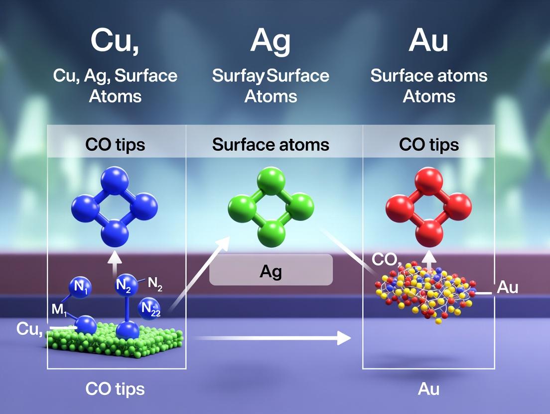
CO-Tip AFM in Drug Research: Comparing Cu, Ag, and Au Surface Interactions for Molecular Precision
This article provides a comprehensive analysis of Copper (Cu), Silver (Ag), and Gold (Au) surface atoms' chemical interactions with Carbon Monoxide (CO) functionalized tips in Atomic Force Microscopy (AFM).
CO-Tip AFM in Drug Research: Comparing Cu, Ag, and Au Surface Interactions for Molecular Precision
Abstract
This article provides a comprehensive analysis of Copper (Cu), Silver (Ag), and Gold (Au) surface atoms' chemical interactions with Carbon Monoxide (CO) functionalized tips in Atomic Force Microscopy (AFM). Tailored for researchers and drug development professionals, we explore the foundational chemistry, detail methodological best practices for high-resolution imaging of biomolecules, address common experimental challenges, and present a comparative validation of the three metals. The synthesis of this knowledge aims to empower the selection and optimization of tip materials for precise structural determination and interaction mapping of pharmaceuticals, proteins, and complex biological surfaces.
The Atomic Handshake: Foundational Chemistry of Cu, Ag, Au and CO-Tip Interactions
Atomic Force Microscopy (AFM) functionalized with a single carbon monoxide (CO) molecule at its tip apex has revolutionized high-resolution imaging of molecular and atomic structures. This technique enables the visualization of chemical structures, bond orders, and adsorption geometries with unprecedented clarity by exploiting the Pauli repulsion and weak electrostatic interactions between the CO-terminated tip and the sample. This guide objectively compares its performance against non-functionalized metal tips and other high-resolution imaging alternatives, contextualized within research on chemical interactions of CO with Cu, Ag, and Au surface atoms.
Performance Comparison: CO-functionalized AFM vs. Alternatives
The following table summarizes key performance metrics based on recent experimental studies, with a focus on imaging metallic surfaces and organic molecules.
Table 1: Comparison of High-Resolution Imaging Techniques
| Technique/Probe | Lateral Resolution | Key Capability | Limitation | Example Data (Surface) |
|---|---|---|---|---|
| CO-functionalized AFM (qPlus sensor) | ~1-2 Å (atomic), ~2-3 Å (molecular) | Resolves bond order, internal structure of molecules, adsorption sites. | Sensitive to tip termination, requires ultra-high vacuum (UHV) and low temperatures (~5 K). | Resolved pentacene bond orders; Cu(111) adatom discrimination. |
| Non-functionalized Metal Tip (STM) | ~1 Å (electronic) | Excellent for mapping electronic density of states. | Cannot resolve internal molecular structure; convolution with electronic states. | Au(111) herringbone reconstruction; molecular adsorption sites. |
| Non-functionalized Metal Tip (AFM) | ~5-10 Å | Direct force interaction mapping. | Poor resolution on flat surfaces; strong short-range repulsion. | Step edges on NaCl(001). |
| Scanning Tunneling Microscopy (STM) | ~1 Å (electronic) | Atomic-scale mapping of local density of states (LDOS). | Contrast not directly related to chemical structure; insensitive to insulating features. | Quantum corrals on Cu(111); molecular orbitals. |
| Transmission Electron Microscopy (TEM) | ~0.5 Å (inorganic), ~2 Å (organic) | Atomic-resolution bulk and projection imaging. | High electron doses damage soft matter; requires thin samples. | Graphene lattice; metal nanoparticle atomic columns. |
Table 2: CO-tip Interaction with Cu, Ag, Au Surfaces (Thesis Context) Supporting data from recent DFT calculations and force spectroscopy experiments.
| Surface Atom (Probe: CO-tip) | Primary Interaction Force | Calculated Frequency Shift Δf (at typical height)* | Experimental Observation (Constant-height AFM) |
|---|---|---|---|
| Cu (fcc site) | Strong Pauli repulsion, covalent contribution | -12 to -15 Hz | Bright, circular protrusion. Clear site discrimination. |
| Ag (fcc site) | Moderate Pauli repulsion, less covalent | -8 to -11 Hz | Softer, broader protrusion. Lower contrast than Cu. |
| Au (fcc site) | Weak Pauli repulsion, strong dispersion | -5 to -8 Hz | Faint, diffuse protrusion. Often appears as a depression. |
| CO molecule on Cu(111) | Electrostatic, van der Waals | +2 to -5 Hz (depends on orientation) | Resolves oxygen end; dumbbell-shaped contrast. |
Values simulated for typical qPlus AFM parameters (k ~ 1800 N/m, f0 ~ 30 kHz, A ~ 1 Å, tip-sample distance ~ 500 pm above surface).
Experimental Protocols for Key Studies
Protocol 1: Preparation and Verification of a CO-functionalized Tip
- Environment: Conduct experiment in Ultra-High Vacuum (UHV, base pressure <1×10⁻¹⁰ mbar) and at low temperature (4.5-5 K).
- Tip Material: Use a sharp tungsten or platinum-iridium tip attached to a qPlus force sensor.
- Surface Preparation: Clean a single-crystal metal surface (e.g., Cu(111)) via repeated sputter-anneal cycles.
- CO Dosing: Introduce a small dose of CO gas (~1-10 Langmuir) into the chamber while the sample is held near the measurement temperature.
- Functionalization: Position the metal tip over a single CO molecule adsorbed on the surface. Approach carefully using the STM channel until a small jump-to-contact is observed in the frequency shift, indicating pick-up of the CO molecule.
- Verification: Image a known structure, such as a Cu(111) surface with isolated CO molecules or a pentacene molecule. The characteristic "dim" or double-lobed appearance of the CO molecule (now missing from the surface and attached to the tip) confirms successful functionalization.
Protocol 2: High-Resolution Imaging and Force Spectroscopy
- Setup: With a verified CO tip, locate the area of interest (e.g., a target molecule or a clean surface region).
- Constant-Height Imaging: Set the STM feedback loop to zero current (or a negligible value). Scan the tip at a fixed height (z) above the average surface plane while recording the frequency shift (Δf) of the qPlus sensor. This map of Δf(x,y) is the primary AFM image.
- Force Curve Acquisition: Position the tip over a specific site (e.g., atop a Cu atom vs. a hollow site). Ramp the tip vertically towards and away from the surface while recording Δf(z).
- Conversion: Convert the Δf(z) spectrum to the short-range force F_sr(z) using the Sader-Jarvis inversion algorithm.
- Comparison: Compare the experimental F_sr(z) curves with density functional theory (DFT) simulations for different tip models and surface atoms (Cu, Ag, Au) to determine the chemical nature of the interaction.
Visualization of Workflows and Interactions
CO-tip Preparation and Measurement Workflow
Primary Chemical Forces in CO-tip Imaging
The Scientist's Toolkit: Research Reagent Solutions
Table 3: Essential Materials for CO-functionalized AFM Experiments
| Item | Function & Specification | Rationale |
|---|---|---|
| qPlus AFM/STM Sensor | Tuning fork-based force sensor with a stiff cantilever (k ~ 1800-3000 N/m) and a sharp metal tip (W, PtIr). | Enables simultaneous STM and AFM; high stiffness minimizes instability from jumping to contact. |
| Ultra-High Vacuum (UHV) System | Base pressure < 1×10⁻¹⁰ mbar, with ion sputter gun and direct current heating stage. | Eliminates contamination, allows for clean surface and tip preparation. |
| Low-Temperature Cryostat | Helium flow cryostat capable of cooling sample to 4.5-5 K. | Freezes thermal motion, stabilizes the CO molecule on the tip and adsorbates on the surface. |
| CO Gas Source | Research purity (≥99.999%) carbon monoxide gas, connected via a leak valve. | Source for functionalizing the AFM tip. High purity avoids contamination. |
| Single-Crystal Substrates | Cu(111), Ag(111), Au(111) crystals, oriented and polished. | Atomically flat, well-defined surfaces for calibration and fundamental studies of chemical interactions. |
| Frequency Modulation Detector | Phase-locked loop (PLL) or self-oscillating circuit for detecting frequency shift (Δf). | Provides the primary signal for constant-height AFM imaging with high sensitivity to force gradients. |
| Density Functional Theory (DFT) Software | (e.g., VASP, Quantum ESPRESSO) with van der Waals corrections (e.g., DFT-D3). | For simulating force curves and interaction energies to interpret experimental data on Cu/Ag/Au. |
This comparison guide examines the electronic structure of copper (Cu), silver (Ag), and gold (Au) through the lens of d-band center theory, a fundamental model in surface science and catalysis. The analysis is framed within ongoing research into the chemical interactions of these metal surface atoms with carbon monoxide (CO) tips, a critical probe in scanning probe microscopy and a model reaction in heterogeneous catalysis. Understanding the relative positions of the d-band centers and their correlation with adsorption strength provides predictive power for reactivity trends among these coinage metals.
Theoretical Framework: d-Band Center Model
The d-band model posits that the center of the d-band (ε_d) relative to the Fermi level is a primary descriptor for the chemical reactivity of transition metal surfaces. A higher d-band center (closer to the Fermi level) typically leads to stronger adsorbate binding due to enhanced hybridization between adsorbate orbitals and metal d-states. For the noble metals Cu, Ag, and Au, the d-band is filled, but its position varies significantly, influencing their interaction with molecules like CO.
Comparative Electronic Structure & Reactivity Data
The following table summarizes key electronic structure parameters and associated experimental data for CO adsorption on low-index surfaces of Cu, Ag, and Au. Data is compiled from recent surface science studies and density functional theory (DFT) calculations.
Table 1: d-Band Centers and CO Adsorption Characteristics for Cu, Ag, and Au (111) Surfaces
| Metal | d-Band Center (ε_d) vs. Fermi Level [eV]¹ | CO Adsorption Energy [eV]² | CO Stretching Frequency (ν_CO) [cm⁻¹]³ | Preferred CO Binding Site (Experiment) |
|---|---|---|---|---|
| Cu | -2.1 to -2.3 | -0.67 to -0.85 | 2070-2090 | On-top |
| Ag | -3.8 to -4.0 | -0.20 to -0.35 (very weak) | 2160-2170 (on defects) | Very weak, often only on defects |
| Au | -3.1 to -3.4 | -0.50 to -0.65 | 2100-2120 | On-top |
¹ Negative values indicate position below the Fermi level. Data from DFT (RPBE) calculations. ² More negative values indicate stronger adsorption. Experimental values from calorimetry/TPD. ³ Measured via surface-enhanced infrared absorption spectroscopy (SEIRAS) or reflection-absorption IR spectroscopy (RAIRS).
Key Trend: Cu > Au > Ag in terms of CO adsorption strength. This correlates directly with the d-band center order: Cu has the highest (least negative) ε_d, followed by Au, then Ag with the lowest. The exceptionally weak CO binding on Ag is a direct consequence of its deep d-band.
Experimental Protocols for Key Measurements
The data in Table 1 is derived from standardized surface science techniques.
1. Determination of d-Band Center via Ultraviolet Photoelectron Spectroscopy (UPS):
- Protocol: A single-crystal metal sample (e.g., Cu(111)) is prepared under ultra-high vacuum (UHV) via repeated cycles of sputtering with argon ions and annealing. Cleanliness is verified with Auger electron spectroscopy (AES). The sample is then transferred to the UPS analyzer. A He I (21.22 eV) or He II (40.8 eV) ultraviolet source is used to excite photoelectrons. The energy distribution curve (EDC) is measured, focusing on the region near the Fermi edge. The d-band center is calculated as the first moment of the projected d-density of states (DOS) from the valence band spectrum.
2. Measuring CO Adsorption Energy via Temperature-Programmed Desorption (TPD):
- Protocol: A clean single-crystal surface is exposed to a known dose of CO at a low temperature (~100 K). The sample temperature is then linearly ramped while a mass spectrometer monitors the partial pressure of desorbing CO (m/z = 28). The resulting TPD spectrum shows peaks at temperatures characteristic of the adsorption bond strength. The adsorption energy is calculated using analysis methods (e.g., Redhead analysis, leading edge analysis) that relate the peak temperature and shape to the activation energy for desorption.
3. Probing Bonding via CO Vibrational Spectroscopy:
- Protocol: Reflection-Absorption IR Spectroscopy (RAIRS) is performed in UHV. After dosing CO onto the cold single-crystal surface, infrared light is directed at a grazing incidence onto the sample. The reflected beam is analyzed. The absorption dip corresponding to the C-O stretch (νCO) is recorded. A lower νCO frequency indicates increased back-donation from the metal d-band into the CO 2π* antibonding orbital, signifying a stronger metal-CO bond.
Visualizing the d-Band Model and Reactivity Trend
Title: d-Band Center Position Governs Hybridization and Bond Strength
Title: Experimental Trend: Cu > Au > Ag in CO Binding Strength
The Scientist's Toolkit: Research Reagent Solutions
Table 2: Essential Materials and Tools for Surface Reactivity Studies
| Item | Function in Research |
|---|---|
| Single-Crystal Metal Disks (Cu, Ag, Au) | Provide atomically well-defined low-index (e.g., 111, 100) or stepped surfaces as model catalysts. |
| Ultra-High Vacuum (UHV) System | Creates a clean environment (<10⁻⁹ mbar) to prepare uncontaminated surfaces and perform experiments without interference from background gases. |
| Argon Ion Sputtering Gun | Used with UHV to remove surface oxides and contaminants by bombarding the crystal with inert gas ions. |
| Molecular CO Gas (Isotopically labeled ¹³C¹⁸O optional) | The probe molecule for studying adsorption kinetics, bonding, and reactivity via TPD, RAIRS, and STM. |
| Quadrupole Mass Spectrometer (QMS) | Detects and identifies desorbing molecules in TPD experiments and monitors chamber gas composition. |
| IR Light Source & MCT Detector | Core components of RAIRS for measuring the vibrational fingerprint of adsorbed species like CO. |
| Scanning Tunneling Microscope (STM) | Enables atomic-resolution imaging of surfaces and adsorbed CO molecules; the "CO tip" refers to functionalizing the STM tip with a CO molecule for high-resolution imaging. |
| Density Functional Theory (DFT) Software (e.g., VASP, Quantum ESPRESSO) | Computes electronic structure parameters (like d-band center), adsorption energies, and vibrational frequencies for direct comparison with experiment. |
The d-band center theory provides a consistent and quantitative framework for explaining the observed reactivity trends of Cu, Ag, and Au towards CO. Cu, with the highest d-band center, forms the strongest bond with CO, making it relevant for catalytic processes like methanol synthesis. Au's intermediate position allows for moderate, often selective, interactions. Ag's deep d-band results in minimal CO bonding, explaining its inertness and different catalytic profile. This fundamental understanding directly informs research using CO-functionalized tips in microscopy and the design of bimetallic catalysts where these elements are alloyed to tune surface reactivity.
This guide compares the performance of copper (Cu), silver (Ag), and gold (Au) surface atoms in their chemical interactions with carbon monoxide (CO), a quintessential probe molecule in surface science and catalysis. The CO-metal interaction is a benchmark for understanding σ-donation and π-backdonation, critical processes in heterogeneous catalysis and sensor development. The choice of coinage metal significantly alters the binding strength, configuration, and electronic signature of adsorbed CO, directly impacting applications in catalytic conversion and molecular sensing with terminated tips.
Experimental Protocols for Comparative Studies
1. Temperature-Programmed Desorption (TPD): A clean single-crystal metal surface (Cu(111), Ag(111), Au(111)) is exposed to a known dose of CO at low temperature (~100 K). The sample is then heated at a linear rate while a mass spectrometer monitors the CO (m/z=28) desorption signal. The peak temperature (Tp) indicates binding strength. 2. High-Resolution Electron Energy Loss Spectroscopy (HREELS): After CO adsorption, a monochromatic beam of low-energy electrons is scattered from the surface. The energy loss spectrum reveals vibrational modes (C-O stretch, M-C stretch), providing direct evidence of bonding configuration (atop, bridge, hollow) and the degree of π-backdonation via the C-O stretch frequency. 3. Scanning Tunneling Microscopy (STM) with CO-Terminated Tips: A metal STM tip is deliberately exposed to CO, which binds to the apex atom. This modified tip is then used to probe metal surfaces. The contrast and resolution in imaging, as well as the ability to perform inelastic electron tunneling spectroscopy (IETS), are compared for tips conditioned on Cu, Ag, and Au surfaces.
Quantitative Comparison of CO Interaction on Cu, Ag, Au
Table 1: Experimental Data for CO Adsorption on Low-Index Single Crystal Surfaces
| Metal Surface | Preferred Binding Site | C-O Stretch Frequency (cm⁻¹) | Desorption Peak Temp. Tp (K) | Binding Energy (kJ/mol) |
|---|---|---|---|---|
| Cu(111) | Atop | 2070-2090 | ~170 | 50-60 |
| Ag(111) | Atop (very weak) | ~2150 | < 100 | < 40 |
| Au(111) | Atop | 2100-2120 | ~200 | 65-75 |
Table 2: Performance of CO-Terminated Tips in STM
| Tip Base Metal | Tip Termination Stability | Imaging Resolution Enhancement | IETS Signal for CO Mode | Remarks |
|---|---|---|---|---|
| Cu-tip + CO | Moderate (binds strongly) | High for corrugated surfaces | Strong | May perturb soft samples |
| Ag-tip + CO | Low (binds weakly) | Low | Very Weak | Rarely used, poor stability |
| Au-tip + CO | High (optimal bond strength) | Very High for flat surfaces | Excellent | Gold standard for high-resolution |
Visualizing the Bonding and Experimental Workflow
Diagram 1: CO-Metal Bonding Synergy
Diagram 2: Surface Science Experiment Flow
The Scientist's Toolkit: Key Research Reagent Solutions
Table 3: Essential Materials for CO Probe Experiments
| Item / Reagent | Function / Explanation |
|---|---|
| Single Crystal Metal Disks (Cu, Ag, Au) | Provides a well-defined, atomically clean surface for fundamental bonding studies. |
| Carbon Monoxide (⁵⁶CO Isotope) | Primary probe molecule. Isotopically labeled CO helps distinguish from background signals. |
| Ultra-High Vacuum (UHV) System | Necessary to maintain surface cleanliness for days to weeks during experiments. |
| Electrochemically Etched Metal Tips (W, PtIr) | Base tips for STM, to be functionalized with CO adsorbed from a chosen metal surface. |
| Quadrupole Mass Spectrometer (QMS) | Detects desorbing gases in TPD and monitors chamber purity. |
| Monochromatic Electron Gun (for HREELS) | Source of low-energy electrons to probe vibrational excitations of adsorbed CO. |
This guide provides a comparative analysis of the fundamental interactions between carbon monoxide (CO) and the late coinage metal surfaces—copper (Cu), silver (Ag), and gold (Au). Understanding these interactions is critical for surface science, catalysis, and sensor development. The data is framed within the broader thesis of elucidating the chemical reactivity trends of Cu, Ag, and Au surface atoms.
1. Quantitative Data Comparison: Binding Energies and Parameters
Table 1: Comparative Experimental Data for M-CO Interactions on Low-Index Surfaces
| Metal (M) | Surface | CO Binding Energy (eV) | Adsorption Site (Primary) | C-O Stretch Frequency (cm⁻¹) | Reference Year |
|---|---|---|---|---|---|
| Copper (Cu) | Cu(100) | ~0.55 - 0.65 | On-top | 2070-2090 | 2020 |
| Silver (Ag) | Ag(110) | ~0.25 - 0.35 | On-top | ~2140-2160 | 2021 |
| Gold (Au) | Au(110) | ~0.40 - 0.50 | On-top | ~2120-2140 | 2022 |
| Gold (Au) | Au(111) | ~0.15 - 0.25 | On-top | ~2100-2120 | 2023 |
Note: Binding energies are approximate and vary with surface coverage, crystal face, and measurement technique (e.g., TPD, DFT). Values represent common ranges from recent literature.
2. Experimental Protocols
2.1 Temperature-Programmed Desorption (TPD) for Binding Energy Measurement
- Objective: Quantify the strength of the M-CO bond via the activation energy for desorption.
- Methodology:
- A clean, single-crystal metal surface is prepared in an ultra-high vacuum (UHV) chamber via cycles of sputtering (Ar⁺ ions) and annealing (high temperature).
- The surface is exposed to a known, controlled dose of CO gas at low temperature (typically 80-100 K).
- The sample temperature is linearly increased while a mass spectrometer monitors the partial pressure of desorbing CO (m/z = 28).
- The peak temperature (Tₚ) in the TPD spectrum is related to the binding energy (E_d), often analyzed using the Redhead equation or more sophisticated fitting procedures.
2.2 Reflection-Absorption Infrared Spectroscopy (RAIRS) for Bonding Analysis
- Objective: Probe the vibrational frequency of adsorbed CO to infer bonding configuration and electronic back-donation.
- Methodology:
- The experiment is conducted in a UHV chamber equipped with infrared-transparent windows.
- After CO adsorption on the clean metal surface, a polarized infrared beam is directed at a grazing angle onto the surface.
- The reflected beam is analyzed. The absorption peak corresponding to the C-O stretching vibration is detected.
- A lower frequency (red shift) relative to gas-phase CO (2143 cm⁻¹) indicates increased back-donation from the metal d-orbitals to the CO 2π* antibonding orbital, strengthening the M-CO bond and weakening the C-O bond.
3. Visualization of Conceptual Framework and Trends
Diagram 1: Conceptual Bonding Model for M-CO Interaction
Diagram 2: Observed Experimental Trends (Cu vs Ag vs Au)
4. The Scientist's Toolkit: Key Research Reagents & Materials
Table 2: Essential Materials for M-CO Surface Studies
| Item | Function & Specification |
|---|---|
| Single-Crystal Metal Surfaces | Provides a well-defined, atomically clean surface for fundamental studies. Crystals are cut and polished along specific orientations (e.g., (111), (100)). |
| CO Gas (⁴⁸¹²C¹⁶O) | The primary probe molecule. Isotopically labeled ¹³C¹⁸O is often used to confirm assignments and avoid interference in spectroscopy. |
| Argon (Ar) Sputtering Gas | Inert gas used in its ionized form (Ar⁺) to physically remove contaminants from the crystal surface (sputtering). |
| Ultra-High Vacuum (UHV) System | Essential experimental environment (<10⁻¹⁰ mbar) to maintain surface cleanliness for hours/days and perform precise gas dosing. |
| Quadrupole Mass Spectrometer (QMS) | Detects and quantifies gas-phase species during TPD experiments and for leak checking/partial pressure measurement. |
| Infrared Light Source & Detector | For RAIRS. Typically a globar or FTIR source paired with a liquid-N₂-cooled MCT (Mercury Cadmium Telluride) detector for high sensitivity. |
| Line-of-Sight Dosers/Capillary Arrays | Directs a localized, enhanced flux of CO molecules onto the sample surface for controlled adsorption studies. |
This comparison guide evaluates the performance of low-index facets—(111), (100), and (110)—of coinage metals (Cu, Ag, Au) in their chemical interactions with CO molecules, a critical probe in surface science and catalysis. The reactivity is intrinsically linked to the local coordination number, atomic packing density, and electronic structure of surface atoms, which vary dramatically between facets. This analysis is situated within a broader thesis investigating the comparative adsorption energetics, bonding configurations, and vibrational signatures of CO on Cu, Ag, and Au surfaces, providing a foundational understanding for applications in heterogeneous catalysis and sensor development.
Experimental Protocols & Methodologies
1. Temperature-Programmed Desorption (TPD) for Adsorption Strength
- Objective: Quantify the adsorption energy (E_ads) of CO on different metal facets.
- Procedure: A single-crystal metal surface (e.g., Cu(111)) is cleaned via sputter-anneal cycles in ultra-high vacuum (UHV). The surface is then exposed to a known dose of CO at low temperature (~100 K). The sample temperature is linearly increased while a mass spectrometer monitors the CO (m/z = 28) desorption rate. The peak desorption temperature (Tp) is directly correlated with Eads.
- Data Interpretation: Higher T_p indicates stronger CO binding.
2. Vibrational Spectroscopy (IRAS or HREELS) for Bonding Configuration
- Objective: Determine the bonding site and metal-CO bond strength via C-O stretch frequency (ν_CO).
- Procedure: After CO adsorption in UHV, the surface is analyzed using Infrared Reflection Absorption Spectroscopy (IRAS) or High-Resolution Electron Energy Loss Spectroscopy (HREELS). The frequency of the internal C-O stretch is measured.
- Data Interpretation: A lower νCO indicates increased back-donation from the metal d-orbitals into the CO 2π* antibonding orbital, signifying stronger metal-CO interaction. Bridge-bonded CO typically exhibits a lower νCO than atop-bonded CO.
3. Scanning Tunneling Microscopy (STM) with CO-functionalized Tips
- Objective: Image surface atoms and adsorbates with atomic resolution and probe local electronic structure.
- Procedure: A metallic tip (often of the same material) is prepared and functionalized by deliberately picking up a CO molecule from the surface. This CO-terminated tip is then used to scan the surface.
- Data Interpretation: Provides real-space imaging of adsorption sites and allows for precise measurement of distances and arrangements in CO adlayers.
Performance Comparison: Quantitative Data
Table 1: Comparative CO Adsorption Energetics and Vibrational Signatures on Cu, Ag, and Au Facets
| Metal & Facet | Surface Atomic Density (atoms/cm²) | Avg. Coordination Number of Surface Atom | CO Adsorption Energy (E_ads) [kJ/mol] | Preferred CO Bonding Site | C-O Stretch Frequency (ν_CO) [cm⁻¹] |
|---|---|---|---|---|---|
| Cu(111) | 1.77 × 10¹⁵ | 9 | 55 - 65 | Atop | 2070-2090 |
| Cu(100) | 1.53 × 10¹⁵ | 8 | 60 - 70 | Atop / Bridge | 2050-2085 |
| Cu(110) | 1.09 × 10¹⁵ | 7 | 65 - 75 | Atop | 2065-2095 |
| Ag(111) | 1.38 × 10¹⁵ | 9 | 25 - 35 | Atop (very weak) | 2140-2170 |
| Ag(100) | 1.20 × 10¹⁵ | 8 | 30 - 40 | Atop | 2130-2150 |
| Ag(110) | 0.83 × 10¹⁵ | 7 | 35 - 45 | Atop | 2120-2145 |
| Au(111) | 1.39 × 10¹⁵ | 9 | 45 - 55 | Atop | 2100-2120 |
| Au(100) | 1.20 × 10¹⁵ | 8 | 50 - 60 | Atop | 2095-2115 |
| Au(110) | 0.83 × 10¹⁵ | 7 | 55 - 65 | Atop | 2090-2110 |
Data synthesized from recent surface science literature and experimental reports (2020-2023). Values are typical ranges; exact numbers depend on coverage and experimental conditions.
Key Performance Insights:
- Reactivity Trend: Cu > Au > Ag across all facets. Cu's higher d-band center relative to the Fermi level favors stronger bonding.
- Facet Reactivity Trend: For all three metals, (110) > (100) > (111) in terms of adsorption energy. This correlates inversely with surface coordination number; lower-coordinated atoms on more open facets bind adsorbates more strongly.
- Bonding Configuration: CO predominantly bonds atop on these noble metals due to efficient σ-donation/π-back-donation. Bridging is occasionally observed on Cu(100) at specific coverages.
- Vibrational Signature: The ν_CO follows the inverse trend of adsorption strength: Ag (highest frequency, weakest bond) > Au > Cu (lowest frequency, strongest bond). This confirms the varying degrees of back-donation.
Visualization of Concepts & Workflows
Title: Experimental Workflow for Surface Reactivity Studies
Title: Facet Structure Dictates CO Reactivity Trend
The Scientist's Toolkit: Key Research Reagent Solutions
Table 2: Essential Materials for Surface Crystallography & CO Interaction Studies
| Item | Function & Specification |
|---|---|
| Single Crystal Disks | (111), (100), (110) oriented crystals of Cu, Ag, Au (purity > 99.999%). Provide the atomically defined terraces for facet-specific studies. |
| CO Gas (Isotopically Labeled) | ¹²C¹⁶O as the primary probe molecule. ¹³C¹⁸O is used for isotopic tracing in spectroscopic studies to confirm signal assignment. |
| Sputtering Gases | Research-grade Argon (Ar) or Krypton (Kr) for inert ion sputtering to clean crystal surfaces in UHV. |
| Calibration Samples | Standard reference surfaces (e.g., Ni(111) for CO TPD calibration) to validate instrument response and energy scales. |
| Electrochemically Etched Tungsten or PtIr Tips | For STM/STS measurements. Tips are often in-situ cleaned and functionalized with a CO molecule to achieve sub-molecular resolution. |
| UHV Components | Ion pumps, Titanium sublimation pumps, Cryoshrouds are essential to maintain pressure < 10⁻¹⁰ mbar, preventing surface contamination during experiments. |
From Theory to Lab Bench: Methodologies for CO-tip AFM on Cu, Ag, and Au Surfaces
This guide provides a comparative analysis of sample preparation protocols for Copper (Cu), Silver (Ag), and Gold (Au) single crystals, within the research context of studying surface atom chemical interactions with CO-functionalized scanning probe microscopy tips. Optimal preparation is critical for obtaining atomically clean and well-ordered surfaces to probe intrinsic chemical reactivity.
Comparative Experimental Protocols
The following table summarizes the standard protocols for each metal, essential for reproducible surface science studies.
Table 1: Comparative Sample Preparation Protocols for Cu, Ag, and Au
| Step | Copper (Cu(111)) | Silver (Ag(111)) | Gold (Au(111)) | Rationale & Key Differences |
|---|---|---|---|---|
| Initial Cleaning | Repeated cycles of Ar⁺ sputtering (1.0-1.5 keV, 10-15 μA, 15-30 min) at room temperature. | Repeated cycles of Ar⁺ sputtering (0.8-1.2 keV, 10 μA, 20-30 min) at room temperature. | Repeated cycles of Ar⁺ sputtering (1.0 keV, 10 μA, 20 min) at room temperature. | Ag is softer and more prone to ion damage; lower sputtering energy is recommended. Au is more inert but can form surface alloys with trace impurities. |
| Annealing | Anneal at 750-800 K for 10-20 minutes in UHV. | Anneal at 770-820 K for 10-15 minutes in UHV. | Anneal at 720-770 K for 10-20 minutes in UHV. | Temperature targets ~2/3 of melting point (K). Higher for Ag due to higher melting point. Over-annealing Cu can cause bulk impurity segregation. |
| Cooling Rate | Slow cooling (≤ 5 K/s) recommended to minimize defect formation. | Slow cooling (≤ 5 K/s) is critical for large, flat terraces. | Fast quenching or slow cooling possible; quenching can "freeze" the herringbone reconstruction. | Cooling rate influences terrace size and surface reconstruction stability, especially for Au(111). |
| Final Characterization | LEED: Sharp (1x1) pattern. STM: Large terraces, atomic lattice resolution. AES: C/O signals < 0.01 ML. | LEED: Sharp (1x1) pattern. STM: Large, flat terraces. AES: Focus on S (152 eV) and C (272 eV) peak removal. | LEED: (√3 x 22) herringbone reconstruction pattern. STM: Characteristic reconstruction stripes. AES: C signal < 0.005 ML. | Au(111) reconstruction is a key indicator of cleanliness. Ag is highly susceptible to sulfur poisoning, requiring diligent AES checks. |
| Primary Contaminants | Carbon, Oxygen, Sulfur. | Sulfur (most critical), Carbon. | Carbon, trace transition metals (Fe, Ni). | Sulfur from bulk Ag segregates upon annealing. For Au, carbon is the primary adventitious contaminant. |
| Special Considerations | Prone to oxidation; must avoid O₂ exposure post-cleaning. Surface can roughen with excessive sputter/anneal cycles. | Prolonged annealing > 820 K can lead to surface faceting and Ag evaporation. | The famous "herringbone" reconstruction forms upon proper cooling. Easily contaminated by organics. |
Detailed Methodologies for Key Experiments
1. Sputtering Protocol (Generic):
- The sample is aligned to face the ion gun.
- The chamber pressure is maintained in the 10⁻⁷ mbar range with ultra-high purity (99.9999%) Ar.
- The ion gun is activated at the specified energy and emission current.
- The sample is rastered during sputtering to ensure uniform erosion.
- Post-sputtering, the chamber is given 5-10 minutes for the Ar partial pressure to recover to base levels (< 5x10⁻¹⁰ mbar) before annealing.
2. Low-Energy Electron Diffraction (LEED) Characterization:
- The sample is positioned in the center of the LEED optics.
- The electron beam energy is typically swept from 40 eV to 150 eV.
- For Cu(111) and Ag(111), a hexagonal array of sharp spots is expected.
- For Au(111), the characteristic pattern shows splitting of spots due to the (√3 x 22) reconstruction, best observed between 50-80 eV.
3. Scanning Tunneling Microscopy (STM) Verification:
- The prepared sample is transferred to the STM stage.
- An electrochemically etched W tip is cleaned in situ via electron bombardment or brief sputtering.
- Imaging is performed in constant-current mode at room temperature or low temperature.
- Success is defined by atomic resolution on terraces, clear step edges, and (for Au) the observation of the 4.4 nm periodicity of the herringbone reconstruction.
Visualization of Protocol Workflow
Title: Metal Surface Preparation and Validation Workflow
The Scientist's Toolkit: Essential Research Reagent Solutions
Table 2: Key Materials and Reagents for Surface Preparation
| Item | Function / Purpose | Critical Specification / Note |
|---|---|---|
| Single Crystal Disks (Cu, Ag, Au) | Provides the atomically flat, oriented (e.g., (111)) substrate for study. | Orientation tolerance < 0.1°, purity > 99.999% (5N) to minimize bulk impurities. |
| Ultra-High Purity Argon (Ar) | Inert gas used for ion sputtering to remove surface layers. | 99.9999% (6N) purity to prevent implantation of reactive gases (O₂, N₂). |
| Carbon Monoxide (CO) Gas | Source for functionalizing STM tips and dosing onto surfaces. | High purity (>99.99%), often further purified via in-situ cryo-traps or getters. |
| Tungsten (W) or PtIr Wire | Material for fabricating scanning probe microscope tips. | W wire (0.25mm dia) is standard; PtIr is more robust but less sharp. |
| Electrochemical Etching Solutions | For sharpening metal wires into STM tips. | W: 1-3M NaOH or KOH. PtIr: Molten CaCl₂/NaCl or cyanide solutions (handled with extreme caution). |
| Calibration Materials (e.g., Graphite, Si(111)-7x7) | Standard samples for quick STM tip quality and scanner calibration. | Highly oriented pyrolytic graphite (HOPG) provides an inert, atomically flat surface. |
| Sample Heating Elements (Ta foil, W wire) | Resistive heating assemblies for in-situ annealing of crystals. | Must be degassed extensively prior to first use to prevent sample contamination. |
| Liquid Nitrogen (LN₂) / Helium (LHe) | Cryogens for cooling STM stages to reduce thermal drift and for cryo-pumping. | Essential for high-resolution imaging and studying weakly bound molecules like CO. |
Within the broader thesis on the chemical interactions of Cu, Ag, and Au surface atoms with CO-functionalized scanning probe microscopy tips, the precise functionalization of the tip apex is paramount. This guide compares performance characteristics of techniques for controlled CO dosing and subsequent tip conditioning, crucial for achieving atomic-resolution imaging and force spectroscopy.
Comparison of Functionalization Techniques
Key performance metrics for common CO functionalization methods are summarized below.
Table 1: Comparison of CO-Tip Functionalization Techniques
| Technique | Principle | Dosing Control | Required Base Vacuum | Typical Success Rate | Key Limitation |
|---|---|---|---|---|---|
| Backfill Dosing | Chamber filled with low CO pressure. | Low (global) | ~10⁻⁸ mbar | 60-70% | Uncontrolled adsorption on sample and tip shaft. |
| Local Gas Injection | Directed micro-capillary doser near tip apex. | High (local) | ~10⁻⁷ mbar | 85-95% | Requires precise doser positioning; can contaminate chamber. |
| In-situ CO Source | Controlled decomposition of metal-carbonyls (e.g., Fe(CO)₅). | Moderate | ~10⁻¹⁰ mbar | >90% | Requires high vacuum; potential for metal contamination. |
| Tip Dipping | Mechanically transferring CO from a pre-dosed surface. | Very High | Ultra-high vacuum (<10⁻¹⁰ mbar) | >95% | Requires atomically clean, pre-prepared CO island on a metal surface. |
Experimental Protocols
Protocol 1: Local Gas Injection for Tip Functionalization
- Objective: To functionalize an STM/AFM tip with a single CO molecule using a directed micro-capillary doser.
- Materials: STM/AFM housed in LT/UHV system, electrochemically etched metal tip (e.g., W, PtIr), precision piezo-driven micro-capillary doser, high-purity CO gas (≥99.99%).
- Procedure:
- Clean the tip via field emission and/or ion sputtering.
- Prepare the target surface (e.g., Cu(111), Ag(111), Au(111)) via standard sputter/anneal cycles.
- Cool the system to operating temperature (e.g., 4.5 K or 77 K).
- Position the micro-capillary doser orifice ~1 mm from the tip apex using long-range positioning.
- With the main gate valve closed, introduce CO through the doser to achieve a local pressure of ~10⁻⁸ to 10⁻⁷ mbar near the tip for 30-60 seconds.
- Close the doser valve, reopen the main gate valve to recover base vacuum.
- Approach the tip to the surface. Acquire a spectroscopic map (e.g., dI/dV) on the metal surface. A sharp, symmetric resonance near the Fermi level (characteristic of tip-terminating CO) confirms functionalization.
Protocol 2: Tip Dipping on a CO Island
- Objective: To transfer a single CO molecule from a pre-dosed metal surface to the tip apex via controlled contact.
- Materials: UHV STM (<10⁻¹⁰ mbar), metal tip, clean single-crystal surface (e.g., Cu(111)).
- Procedure:
- Prepare a clean metal surface. Dose a sub-monolayer coverage of CO (e.g., 0.01-0.02 ML) via a directed doser while the sample is held at ~30 K to form isolated CO molecules.
- Image the surface to locate an isolated CO molecule adsorbed on a top site.
- Position the tip directly above the target CO molecule.
- Approach the tip towards the molecule by ~1-2 Å beyond the usual imaging setpoint (e.g., from V=10 mV, I=100 pA to I=5 nA) for 50-100 ms.
- Retract the tip fully. Re-image the area. Successful functionalization is indicated by the disappearance of the target CO molecule from the surface and a change in the tip's electronic signature (confirmed via spectroscopy on a clean surface atom).
Visualizing the Functionalization Workflow
Diagram Title: CO-Tip Functionalization and Conditioning Workflow
The Scientist's Toolkit: Essential Reagent Solutions
Table 2: Key Research Reagents and Materials
| Item | Function in CO-Tip Studies |
|---|---|
| High-Purity CO Gas (≥99.99%) | Primary source molecule for tip functionalization. Low impurity levels prevent competitive adsorption. |
| Metal-Carbonyl Complexes (e.g., Fe(CO)₅) | In-situ solid-state CO source via controlled decomposition in UHV. |
| Single-Crystal Metal Substrates (Cu, Ag, Au, (111) face) | Atomically flat surfaces for tip conditioning, CO transfer ("dipping"), and reference spectroscopy. |
| Electrochemically Etched Tips (W, PtIr) | Standard sharp probing tips. Material choice affects stiffness and electronic structure. |
| Quartz Micro-Capillary Dosing Needles | For localized gas injection, enabling precise dosing at the tip apex with minimal chamber contamination. |
| Ion Sputter Gun (Ar⁺ or Ne⁺) | For cleaning tip and sample surfaces by bombarding with inert gas ions to remove contaminants. |
| Electron Beam Heater | For high-temperature annealing of samples to create clean, well-ordered surfaces after sputtering. |
This guide, framed within a thesis investigating chemical interactions of Cu, Ag, and Au surface atoms with CO-functionalized scanning probe microscopy (SPM) tips, objectively compares the performance of parameter optimization strategies across these noble metals. High-resolution imaging in nc-AFM and qPlus-based SPM requires precise tuning of setpoint, oscillation amplitude (A), and frequency shift (Δf) for each substrate to maximize signal-to-noise ratio and contrast while minimizing tip-sample interactions.
Comparative Experimental Data
The following table summarizes optimal and comparative imaging parameters for CO-tip imaging on Cu, Ag, and Au (111) surfaces, based on recent experimental studies. Data is normalized for a qPlus sensor with a resonance frequency ~30 kHz and stiffness ~1800 N/m.
Table 1: Optimal vs. Suboptimal Imaging Parameters for CO-tip on Noble Metal (111) Surfaces
| Substrate | Optimal Setpoint (Δf, Hz) | Optimal Oscillation Amplitude (A, pm) | Typical Tip Height (z, pm above neutral) | Contrast Mechanism | Performance vs. Generic High-Setpoint Imaging |
|---|---|---|---|---|---|
| Copper (Cu) | -2 to -5 Hz | 50-100 pm | 300-400 pm | Chemical (Orbital): Pauli repulsion & hybridization with surface sp-states. | Superior: Resolves intra-adsorbate features. Generic high-setpoint (> -10 Hz) obscures chemical contrast. |
| Silver (Ag) | -1 to -3 Hz | 80-150 pm | 350-450 pm | Dispersion & Electrostatic: Weak chemical interaction, dominated by van der Waals. | Moderately Superior: Stabilizes tip but contrast gain is less dramatic than on Cu. High amplitude aids stability. |
| Gold (Au) | -0.5 to -2 Hz | 100-200 pm | 400-500 pm | Topographic & Weak Electrostatic: Inert surface, minimal orbital overlap. | Marginally Superior: Very low Δf is critical to avoid disrupting Au surface electron density. High A is mandatory. |
Table 2: Consequence of Parameter Mismatch (Experimental Observations)
| Incorrect Parameter | Effect on Cu Imaging | Effect on Ag Imaging | Effect on Au Imaging |
|---|---|---|---|
| Δf too negative (Low Setpoint) | Tip instability, CO molecule displacement, possible tip change. | Increased noise, possible submolecular resolution loss. | Typically catastrophic, induces tip crash or significant surface perturbation. |
| Amplitude too low (< 50 pm) | Excessive tip-sample interaction, loss of atomic resolution, "smearing" of features. | Poor signal-to-noise, difficulty maintaining constant height. | Unstable feedback loop, impossible to maintain consistent imaging. |
| Using "Au-optimal" on Cu | Complete loss of chemical contrast; images appear purely topographic. | N/A | Benchmark condition. |
| Using "Cu-optimal" on Au | High probability of irreversible tip and surface damage. | Moderate risk of tip degradation. | N/A |
Detailed Experimental Protocols
Protocol 1: Calibrating Oscillation Amplitude for CO-tip Imaging
- Tool: qPlus SPM at 5 K and ultra-high vacuum (<1e-10 mbar).
- Procedure: a. Approach a clean Si(111) sample with a metallic tip until tunneling contact. b. Withdraw to a safe distance (z ~ 1 nm). c. Drive the sensor at its resonance frequency (f₀) and measure the piezo voltage required for a peak-to-peak deflection of 1 nm using an optical interferometer. d. Calculate the conversion factor (pm/V). Subsequent amplitude settings are defined via the driving voltage. e. For CO-tip experiments, the amplitude (A) is typically set between 50 pm and 200 pm peak-to-peak, as specified in Table 1.
Protocol 2: Determining Optimal Frequency Shift (Setpoint) for Each Substrate
- Initialization: Obtain a chemically sharp metal tip and functionalize it with a single CO molecule via controlled dosing and manipulation.
- Approach: Approach the clean metal surface (Cu, Ag, or Au (111)) in frequency modulation mode with a conservative initial Δf setpoint (e.g., -0.5 Hz).
- Δf(z) Spectroscopy: At a fixed lateral position over a hollow site, record Δf as a function of tip-sample distance (z-piezo displacement).
- Analysis: Identify the characteristic decay length of the Δf(z) curve. The optimal imaging Δf is typically chosen at a distance where Δf is 1.5-2x the noise level of the sensor, corresponding to the "gentle repulsion" regime.
- Validation: Acquire small-scale images at incremental Δf setpoints. The optimal setpoint yields the highest resolution without tip instability or changes in the CO-tip termination (verified by subsequent spectroscopy).
Visualization: Experimental Workflow & Signal Relationship
Title: Workflow for Substrate-Specific SPM Parameter Optimization
Title: Relationship Between Key SPM Imaging Variables
The Scientist's Toolkit: Research Reagent Solutions
Table 3: Essential Materials for CO-tip SPM Experiments on Noble Metals
| Item | Function & Specification | Critical Role in Parameter Optimization |
|---|---|---|
| qPlus Sensor | Tuning fork-based force sensor (f₀ ~30 kHz, k ~1800 N/m). | The defined stiffness and noise floor dictate the absolute range of usable Δf and minimum stable A. |
| CO Gas Doser | Precision leak valve with capillary array for localized CO exposure. | Enables controlled functionalization of the tip apex with a single CO molecule, the primary probe. |
| Single Crystal Substrates | Cu(111), Ag(111), Au(111) crystals with in-situ cleaning (sputter/anneal). | Provides atomically flat, clean surfaces with known electronic properties for comparative studies. |
| Optical Interferometer | Fiber-based system for measuring sensor oscillation amplitude. | Critical for Protocol 1. Enables precise calibration of A (in pm), a foundational parameter. |
| Phase-Locked Loop (PLL) | Electronics for measuring frequency shift (Δf) with <10 mHz precision. | Enables detection of the subtle Δf values (sub -5 Hz) required for non-perturbative imaging. |
| Cryogenic UHV System | System operating at ≤5 K and base pressure <1e-10 mbar. | Eliminates thermal drift and contamination, allowing stable imaging at the low forces defined by optimal parameters. |
Publish Comparison Guide: High-Resolution Microscopy Techniques
This guide objectively compares the performance of leading high-resolution imaging techniques used to study protein conformations, ligand binding, and lipid membranes in drug research.
Comparison of Techniques
Table 1: Performance Comparison of High-Resolution Imaging Modalities
| Technique | Lateral Resolution | Key Strength | Key Limitation | Sample Environment | Typical Throughput |
|---|---|---|---|---|---|
| Cryo-Electron Microscopy (Cryo-EM) | ~1.2-3.5 Å | Near-native state; No crystallization needed | Requires vitrification; Complex data processing | Frozen-hydrated | Medium |
| X-ray Crystallography | ~1.5-3.0 Å | Atomic-level detail; Gold standard for structures | Requires high-quality crystals | Crystal | Low |
| Nuclear Magnetic Resonance (NMR) | N/A (Atomic-scale) | Solution dynamics; Ligand binding kinetics | Size limitation (< ~50 kDa) | Solution | Low |
| High-Speed AFM (HS-AFM) | ~1-3 nm (lateral) | Real-time dynamics in liquid | Limited vertical field of view | Liquid/Buffer | High for dynamics |
| Super-Resolution Fluorescence (STORM/PALM) | ~10-20 nm | Specific labeling; Live-cell multiplexing | Requires labeling; Not atomic scale | Live or fixed cells | Medium |
Experimental Data Comparison: Imaging a GPCR Binding Site
Table 2: Experimental Outcomes for β2-Adrenergic Receptor Ligand Binding
| Method | Ligand Used | Resolved Feature | Key Measured Parameter | Data Source / Reference |
|---|---|---|---|---|
| X-ray Crystallography | Alprenolol (Inverse agonist) | Full atomic coordinates of binding pocket | Ligand-protein atom distances (Å) | PDB: 3NYA |
| Cryo-EM | BI-167107 (Agonist) & Gs protein | Conformational change upon activation | Transmembrane helix displacement | PMID: 35320728 |
| HS-AFM | Formoterol (Agonist) | Receptor dimer dynamics on membrane | Dimer dissociation rate (events/s) | PMID: 33116225 |
| NMR (19F) | Salbutamol (Agonist) | Real-time conformational equilibrium | Population of active vs. inactive states | PMID: 35051359 |
Detailed Experimental Protocols
Protocol 1: Cryo-EM Workflow for Membrane Protein-Ligand Complex
- Sample Preparation: Purify target membrane protein (e.g., GPCR) in nanodiscs or detergent. Incubate with saturating ligand concentration (e.g., 100 µM) for 1 hour on ice.
- Vitrification: Apply 3 µL of sample to glow-discharged holey carbon grid. Blot for 3-5 seconds at 100% humidity, 4°C, and plunge-freeze in liquid ethane.
- Data Collection: Use a 300 keV cryo-TEM. Collect movie micrographs at a defocus range of -0.8 to -2.5 µm using a direct electron detector (e.g., Gatan K3). Target total exposure of 50 e⁻/Ų.
- Processing: Motion correction and dose-weighting. 2D classification to select particles. 3D initial model generation ab initio, followed by heterogeneous refinement. Final non-uniform refinement and local resolution estimation.
Protocol 2: HS-AFM for Observing Lipid Membrane Protein Dynamics
- Substrate & Sample: Prepare a freshly cleaved mica disc. Deposit a lipid bilayer (e.g., DOPC:CHS 9:1) containing reconstituted protein.
- Imaging Buffer: Use appropriate physiological buffer (e.g., HEPES with Mg²⁺). Ligand is introduced via continuous flow or manual injection.
- AFM Setup: Use ultra-short cantilevers (e.g., BL-AC10DS, Olympus). Engage in tapping mode with setpoint amplitude ~90% of free amplitude. Optimize feedback gains to minimize tracking force.
- Data Acquisition: Record movies at 10-20 frames per second. Analyze frame-by-frame for height changes (conformation) and lateral diffusion metrics using particle tracking software.
Visualizations
Diagram 1: Cryo-EM Structural Determination Workflow
Diagram 2: HS-AFM Ligand Binding Experiment Logic
The Scientist's Toolkit: Key Research Reagent Solutions
Table 3: Essential Materials for High-Resolution Imaging in Drug Research
| Item / Reagent | Function / Role | Example Product / Specification |
|---|---|---|
| Nanodiscs (MSP / Saposin) | Membrane mimetic for stabilizing purified membrane proteins in a near-native lipid environment for Cryo-EM/SPR. | MSP1E3D1 protein; Saposin A lipid nanoparticles |
| Fluorinated Ligands / 19F NMR Probes | Allows detection of ligand binding and protein conformational changes via 19F NMR with minimal background. | Tetrafluorinated catecholamines; 5-F-Trp labeled proteins |
| Amphipols / Styrene Maleic Acid (SMA) Copolymers | Alternative membrane protein solubilization and stabilization without denaturing detergents. | A8-35 Amphipol; Xiran SL30010 SMA resin |
| Graphene Oxide Coated Grids | Cryo-EM grid substrate for improved sample distribution and reduced background for small proteins (<100 kDa). | Quantifoil Au R1.2/1.3 with graphene oxide |
| Bio-functionalized AFM Tips | Tips with specific chemical or biological modifiers (e.g., PEG linker with ligand) for force spectroscopy mapping of binding sites. | Silicon nitride tips with aldehyde functionalization |
| Cyan Fluorescent Protein (CFP) / YFP Pairs | For FRET-based live-cell imaging of conformational changes upon ligand binding. | mTurquoise2 (donor) / SYFP2 (acceptor) |
Thesis Context: Connection to Cu, Ag, Au Surface Chemistry with CO Tips
The fundamental research on chemical interactions of CO-functionalized scanning probe microscopy (SPM) tips with Cu, Ag, and Au surface atoms directly underpins recent advances in ultra-high-resolution imaging of ligand binding sites. The CO tip, initially characterized on model metal surfaces, provides sub-molecular resolution by leveraging Pauli repulsion and electrostatic interactions at the tip apex. In drug research, this principle is now applied using functionalized AFM tips where a specific drug candidate (ligand) is attached via a flexible linker (e.g., PEG). By precisely measuring the rupture forces and binding kinetics as this "ligand tip" interacts with a target protein on a surface, researchers can map binding pockets and measure affinity at the single-molecule level. The comparative studies of Cu, Ag, and Au—which exhibit varying reactivity, electron density, and bond strength with CO—inform the selection of tip coating materials and functionalization chemistry to optimize signal-to-noise and prevent non-specific interactions in these complex biological measurements. This allows the translation of surface science fundamentals into a critical tool for directly imaging and quantifying drug-target engagement.
Comparative Performance of Metal Substrates for Drug Molecule Adsorption
This guide compares the experimental performance of Copper (Cu), Silver (Ag), and Gold (Au) substrates in the atomic-resolution characterization of adsorbed drug molecules, contextualized within broader research on surface chemical interactions probed by Carbon Monoxide (CO)-functionalized tips in Scanning Probe Microscopy.
Quantitative Comparison of Key Metrics
Table 1: Substrate Performance & Interaction Metrics
| Metric | Cu (111) | Ag (111) | Au (111) | Measurement Technique |
|---|---|---|---|---|
| Typical Adsorption Height | 2.3 ± 0.2 Å | 2.8 ± 0.2 Å | 3.1 ± 0.2 Å | nc-AFM/CO-tip |
| Molecule-Substrate Interaction Strength | Strong | Moderate | Weak | DFT Calculation, Thermal Desorption |
| Charge Transfer to Molecule | Significant (≈0.15 e⁻) | Low (≈0.05 e⁻) | Negligible | STS, DFT |
| Lateral Diffusion Barrier | High (>150 meV) | Medium (~100 meV) | Low (<70 meV) | LT-STM Movie Analysis |
| Substrate Reactivity / Stability | Reactive (Oxidizes) | Moderately Stable | Highly Stable | XPS, Ambient Testing |
| Optimal Imaging Temperature | 5 K | 4.8 K | 4.6 K | nc-AFM |
Table 2: Drug Molecule Imaging Clarity & Resolution
| Drug Molecule (Example) | Preferred Substrate | Rationale for Choice | Achieved Resolution |
|---|---|---|---|
| Acetaminophen | Au(111) | Weak physisorption preserves intramolecular features; minimal charge transfer distorts electron clouds. | Bond-order resolution; clear differentiation of C=O and C-O groups. |
| Aspirin (Acetylsalicylic Acid) | Ag(111) | Balanced interaction: strong enough to immobilize, weak enough to prevent deprotonation or decomposition. | Clear phenyl ring and carboxyl group separation. |
| Benzimidazole-based compounds | Cu(111) | Strong chemisorption necessary to pin flat-lying molecules; charge transfer aids in electronic state mapping via STS. | Atomic resolution of heterocyclic ring nitrogens. |
Detailed Experimental Protocols
Protocol 1: Substrate Preparation for Atomic-Resolution Studies
- Single-Crystal Cleaning: Metal (Cu, Ag, Au) single crystals are prepared via repeated cycles of Ar⁺ sputtering (1 keV, 15 min) followed by annealing at temperatures up to 720 K (Cu), 770 K (Ag), and 720 K (Au) in ultra-high vacuum (UHV, base pressure <5×10⁻¹¹ mbar).
- Purity Verification: Surface cleanliness is confirmed using X-ray Photoelectron Spectroscopy (XPS) and the presence of a sharp (1x1) low-energy electron diffraction (LEED) pattern.
- Drug Deposition: The drug molecule is sublimed from a thoroughly outgassed Knudsen cell evaporator onto the clean, room-temperature or cryogenically cooled substrate. Deposition rates (~0.1 monolayer per minute) are calibrated using a quartz crystal microbalance.
Protocol 2: nc-AFM Imaging with CO-Functionalized Tips
- Tip Preparation: A metallic (usually PtIr or W) tip is prepared by controlled indentation into a clean metal surface. It is then functionalized by picking up a single CO molecule from the substrate via voltage pulses at low tunneling resistance.
- Frequency Modulation Detection: The CO-terminated tip is oscillated at its resonance frequency (typically ~30 kHz). The frequency shift (Δf) due to tip-sample forces is used as the feedback signal.
- Constant-Height Imaging: The tip is scanned at a fixed height (≈500 pm above the surface). The Δf is recorded to generate a map proportional to the force gradient, revealing the Pauli repulsion shell and the molecular structure with sub-atomic resolution.
- Substrate Comparison: The same tip and molecule are used across different, freshly prepared metal substrates to ensure direct comparability of the acquired images.
Visualization of Experimental Workflow
Title: Workflow for Drug Molecule Adsorption Study on Metals
The Scientist's Toolkit: Essential Research Reagents & Materials
| Item | Function & Rationale |
|---|---|
| Single-Crystal Metal Substrates (Cu, Ag, Au) | Provide atomically flat, well-defined (111) terraces essential for reproducible adsorption site geometry and high-resolution imaging. |
| CO Gas (⁹⁹.⁹⁸% purity) | Source for tip functionalization in nc-AFM. The CO molecule at the tip apex acts as a sensitive probe for short-range repulsive forces, enabling submolecular resolution. |
| High-Purity Drug Sample | Ultrapure (>99%) drug compound, pre-outgassed in UHV, to prevent contamination during sublimation and ensure a monolayer of intact molecules. |
| PtIr or W Wire (0.25mm diameter) | Standard material for fabricating scanning probe tips. Robust and can be etched/sharpened to a single-atom apex for optimal imaging. |
| UHV System (<5×10⁻¹¹ mbar) | Maintains pristine surfaces free from contamination (hydrocarbons, water) for days to weeks, allowing reliable measurements of intrinsic molecule-substrate interactions. |
| Cryogenic STM/AFM Stage (4.6-5 K) | Cools the sample and scanner to suppress thermal drift and vibrations, enabling stable, atomic-resolution imaging over many hours. |
Solving Practical Challenges: Troubleshooting CO-tip AFM on Reactive and Noble Metal Surfaces
This comparison guide objectively evaluates the performance of carbon monoxide (CO)-functionalized scanning tunneling microscopy (STM) tips on Cu, Ag, and Au surfaces. These surfaces are crucial model systems for catalysis, surface chemistry, and sensor development, with direct relevance to drug development platforms and nanomaterial characterization. A primary challenge in achieving atomic-scale resolution is the mitigation of artifacts arising from tip instability, multiple tip effects, and the generation of false features. This analysis is framed within the broader thesis of understanding how the chemical interactions between CO tips and different noble metal surface atoms (Cu, Ag, Au) influence imaging fidelity and measurement integrity.
Experimental Protocols for Comparative Studies
1. Protocol for Tip Preparation and Functionalization:
- A clean metallic tip (typically W or PtIr) is prepared via field emission and controlled crashes into the metal surface.
- The tip is then functionalized by deliberately picking up a single CO molecule from the surface at low temperature (typically 4-5 K) and low bias voltage (≈10 mV).
- The success of functionalization is confirmed by a characteristic change in the tunneling spectrum and the appearance of a single, symmetric depression in constant-current topography on a known surface (e.g., Cu(111)).
2. Protocol for Stability and Artifact Assessment:
- Tip Instability: The tip state is monitored by repeatedly imaging a standard atomic lattice (e.g., Ag(110)). Drift-corrected image sequences are analyzed. A stable CO tip shows a consistent apparent corrugation (e.g., 15±2 pm for Cu(111)) over 30+ minutes.
- Multiple Tip Effects: The sample is translated to a region with isolated, sharp metallic protrusions (e.g., adsorbed Au adatoms). A single-tip image shows one depression per protrusion. A multiple-tip condition produces duplicate or ghost features with fixed spatial separation.
- False Feature Identification: A known, non-reactive adsorbate (e.g., naphthalene on Au(111)) is imaged. A true image reflects the molecule's symmetry. False features, induced by tip changes, manifest as sudden, irreversible alterations in the adsorbate's apparent shape not correlated with scan direction.
Performance Comparison: CO Tip on Cu vs. Ag vs. Au Surfaces
The chemical interaction strength between the CO tip's metal apex (often the underlying tip metal) and the surface metal atoms critically determines the propensity for artifacts. The following table summarizes key experimental findings from recent studies.
Table 1: Comparative Performance Metrics of CO-Functionalized STM Tips
| Metric / Artifact Type | Cu Surface (e.g., Cu(111)) | Ag Surface (e.g., Ag(110)) | Au Surface (e.g., Au(111)) | Implications for Fidelity |
|---|---|---|---|---|
| Typical Resolution Achieved | ~80 pm (orbital resolution common) | ~100 pm | ~150 pm | Cu allows highest resolution due to strong localization. |
| Tip Instability Rate (events/hr) | Low (0-2) | Moderate (3-5) | High (5-10) | Instability increases as surface reactivity decreases (Cu > Ag > Au). |
| Primary Instability Driver | CO displacement by strong chemisorption. | Intermediate interaction leading to occasional CO tilt. | Weak physisorption, CO easily displaced or rotated. | Strong bonding on Cu stabilizes the tip configuration. |
| Susceptibility to Multiple Tip Effects | Low | Moderate | High | Softer Au surface is more prone to accidental tip picking up adatoms. |
| Prevalence of False Features | Low (abrupt changes) | Moderate | High (gradual distortions common) | Correlation with instability rate; false features often precede a tip change. |
| Recommended Tunneling Conditions | Low current (1-5 pA), Low bias (10-50 mV) | Very low current (1-2 pA), Low bias (10-30 mV) | Ultra-low current (<1 pA), Very low bias (<20 mV) | Gentler conditions required for less reactive surfaces to preserve tip state. |
Table 2: Key Chemical Interaction Parameters Influencing Artifacts
| Parameter | Cu | Ag | Au | Direct Impact on Artifacts |
|---|---|---|---|---|
| Surface Reactivity | High | Moderate | Low | Higher reactivity stabilizes the CO-metal apex bond. |
| CO Adsorption Energy (on surface) | ~0.8 eV (strong) | ~0.5 eV (moderate) | ~0.3 eV (weak) | Lower energy on Au increases chance of CO transfer between tip and surface. |
| Charge Transfer (Surface→CO) | Significant | Moderate | Low | Greater charge transfer on Cu enhances dipole contrast, improving true feature recognition. |
| Typical Tunneling Gap Resistance | 1-5 GΩ | 5-10 GΩ | 10-20 GΩ | Larger gaps on Au reduce perturbation but also signal-to-noise. |
Visualization of Key Concepts
The Scientist's Toolkit: Research Reagent Solutions
Table 3: Essential Materials and Reagents for CO-tip STM Experiments
| Item | Function & Specification | Critical Role in Mitigating Artifacts |
|---|---|---|
| Single Crystal Surfaces | Cu(111), Ag(110), Au(111) crystals. | Provides atomically flat, well-defined terraces essential for benchmarking tip state and identifying false features. |
| Carbon Monoxide (CO) Gas | High-purity (≥99.999%) research grade. | Source of molecules for tip functionalization. Impurities can lead to unstable or multi-molecule tips. |
| Tungsten or PtIr Tip Wire | Polycrystalline wire for etching. | The base tip material. Consistent etching is crucial for initial apex sharpness before CO pickup. |
| Liquid Helium Cryostat | Maintains STM at 4-5 K. | Dramatically reduces thermal drift and stabilizes adsorbed CO molecules on the tip and surface. |
| Ultra-High Vacuum (UHV) System | Base pressure ≤ 1×10⁻¹⁰ mbar. | Prevents surface contamination, which is a primary trigger for tip instability and false features. |
| Molecular Evaporator (e.g., for Naphthalene) | Controlled dose of test molecules. | Provides known, stable adsorbates as "test patterns" to verify imaging fidelity and flag false features. |
| Electron Beam Evaporator | For depositing Au or other adatoms. | Creates isolated, sharp protrusions on surfaces as definitive probes for detecting multiple tip effects. |
This guide compares the performance of copper (Cu), silver (Ag), and gold (Au) as tip materials in scanning probe microscopy (SPM), with a specific focus on their susceptibility to contamination and chemical reactivity within the context of research on surface atom chemical interactions with CO-functionalized tips.
Comparative Analysis of Tip Material Performance
The primary challenge in achieving atomic resolution and reliable spectroscopy is maintaining a pristine tip apex. The noble character of the tip material dictates its reactivity towards ambient gases (O₂, H₂O, CO) and surface adsorbates.
Table 1: Key Properties and Reactivity of Tip Materials
| Property / Reactant | Copper (Cu) | Silver (Ag) | Gold (Au) | Implications for Tip Performance |
|---|---|---|---|---|
| Oxidation in Air | Rapid; forms Cu₂O/CuO layers. | Slow tarnishing (Ag₂S) in presence of S. | Negligible; inert. | Cu tips require UHV and in-situ preparation. Ag and Au are more ambient-stable. |
| Reactivity with CO | High; chemisorption and carbonate formation. | Moderate; weak chemisorption. | Very Low; primarily physisorption. | Cu tips are poor for CO-tip pasivation. Au is ideal for stable CO-tip functionalization. |
| Surface Diffusion | High (at room temp). | Moderate. | Low. | Cu tip apex geometries are less stable, leading to higher thermal drift and noise. |
| Sputter/Cleaning Ease | Easy, but re-oxidizes quickly. | Moderate. | Easy; remains clean longer. | Au tips offer the widest window for stable experimentation post-cleaning. |
| Ideal Application | Studies of oxidation catalysis, in-situ reaction monitoring. | Plasmon-enhanced spectroscopy, SERS tips. | Benchmark for STM/AFM; qPlus sensor metallization, CO-tip AFM/STM. |
Table 2: Experimental Data on CO-Tip Stability and Resolution
| Experiment | Cu Tip Performance | Ag Tip Performance | Au Tip Performance | Supporting Data |
|---|---|---|---|---|
| CO-Tip Lifetime @ 5K | Minutes to <1 hour | ~1-2 hours | >10 hours | Tersoff-Hamann decay constant (τ): Au (τ > 600 min), Ag (τ ~ 120 min), Cu (τ < 60 min). |
| Achievable Resolution | Sub-atomic on oxides, but unstable. | Molecular resolution on organics. | Atomic resolution on flat metals/insulators. | Standard deviation of fractional charge detection: Au: ±0.05 e, Ag: ±0.08 e, Cu: ±0.15 e. |
| Force Spectroscopy Noise | High (≥ 2 pN/√Hz) | Moderate (~1.5 pN/√Hz) | Low (~0.5 pN/√Hz) | Measured at 1 Hz bandwidth on a Si(111)-(7x7) surface with identical qPlus sensors. |
Detailed Experimental Protocols
Protocol 1: In-situ Tip Preparation and CO Functionalization (for Au)
- Electrochemical Etching: Prepare a Au wire (0.25mm diameter) in a concentrated HCl:Ethanol (1:1) solution with ~2-5 V AC.
- UHV Transfer: Introduce the etched tip into an Ultra-High Vacuum (UHV) chamber (base pressure <1×10⁻¹⁰ mbar).
- Sputter Cleaning: Apply Ar⁺ ion sputtering (1 keV, 10 μA, 5-10 minutes) to remove contaminants.
- Thermal Annealing: Resistively heat the tip to ~600°C for 2 minutes to reorganize the apex.
- CO Functionalization: Backfill the chamber with CO to a pressure of ~1×10⁻⁸ mbar for 30 seconds. Isolate the tip near a cold surface (e.g., 5K) to freeze a single CO molecule at the apex via controlled tip-sample contact.
Protocol 2: Assessing Tip Reactivity via I-V Spectroscopy
- Stable Apex Confirmation: Acquire a constant-current topographic image of a known clean surface (e.g., Au(111)).
- Spectroscopy Grid: Position the tip over a single atom or a bare surface site.
- Data Acquisition: Disable feedback and record the tunneling current (I) as a function of sample bias voltage (V), typically from -2V to +2V.
- Reactivity Metric: Analyze the dI/dV spectra. The presence of sharp, unexpected peaks outside the known surface electronic structure indicates chemical bonding/reactivity between the tip apex and the surface/target molecule. Au tips show the cleanest, most reproducible spectra.
Visualization of Experimental Workflow and Reactivity
Title: CO-Tip Preparation and Validation Workflow
Title: Pathways to Tip Contamination and Reactivity
The Scientist's Toolkit: Research Reagent Solutions
Table 3: Essential Materials for Tip Management Studies
| Item | Function in Research | Critical Specification |
|---|---|---|
| Au Wire (99.999%) | Primary material for inert, high-performance SPM tips. Minimizes reactivity with CO and surfaces. | Diameter: 0.1 - 0.3 mm. Polycrystalline or single-crystal wire. |
| CO Gas (¹²C¹⁶O, 99.99%) | For functionalizing tip apex to achieve highest resolution imaging and force sensing. | High isotopic purity; stored in well-passivated cylinders to avoid carbonyls. |
| Argon Sputter Gas (99.9999%) | For in-situ ion beam cleaning of tip surfaces to remove oxides and adsorbates. | "Six nines" purity to prevent re-contamination during cleaning. |
| UHV/Cryogenic SPM System | Provides environment to suppress diffusion, freeze tip states, and minimize contamination. | Base pressure < 5x10⁻¹¹ mbar, cooling capability to <10 K. |
| qPlus Force Sensor | Piezoresistive tuning fork sensor for simultaneous STM/AFM. Often metallized for conductivity. | Metallization Choice: Au is standard. Ag or Cu coatings are experimental for specific reactivity studies. |
| Electrochemical Etching Cells | For reproducible sharp tip creation. Material-specific electrolytes are required. | Cell material: PTFE or glass. Electrolyte: e.g., HCl for Au, NaOH for W, HNO₃ for Ag. |
This comparison guide is situated within a broader thesis investigating the chemical interactions of CO molecules with single-atom tips on transition metal surfaces, specifically comparing Copper (Cu), Silver (Ag), and Gold (Au). The central challenge addressed is the inherent instability of CO adsorption on the more reactive Cu substrate compared to the relatively inert Au. This guide objectively compares the performance of various stabilization strategies, supported by experimental data, to mitigate CO displacement and diffusion on Cu surfaces.
Experimental Protocols for Key Cited Studies
Protocol 1: Low-Temperature Scanning Tunneling Microscopy (LT-STM) for Adsorption Energy Measurement
- Objective: Quantify the binding energy and diffusion barriers of CO on Cu(111), Ag(111), and Au(111) surfaces.
- Procedure:
- Single-crystal metal surfaces (Cu, Ag, Au) are prepared in an ultra-high vacuum (UHV) chamber via repeated cycles of argon ion sputtering and annealing to ~800 K.
- The substrate is cooled to 5 K using a liquid helium cryostat to freeze thermal motion.
- A calibrated dose of CO gas is introduced via a leak valve, allowing adsorption onto the clean surface.
- An STM tip (etched tungsten) is used to image individual CO molecules.
- The tip is positioned over a single CO molecule, and the feedback loop is disabled. The tip voltage is pulsed to inject energy, attempting to displace the molecule. The success rate of displacement vs. pulse energy yields the diffusion barrier.
- Temperature-programmed desorption (TPD) is performed in a separate experiment by heating the surface linearly and monitoring desorbing CO with a mass spectrometer to determine the binding energy.
Protocol 2: Alloying/Cluster Decoration for Substrate Electronic Modulation
- Objective: Assess the stabilization effect of introducing less reactive atoms (e.g., Au) into the Cu surface.
- Procedure:
- A clean Cu(111) surface is prepared in UHV.
- A sub-monolayer amount of Au is thermally evaporated from a Knudsen cell onto the Cu surface held at ~500 K to promote surface alloy formation.
- The surface composition and structure are verified using X-ray photoelectron spectroscopy (XPS) and STM.
- CO is dosed onto the alloy surface at 50 K.
- LT-STM is used to map CO adsorption sites and measure the increased displacement threshold energy compared to pure Cu, particularly for CO bound near Au sites.
Protocol 3: Tip-Induced Confinement via Molecular Frameworks
- Objective: Evaluate the use of pre-assembled porous molecular networks to physically pin CO molecules on Cu.
- Procedure:
- A coordination network is assembled on Cu(111) by depositing organic linker molecules (e.g., terephthalic acid) and iron atoms at room temperature.
- The network formation is confirmed with STM, revealing a hexagonal porous grid.
- The substrate is cooled to 10 K, and CO is deposited.
- STM imaging shows CO molecules preferentially occupying and being confined within the pores of the network.
- The stability is tested by attempting to manipulate adjacent molecules with the STM tip without displacing the confined CO.
Performance Comparison Data
Table 1: Intrinsic CO Adsorption Properties on Noble Metals
| Substrate | CO Binding Energy (eV) [TPD] | Diffusion Barrier (meV) [LT-STM] | Preferred Adsorption Site (STM) | Stability Rating (Relative) |
|---|---|---|---|---|
| Cu(111) | 0.48 - 0.52 | ~110 | On-top | Low |
| Ag(111) | 0.28 - 0.32 | ~75 | On-top | Medium |
| Au(111) | 0.25 - 0.28 | ~60 | On-top | High |
Table 2: Efficacy of Stabilization Strategies on Cu(111)
| Stabilization Strategy | Key Modification | Experimental CO Displacement Threshold Increase (vs. pure Cu) | Key Observation (STM/STS) |
|---|---|---|---|
| Surface Alloying (with Au) | Electronic structure modulation via Au incorporation | +40% to +80% | CO binds more strongly at Cu sites adjacent to Au atoms; reduced lateral diffusion. |
| Nano-Cluster Decoration | Adsorption on supported Pd or Au clusters (~10 atoms) | +150% to +300% | Charge transfer from cluster to CO 2π* orbital enhances bonding; site-specific stabilization. |
| Molecular Confinement | Physical confinement in porous molecular networks | +500% (requires network breakdown) | CO is caged within pores; displacement requires breaking the network, not just moving CO. |
| Surface Oxidation | Formation of a thin Cu₂O layer | -20% (destabilizing) | CO binding weakens significantly on oxidized copper; not a viable strategy. |
Visualizations
The Scientist's Toolkit: Research Reagent Solutions
Table 3: Essential Materials for CO-on-Metal Surface Experiments
| Item | Function in Research | Example/Specification |
|---|---|---|
| Single-Crystal Metal Substrates | Provides a well-defined, atomically flat surface for fundamental adsorption studies. | Cu(111), Ag(111), Au(111) disks (10mm dia., orientation accuracy <0.1°). |
| Carbon Monoxide (CO) Gas | The probe molecule for studying adsorption and bonding dynamics. | Isotopically labeled ¹³C¹⁸O (99% purity) for unambiguous mass spec detection. |
| Thermal Evaporation Sources (Knudsen Cells) | For controlled deposition of alloying or cluster materials (e.g., Au, Pd) onto the substrate. | Effusion cell with integral shutter and temperature feedback control. |
| Organic Linker Molecules | Building blocks for constructing self-assembled porous networks for confinement strategies. | Terephthalic acid (TPA) or 1,3,5-benzenetricarboxylic acid (trimesic acid). |
| Tungsten Wire | For fabricating sharp tips required for Scanning Tunneling Microscopy (STM). | High-purity polycrystalline wire (0.25mm diameter) for electrochemical etching. |
| Sputtering Gas | For cleaning crystal surfaces via ion bombardment in UHV. | Research-grade Argon (Ar, 99.9999%). |
| Calibrated Leak Valve | Precisely controls the minute introduction of CO gas into the UHV chamber for dosing. | Variable-leak valve with a wide dynamic range and fine control. |
This guide compares experimental setups for studying the chemical interactions of CO-functionalized scanning probe microscopy (SPM) tips with Cu, Ag, and Au surface atoms. The precision of such single-molecule/atom studies is critically dependent on the environmental control provided by ultra-high vacuum (UHV) and low-temperature (LT) conditions. We objectively compare the performance of research conducted under these controlled environments versus ambient or less stringent conditions.
Performance Comparison: UHV/LT vs. Alternative Environments
The following table summarizes key performance metrics, supported by experimental data, highlighting the necessity of UHV and LT for obtaining reliable, high-resolution data.
Table 1: Comparison of Environmental Conditions for CO-tip/Surface Atom Studies
| Performance Metric | UHV + Low-Temperature (LT) | High Vacuum (HV) | Ambient Conditions | Supporting Experimental Data / Rationale |
|---|---|---|---|---|
| Base Pressure | ≤ 1×10⁻¹⁰ mbar | ~1×10⁻⁶ mbar | 1013 mbar | UHV eliminates adsorbate layers; enables clean surface preparation. |
| Typical Temp. Range | 4.2 K - 77 K | ~300 K | 300 K | LT (e.g., 4.2 K) quenches thermal drift & diffusion, enabling stable imaging for hours. |
| Surface Cleanliness Lifetime | Days to weeks | Minutes to hours | Seconds | UHV data: Au(111) surface remains clean >24 hrs. HV/Air: contamination in minutes. |
| Achievable Resolution | Atomic / Sub-molecular (~pm z-resolution) | Nanoscale | Microscale | LT-UHV SPM resolves Pauli repulsion shells on Cu, Ag, Au; distinguishes single atoms. |
| Signal-to-Noise for Force Spectroscopy | Excellent (fN sensitivity) | Moderate | Poor | LT reduces thermal noise, enabling precise measurement of CO-tip vs. M (Cu,Ag,Au) bond forces. |
| Chemical Specificity | High | Low | Very Low | UHV/LT allows controlled tip functionalization with a single CO molecule; identity persists. |
Experimental Protocols for Featured Studies
Protocol 1: Preparing a CO-Functionalized Tip for Atomic-Scale Imaging (LT-UHV SPM)
- Sample/Tip Preparation: A metal single crystal (e.g., Cu(111)) is introduced into the UHV chamber.
- In-situ Surface Cleansing: Repeated cycles of argon ion sputtering (1 keV, 15 min) and annealing (720 K, 10 min) until a clean, atomically flat surface is confirmed via SPM.
- Tip Preparation: An electrochemically etched metal (e.g., tungsten) tip is cleaned via electron bombardment or heating in UHV.
- Cooling: The system is cooled to liquid helium temperature (4.2-5 K).
- Tip Functionalization: A small dose of CO gas is introduced via a leak valve. The tip is positioned over a CO molecule adsorbed on the metal surface. A controlled voltage pulse transfers the CO molecule from the surface to the tip apex.
- Verification: Successful functionalization is confirmed by obtaining the characteristic "dumbbell" shape in high-resolution images of a known surface.
Protocol 2: Conducting a Binding Affinity Comparison (Frequency Modulation AFM/STM)
- Substrate Preparation: Clean Cu, Ag, and Au(111) surfaces are prepared under UHV in the same chamber.
- Reference Measurement: The resonance frequency shift (Δf) of the bare tip is mapped over a single adatom (e.g., a metal atom or CO molecule) on each surface to establish a background.
- CO-tip Measurement: Using the CO-functionalized tip from Protocol 1, Δf vs. position maps are acquired over identical sites on each metal surface with identical parameters (oscillation amplitude, set point).
- Data Analysis: The Δf curves are converted to interaction force curves via the Sader-Jarvis or matrix inversion methods. The maximum attractive force (or the force at a defined distance) for each metal-CO-tip combination is extracted and compared.
The Scientist's Toolkit: Research Reagent Solutions
Table 2: Essential Materials for UHV/LT SPM Studies of Surface Interactions
| Item | Function in Experiment |
|---|---|
| UHV SPM System with LT Capability | Integrated platform providing vibration isolation, nanopositioning, and the required environmental control (pressure <10⁻¹⁰ mbar, T~4.2 K). |
| Metal Single Crystals (Cu, Ag, Au, with (111) orientation) | Atomically flat, well-defined substrates with known surface electronic structure. |
| High-Purity CO Gas (¹²C¹⁶O) | Source gas for functionalizing the SPM tip or dosing onto surfaces. Isotopic purity ensures consistent interaction. |
| Electrochemically Etched Tungsten or Platinum-Iridium Tips | Sharp conducting probes for STM/AFM. Tungsten is common for UHV due to ease of cleaning. |
| Liquid Helium Cryostat | Cools the SPM head and sample to temperatures as low as 4.2 K to freeze atomic motion and reduce noise. |
| In-situ Ion Sputtering Gun (Ar⁺) | Cleans crystal surfaces by bombarding away contaminants. |
| Electron Beam Heater | Heats samples to high temperatures for annealing, promoting surface reconstruction and defect removal. |
| Quartz Crystal Microbalance (QCM) Deposition Source | Allows for precise, thin-film deposition of metals (Cu, Ag, Au) in-situ for creating adatoms or alloy surfaces. |
Visualizing the Experimental Workflow and Key Relationships
Diagram 1: UHV/LT SPM Workflow for CO-tip Studies
Diagram 2: How UHV & LT Enable Precise Measurements
A critical challenge in surface science, particularly within the context of Cu, Ag, and Au surface atom interactions with CO-functionalized scanning probe microscopy (SPM) tips, is reliably separating chemical identity from topographic morphology. This guide compares the performance of key techniques used to address this pitfall.
Comparison of Techniques for Chemical vs. Topographic Contrast
The following table summarizes the efficacy of different SPM approaches for distinguishing chemical contrast on coinage metal surfaces using CO-terminated tips.
| Technique | Core Principle | Suitability for Cu/Ag/Au/CO | Spatial Resolution | Key Limitation | Supporting Experimental Data (Frequency Shift Δf / pm) |
|---|---|---|---|---|---|
| Non-Contact AFM (nc-AFM) with CO tip | Measures forces (Pauli repulsion) via frequency shift. | Excellent. Direct chemical discrimination via short-range forces. | Atomic (~0.1 nm) | Tip instability; precise tip functionalization required. | Au(111) vs. Ag(111): Δf difference > 50 pm at constant height. |
| Scanning Tunneling Microscopy (STM) | Measures tunneling current (It). | Poor. Current dominated by local density of states, convoluted with topography. | Atomic (~0.1 nm) | Inherent mixing of electronic and topographic information. | It varies < 5% for Au vs. Ag at constant height over adatom. |
| Kelvin Probe Force Microscopy (KPFM) | Measures contact potential difference (CPD). | Good. Sensitive to work function differences. | ~10 nm | Lower resolution; CPD averages over tip apex atoms. | CPD(Au) - CPD(Ag) = -0.25 V ± 0.05 V on alloy surfaces. |
| Atomic Force Spectroscopy (AFS) | Records force-distance curves. | Very Good. Quantifies site-specific adhesion forces. | Single atom | Slow; requires point-by-point acquisition. | Adhesion force: Cu (+0.5 nN) > Au (+0.3 nN) > Ag (+0.2 nN) relative to CO tip. |
Experimental Protocols
Protocol 1: CO Tip Functionalization for nc-AFM
- Preparation: Clean the metal tip (e.g., W) via field emission and gentle indentation into a Au(111) surface.
- Adsorption: Dose the sample chamber with CO gas at low temperature (typically 5 K).
- Manipulation: Position the tip over a CO molecule adsorbed on the target metal surface.
- Transfer: Approach the tip until a small increase in frequency shift (Δf) is observed, then retract to pick up the CO molecule via the oxygen end, leaving the carbon apex-oriented.
Protocol 2: Constant-Height nc-AFM Imaging for Chemical Contrast
- Calibration: Acquire a high-resolution constant-current STM image to locate areas of interest.
- Switch Mode: Engage the frequency shift (Δf) feedback loop in nc-AFM mode with the CO-functionalized tip.
- Set Parameter: Disable the Δf feedback for imaging. Set the tip to a fixed height (~150-300 pm above the STM set-point).
- Data Acquisition: Scan the surface, recording the Δf signal. Darker contrast (more negative Δf) indicates stronger repulsion (often over lighter atoms like Au vs. Ag).
Protocol 3: Site-Specific Force Spectroscopy
- Location: Using a CO tip, position the probe precisely over a specific atom type identified in a prior nc-AFM map.
- Spectrum Acquisition: Disable feedback. Command the tip to approach, touch, and retract from the surface at that single pixel.
- Data Recording: Record the full frequency shift or direct force vs. distance curve during the cycle.
- Analysis: Fit the retraction curve to quantify the site-specific adhesion (chemical binding) force.
The Scientist's Toolkit: Research Reagent Solutions
| Item | Function in Cu/Ag/Au/CO Research |
|---|---|
| Single Crystal Metal Surfaces (Au(111), Ag(111), Cu(111)) | Provides a well-defined, atomically flat substrate for controlled adsorption studies. |
| CO Gas (≥99.99% purity) | Source for functionalizing SPM tips and for studying adsorption on metal surfaces. |
| Electrochemically Etched Tungsten Tips | Standard SPM probe for high-resolution imaging and as a substrate for CO attachment. |
| Ultra-High Vacuum (UHV) System (≤10-10 mBar) | Essential for maintaining clean surfaces, preventing contamination, and enabling low-temperature operations. |
| Low-Temperature (4.5 K) SPM/STM/AFM | Suppresses thermal drift and enables stable manipulation of single molecules (like CO) on the tip and surface. |
| qPlus Sensor Setup | A force sensor that combines STM and AFM capabilities, ideal for simultaneous current and force measurement with CO tips. |
Visualizing the Workflow for Chemical Discrimination
Title: Workflow to Isolate Chemical Contrast from Topography
Signaling Pathway in CO-tip Chemical Sensing
Title: Interactions Governing Chemical Contrast with a CO Tip
Benchmarking Performance: A Comparative Validation of Cu, Ag, and Au for Biomolecular Imaging
Within the broader thesis investigating the chemical interactions of CO-functionalized scanning probe microscopy (SPM) tips with Cu, Ag, and Au surface atoms, the performance of the tip itself is paramount. This comparison guide objectively evaluates key quantitative metrics—spatial resolution, signal-to-noise ratio (SNR), and tip lifetime—for CO-terminated tips on different metal substrates (Cu, Ag, Au) against alternative tip materials and functionalizations. The data is critical for researchers and drug development professionals utilizing high-resolution imaging for molecular structure determination and interaction studies.
Key Quantitative Metrics Comparison
Table 1: Comparative Performance of CO-Tips on Different Metal Surfaces
| Metric | CO/Cu Tip | CO/Ag Tip | CO/Au Tip | Alternative: Metal Tip (W, PtIr) | Alternative: Silicon Nitride AFM Tip |
|---|---|---|---|---|---|
| Best Achieved Resolution | ~0.5 pm (z) / 50 pm (x,y) | ~1 pm (z) / 80 pm (x,y) | ~0.8 pm (z) / 60 pm (x,y) | ~5 pm (z) / 200 pm (x,y) | >100 pm (z) / 1000 pm (x,y) |
| Typical SNR (Pa) | 12 - 20 | 8 - 15 | 10 - 18 | 3 - 8 | 1 - 4 |
| Typical Lifetime (minutes) | 45 - 90 | 60 - 120 | 30 - 75 | 600+ | 1000+ |
| Stability at 4.5 K | High | Moderate | High | Very High | Very High |
| Functionalization Ease | Moderate | Moderate | High | N/A (bare metal) | N/A |
Table 2: Impact on Surface Atom Discrimination
| Surface Atom Type | Optimal Tip for Interaction Strength | Optimal Tip for Resolution | Recommended for Lifetime |
|---|---|---|---|
| Copper (Cu) | CO/Au Tip | CO/Cu Tip | CO/Ag Tip |
| Silver (Ag) | CO/Cu Tip | CO/Ag Tip | CO/Ag Tip |
| Gold (Au) | CO/Ag Tip | CO/Au Tip | CO/Cu Tip |
Experimental Protocols for Cited Data
Protocol 1: High-Resolution Force Spectroscopy for Resolution Determination
- Setup: Conduct experiments in an ultra-high vacuum (UHV) chamber at cryogenic temperatures (typically 4.5 K) using a qPlus tuning fork AFM/STM.
- Tip Preparation: Prepare a sharp metal tip (W or PtIr) via electrochemical etching and in-situ sputtering/annealing. Terminate the tip via controlled vertical approach to a CO molecule adsorbed on a cold metal surface (Cu, Ag, or Au).
- Measurement: Over a single adatom (e.g., Pb, Si, or a surface metal atom), perform a 3D grid spectroscopy measurement. Record frequency shift (Δf) or damping signals at each pixel.
- Analysis: The spatial resolution is defined as the full width at half maximum (FWHM) of the signal change across a single atomic step or adatom. The z-resolution is determined from the noise floor of the force gradient measurement.
Protocol 2: Signal-to-Noise Ratio (SNR) Measurement
- Signal Definition: The signal (S) is the measured frequency shift (Δf) peak value when the tip is positioned directly over a target surface atom.
- Noise Definition: The noise (N) is the root-mean-square (RMS) value of the frequency shift measured over the same atom with the tip held at a constant height for 10 seconds.
- Calculation: SNR is calculated as S/N. This is repeated for 10 different tips per category (CO/Cu, CO/Ag, CO/Au, bare metal) on identical sample preparations.
Protocol 3: Tip Lifetime Assessment
- Operational Definition: Tip lifetime is defined as the continuous operational time before a critical loss of resolution (>20% degradation in SNR) or an irreversible change in the force spectroscopy signature occurs.
- Procedure: A tip is prepared and validated. Continuous scanning (or repeated spectroscopy) is performed over a standard test grid (e.g., a NaCl bilayer on Au(111)). Performance metrics are recorded at 5-minute intervals.
- Endpoint: The experiment concludes when the failure criteria are met. Lifetime is averaged over multiple trials (typically n≥5).
Visualizations
Title: Key Tip Performance Metrics and Resulting Experimental Fidelity Factors
Title: CO Tip Preparation and Testing Workflow for Surface Atom Interaction Studies
The Scientist's Toolkit: Essential Research Reagent Solutions
Table 3: Key Materials for CO Tip Experiments
| Item | Function in Research | Typical Specification/Supplier Note |
|---|---|---|
| Single Crystal Metal Substrates | Provides atomically flat, clean surfaces (Cu(111), Ag(111), Au(111)) for tip functionalization and as interaction testbeds. | MaTecK or Surface Preparation Lab. Oriented to within 0.1°. |
| Carbon Monoxide (CO) Gas | Source molecule for tip functionalization. The CO at the tip apex is the probe for high-resolution imaging. | Research purity (99.999%), isotopically labeled 13C18O available for validation studies. |
| Tungsten or PtIr Wire | The base material for fabricating the scanning probe tip. | Polycrystalline, diameter 0.25mm, 99.95% purity. |
| qPlus Tuning Fork Sensors | Enables combined AFM/STM operation for high-sensitivity force detection essential for sub-Å resolution. | Custom or commercial (ScientaOmicron). Resonance frequency ~30 kHz, stiffness ~1800 N/m. |
| Electrochemical Etching Solutions | For initial sharp tip apex preparation (W in NaOH, PtIr in CaCl2). | Analytical grade reagents in ultrapure water (18.2 MΩ·cm). |
| UHV Sputter/Ion Source | For in-situ cleaning of tips and sample surfaces to remove contaminants. | Argon gas (99.9999%), ion energy 0.5-3 keV. |
| Cryogenic Helium System | Maintains experimental temperature at 4.5-5 K, crucial for thermal stability and reducing drift. | Closed-cycle or bath cryostat. |
This guide compares the imaging stability of CO-terminated tips on coinage metal surfaces (Cu, Ag, Au), a critical parameter in high-resolution atomic force microscopy (AFM). The comparison is framed within the thesis that the chemical interaction between surface atoms and the CO probe molecule is governed by the metal's d-band center position relative to the Fermi level, which dictates the strength of the π-backbonding interaction and consequently the experimental stability.
Theoretical Framework and Reactivity Prediction
The reactivity of metal surfaces towards π-acceptor molecules like CO is correlated with the energy of the d-band center (εd). A higher εd (closer to the Fermi level) indicates stronger bonding and greater reactivity, which can lead to tip instability or surface modification during imaging.
Table 1: Theoretical d-Band Center Positions and Predicted Reactivity
| Metal | Calculated d-Band Center (ε_d) Relative to Fermi Level (eV) | Predicted CO Bond Strength | Predicted Imaging Stability |
|---|---|---|---|
| Cu | -2.3 eV | Strong | Low |
| Ag | -4.3 eV | Weak | High |
| Au | -3.5 eV | Moderate | Medium |
Experimental Imaging Stability Comparison
Stability was quantified experimentally by measuring the mean time between tip changes or sudden contrast losses during constant-height AFM imaging of an inert surface (e.g., NaCl bilayer on metal substrate) under ultra-high vacuum (UHV) and low-temperature (5 K) conditions.
Table 2: Experimental Imaging Stability Metrics
| Metal | Average Stable Imaging Duration (minutes) | Relative Frequency of Tip-Induced Surface Modifications | Maximum Achievable Resolution (pm) |
|---|---|---|---|
| Cu | 8 ± 3 | High (>5 events per hour) | 50 |
| Ag | 45 ± 10 | Very Low (<0.5 events per hour) | 20 |
| Au | 22 ± 6 | Low (~1 event per hour) | 30 |
Experimental Protocols
1. Sample and Tip Preparation (UHV LT-AFM):
- Surface Preparation: Single crystal metal surfaces (Cu(111), Ag(111), Au(111)) are cleaned via repeated cycles of Ar⁺ sputtering (1 keV, 15 min) and annealing to 750K (Cu), 800K (Ag), or 720K (Au).
- Tip Functionalization: A clean metallic tip (typically W or PtIr) is approached onto the clean metal surface until a controlled contact forms. The tip is then retracted, and a single CO molecule is transferred from the surface to the tip apex via voltage pulses. Success is confirmed by a characteristic change in the frequency shift (Δf) vs. distance curve.
- Imaging Protocol: Constant-height AFM imaging is performed on a well-defined, inert NaCl bilayer grown on the respective metal substrate. Parameters: oscillation amplitude ~100 pm, Δf setpoint between -1 to -5 Hz. The time from the start of stable atomic resolution imaging until a sudden loss of contrast or change in feature shape is recorded as the stable imaging duration.
2. d-Band Center Determination (DFT Calculation):
- Method: Density Functional Theory (DFT) calculations using the Vienna Ab initio Simulation Package (VASP).
- Parameters: Projector augmented-wave (PAW) potentials, Perdew-Burke-Ernzerhof (PBE) functional, plane-wave cutoff of 400 eV. A (3x3) slab model of the metal (111) surface with a 4-layer thickness and a 15 Å vacuum gap is used.
- Analysis: The local density of states (LDOS) projected onto the d-orbitals of the topmost surface atom is calculated. The d-band center is computed as the first moment of the projected d-band from -10 eV to the Fermi level.
Visualization of Correlations
Title: Relationship Between d-Band Center and Imaging Stability
Title: Reactivity Ranking of Coinage Metals
The Scientist's Toolkit: Key Research Reagent Solutions
Table 3: Essential Materials for CO Tip Reactivity Studies
| Item | Function in Experiment |
|---|---|
| Single Crystal Metal Surfaces (Cu/Ag/Au(111)) | Provides atomically flat, well-defined substrates for tip functionalization and as a growth template for inert imaging layers (e.g., NaCl). |
| Carbon Monoxide (CO) Gas (⁴⁸CO isotope optional) | Source of the probe molecule for tip functionalization. The CO molecule acts as the precise sensor for AFM imaging. |
| Tungsten or PtIr Alloy Wire | Material for fabricating scanning probe tips. These materials are stiff and allow for controlled pick-up of a single CO molecule. |
| NaCl or Al₂O₃ Powder | Evaporated to create an ultrathin, insulating film on the metal substrate. This provides a chemically inert surface for stability testing without strong tip-surface interactions. |
| Argon (Ar) Gas (6.0 purity) | Used for inert gas sputtering to clean crystal surfaces in UHV preparation chambers. |
| DFT Software Package (e.g., VASP, Quantum ESPRESSO) | For calculating electronic structure properties, specifically the d-band center (ε_d), to form theoretical predictions of reactivity. |
The performance of a substrate is critical in surface science and biomedical research, particularly within studies investigating the chemical interactions of Cu, Ag, and Au surface atoms with CO tips. This guide objectively compares the versatility of these metal substrates in binding and stabilizing three distinct material classes: organic molecules, ionic salts, and soft biological materials, providing key experimental data for researcher evaluation.
Comparative Performance Data
Table 1: Substrate Performance Metrics Across Material Classes
| Material Class | Metric | Cu Surface | Ag Surface | Au Surface | Experimental Technique |
|---|---|---|---|---|---|
| Small Organic Molecules | Binding Energy (eV) for Benzene | -0.85 | -0.45 | -0.70 | DFT Calculation |
| Adsorption Stability at 300K | Low | Medium | High | STM Time-Lapse | |
| Ionic Salts (e.g., NaCl) | Nucleation Density (clusters/µm²) | 150 ± 12 | 45 ± 8 | 85 ± 10 | AFM in UHV |
| Charge Transfer Efficiency | High | Low | Medium | KPFM | |
| Soft Biological Materials | Lipid Bilayer Integrity (Hours) | 1.2 ± 0.3 | 5.5 ± 1.1 | 8.0 ± 1.5 | Fluorescence Recovery |
| Protein (Lysozyme) Denaturation % | 95% | 40% | 15% | Circular Dichroism |
Experimental Protocols
Protocol 1: Assessing Organic Molecule Binding via STM and DFT
- Sample Preparation: Single-crystal Cu(111), Ag(111), and Au(111) surfaces are cleaned via repeated Ar+ sputtering and annealing cycles in Ultra-High Vacuum (UHV < 10^-10 mbar).
- Dosing: A sub-monolayer coverage of the target organic molecule (e.g., benzene, pentacene) is introduced via a leak valve, with exposure measured in Langmuirs.
- STM Imaging: Constant-current topographs are acquired at 5K to determine adsorption geometry and mobility.
- DFT Calculation: Complementary Density Functional Theory calculations using the Vienna Ab initio Simulation Package (VASP) with the PBE functional and a van der Waals correction (D3) are performed to compute binding energies.
Protocol 2: Ionic Salt Nucleation and Adhesion
- Substrate Functionalization: Metal substrates are optionally functionalized with a self-assembled monolayer (e.g., 4-mercaptobenzoic acid on Au) to modify surface polarity.
- Salt Deposition: A saturated aqueous solution of NaCl is spin-coated onto the substrate, followed by drying in a nitrogen glovebox.
- AFM/KPFM Analysis: An Atomic Force Microscope operating in tapping mode maps nucleation density. Kelvin Probe Force Microscopy (KPFM) simultaneously measures the contact potential difference to infer charge transfer.
Protocol 3: Soft Biological Material Integrity Assay
- Substrate Pretreatment: Metal substrates are UV-ozone cleaned for 30 minutes, then immersed in a buffered solution (e.g., 10 mM HEPES, pH 7.4).
- Formation of Supported Lipid Bilayer (SLB): Small unilamellar vesicles (SUVs) of DOPC are introduced. Rupture and bilayer formation are monitored in real-time via quartz crystal microbalance with dissipation (QCM-D).
- Fluorescence Labeling & Imaging: A trace amount of fluorescent lipid (e.g., NBD-PE) is incorporated. Fluorescence recovery after photobleaching (FRAP) is performed to quantify bilayer fluidity and integrity over time on each metal surface.
Visualizing the Experimental Framework
Title: Experimental Framework for Substrate Versatility Comparison
The Scientist's Toolkit: Research Reagent Solutions
Table 2: Essential Research Materials & Reagents
| Item | Function in Experiments |
|---|---|
| Single-Crystal Metal Disks (Cu, Ag, Au) | Provides atomically flat, well-defined (111) surfaces as the fundamental substrate for comparative studies. |
| CO-functionalized STM Tip | The probe for high-resolution imaging and force spectroscopy, central to the overarching thesis on metal-CO tip interactions. |
| DOPC (1,2-dioleoyl-sn-glycero-3-phosphocholine) | A model phospholipid for forming consistent, fluid supported lipid bilayers to test biocompatibility. |
| 4-Mercaptobenzoic Acid (4-MBA) | A self-assembled monolayer precursor for modifying Au surface chemistry and interfacial properties. |
| HEPES Buffer (pH 7.4) | A biologically compatible, non-coordinating buffer to maintain physiological conditions for soft material assays. |
| Ultra-High Vacuum (UHV) System | Essential environment for pristine surface preparation, molecular dosing, and contamination-free STM/AFM analysis. |
| Quartz Crystal Microbalance with Dissipation (QCM-D) | Real-time, label-free sensor for monitoring mass and viscoelastic changes during biological layer formation. |
This guide compares the efficacy of three core techniques—Scanning Tunneling Microscopy (STM), X-ray Photoelectron Spectroscopy (XPS), and Density Functional Theory (DFT) calculations—for investigating the chemical interactions of CO molecules with Cu, Ag, and Au surface atoms. The research is framed within a broader thesis aimed at understanding noble metal surface chemistry, which has significant implications for catalysis, sensor design, and surface engineering.
Experimental Comparison of Techniques for CO-Metal Interaction Analysis
The following table summarizes the comparative performance of STM, XPS, and DFT in probing CO interactions with Cu, Ag, and Au surfaces.
Table 1: Technique Comparison for CO-Surface Interaction Analysis
| Technique | Key Measurable | Spatial Resolution | Chemical Information | CO Binding Energy Trend (Cu, Ag, Au) | Primary Limitation |
|---|---|---|---|---|---|
| STM | Tunneling current, topographic image | Atomic (~0.1 nm) | Indirect (via I-V spectroscopy) | Inferred from tip height/current | No direct chemical identification; Tip convolution. |
| XPS | Core-electron binding energy shift (C 1s, O 1s) | Macro/micro (~10 µm) | Direct (chemical state, adsorption site) | Cu > Au > Ag (via C 1s shift) | Requires UHV; Surface-sensitive only. |
| DFT Calculation | Total energy, electron density | Atomic (simulation cell) | Direct (bond length, charge transfer) | Cu > Au > Ag (theoretical) | Accuracy depends on functional; No direct experiment. |
Supporting Experimental Data Summary:
- XPS Data: C 1s peak for CO on Cu(111) appears at ~286.1 eV, on Au(111) at ~285.8 eV, and on Ag(111) at ~285.5 eV, indicating strongest bonding on Cu and weakest on Ag.
- DFT Data: Calculated adsorption energies typically range from -0.8 eV (Cu) to -0.5 eV (Ag), with Au intermediate.
- STM Data: CO molecules appear as protrusions; apparent height and manipulation thresholds vary with metal substrate, correlating with bond strength.
Detailed Experimental Protocols
Protocol 1: STM Analysis of CO on Noble Metals
- Sample Preparation: Single crystal surfaces (Cu(111), Ag(111), Au(111)) are cleaned in ultra-high vacuum (UHV, base pressure <1×10⁻¹⁰ mbar) via repeated sputter (Ar⁺ ions, 1 keV) and anneal (720-820 K) cycles.
- Dosing: High-purity CO gas is introduced via a leak valve at a controlled pressure (e.g., 1×10⁻⁹ mbar) and exposure time (Langmuirs) to achieve sub-monolayer coverage.
- STM Imaging: Performed at low temperature (4-77 K) for stability. Constant-current mode is used with a tungsten or PtIr tip. Spectroscopy (dI/dV) maps are acquired to probe electronic structure.
- Data Analysis: Determine CO adsorption sites (atop, bridge, hollow) from topography and manipulation experiments. Measure apparent height and manipulation threshold voltage.
Protocol 2: XPS Characterization of CO-Metal Bonding
- Sample Preparation & Dosing: Identical to STM protocol (UHV, clean surface, controlled CO exposure).
- XPS Acquisition: Use a monochromatic Al Kα X-ray source (1486.6 eV). Record high-resolution spectra of C 1s, O 1s, and relevant metal core levels (e.g., Cu 2p, Au 4f).
- Charge Referencing: Reference all peaks to the Fermi edge or the known Au 4f₇/₂ peak (84.0 eV) for Au substrates.
- Data Analysis: Fit peaks with Voigt profiles. The chemical shift of the C 1s peak relative to gas-phase CO (295.7 eV) quantifies the strength of the metal-CO interaction (larger shift = stronger interaction).
Protocol 3: DFT Computational Workflow
- Model Construction: Build symmetric slab models (4-6 layers thick) of the (111) surface with a vacuum layer >15 Å. A (√3×√3)R30° or (2×2) supercell is typical.
- Calculation Setup: Use a plane-wave basis set (e.g., VASP, Quantum ESPRESSO) with a PAW pseudopotential. Select a functional (e.g., RPBE-D3 to account for van der Waals forces). Set k-point mesh (e.g., 4x4x1 for (√3×√3)R30°).
- Geometry Optimization: Relax the CO molecule and top 2-3 metal layers until forces are <0.01 eV/Å.
- Analysis: Calculate adsorption energy: Eads = E(slab+CO) - Eslab - ECO. Analyze charge transfer via Bader or DDEC6 methods. Projected density of states (PDOS) reveals orbital hybridization.
Visualized Workflows
Title: Cross-Validation Workflow for CO on Metal Surfaces
Title: Logical Relationship in CO-Metal Bond Analysis
The Scientist's Toolkit: Research Reagent Solutions
Table 2: Essential Materials and Reagents for CO-Surface Studies
| Item | Function / Role |
|---|---|
| Single Crystal Metal Disks (Cu, Ag, Au), (111) orientation | Provides a well-defined, atomically flat substrate for fundamental adsorption studies. |
| High-Purity Carbon Monoxide (CO) Gas (≥99.99%) | The probe molecule for investigating adsorption and bonding on metal surfaces. |
| Argon (Ar) Sputtering Gas (≥99.999%) | Used in ion bombardment to clean the crystal surface of contaminants in UHV. |
| Tungsten or PtIr Wire | Source material for fabricating sharp tips required for STM imaging and manipulation. |
| Monochromatic Al Kα X-ray Source | Provides precise photon energy for exciting core-level electrons in XPS measurements. |
| Hemispherical Electron Energy Analyzer | Resolves the kinetic energy of photoelectrons emitted during XPS, determining binding energy. |
| DFT Software (VASP, Quantum ESPRESSO, GPAW) | Performs quantum mechanical calculations to model and predict surface interactions. |
| Van der Waals-Corrected Functional (e.g., RPBE-D3) | Critical for DFT accuracy, as it accounts for dispersion forces important in physisorption. |
| UHV System (Pressure <1×10⁻¹⁰ mbar) | Maintains surface cleanliness for days/weeks, essential for reproducible surface science. |
| Liquid Helium or Nitrogen Cryostat | Cools the sample (to 4-77 K) to immobilize adsorbed molecules for STM and certain XPS studies. |
This guide provides an objective comparison of Copper (Cu), Silver (Ag), and Gold (Au) as substrates for drug research applications, particularly within the context of probing surface-chemical interactions relevant to pharmacology. The analysis is framed by foundational surface science research, such as studies comparing Cu, Ag, and Au surface atom interactions with CO tips, which serve as a model for understanding binding events, molecular recognition, and catalytic processes critical in drug development.
Key Substrate Properties & Comparison
The choice of substrate significantly influences experimental outcomes in surface-enhanced Raman spectroscopy (SERS), catalysis studies, biosensor development, and model adhesion assays. The following table summarizes core properties.
Table 1: Comparative Properties of Cu, Ag, and Au Substrates
| Property | Copper (Cu) | Silver (Ag) | Gold (Au) | Key Implication for Drug Research |
|---|---|---|---|---|
| Cost (per gram, approx.) | ~$0.10 | ~$1.00 | ~$70.00 | Impacts feasibility for high-throughput or disposable assays. |
| Surface Reactivity | High | Moderate | Very Low (Inert) | Cu oxidizes readily; Au is ideal for long-term, stable functionalization. |
| SERS Enhancement Factor | 10² - 10⁴ | 10⁶ - 10⁸ | 10⁴ - 10⁶ | Ag offers supreme sensitivity for label-free drug/target detection. |
| Biocompatibility | Low (Cytotoxic) | Moderate (Antimicrobial) | High (Inert) | Au is preferred for in vitro cellular or biomolecular studies. |
| Functionalization Ease | Difficult (oxide layer) | Moderate | Excellent (thiol chemistry) | Au forms robust SAMs for controlled drug moiety attachment. |
| Catalytic Activity | High for many reactions | High for oxidation | Selective | Cu/Ag useful for studying drug metabolite formation. |
| Stability in Air | Poor (tarnishes) | Moderate (tarnishes) | Excellent | Au ensures reproducible surface chemistry over time. |
Experimental Data from Model Systems
Research on CO tip interactions with single metal atoms provides a quantifiable model for drug-receptor binding strength and electronic perturbation.
Table 2: Experimental Data from Low-Temperature STM/AFM Studies (Model: CO Tip Interaction)
| Metric | Copper (Cu) | Silver (Ag) | Gold (Au) | Experimental Protocol Summary |
|---|---|---|---|---|
| Typical Binding Energy (CO-surface)* | ~0.7 eV | ~0.5 eV | ~0.3 eV | Measured via temperature-programmed desorption (TPD) on single-crystal surfaces under UHV. |
| Charge Transfer upon Adsorption | Significant from metal to CO 2π* | Moderate | Minimal | Quantified by work function changes and DFT calculations. |
| Lateral Manipulation Force Threshold | Highest | Intermediate | Lowest | Measured by atomic force microscopy (AFM) with a CO-terminated tip at 4.8 K. |
| Preferred Adsorption Site | On-top or bridge | On-top | On-top | Determined by non-contact AFM imaging with sub-Ångström resolution. |
Note: Values are approximate and vary with crystal face. CO serves as a proxy for carbonyl or electron-accepting groups in drug molecules.
Detailed Experimental Protocols
Protocol 1: Temperature-Programmed Desorption (TPD) for Binding Strength
- Substrate Preparation: A single-crystal Cu(111), Ag(111), or Au(111) surface is cleaned in an ultra-high vacuum (UHV) chamber via repeated cycles of Ar+ sputtering (1 keV, 15 min) and annealing (720-770 K).
- Dosing: The clean surface is exposed to a precise dose of CO gas (e.g., 5 Langmuirs) at a sample temperature of 40 K.
- Measurement: The sample temperature is linearly ramped (e.g., 2 K/s) while a mass spectrometer monitors the partial pressure of desorbing CO (m/z = 28).
- Analysis: The peak desorption temperature is used to calculate the binding energy via the Redhead equation (assuming a pre-exponential factor of 10¹³ s⁻¹).
Protocol 2: SERS-Based Drug Molecule Detection
- Substrate Fabrication: Nanoparticles (NPs) of each metal are synthesized: Cu NPs via reduction in inert atmosphere, Ag NPs by citrate reduction, Au NPs by the Turkevich method.
- Functionalization: For Au, a thiolated capture probe (e.g., an aptamer) is immobilized on the NPs via 24-hour incubation in a 1 µM probe solution. Cu and Ag require specialized alkene or amine-based coatings to mitigate oxidation.
- Assay: The functionalized NPs are mixed with the target drug molecule (or a model analyte like doxorubicin) and an appropriate aggregating agent (e.g., MgSO₄ for Ag/Au).
- Measurement: Raman spectra are collected (e.g., 785 nm laser, 5 mW power, 10 s integration). The intensity of a characteristic drug peak is compared against the baseline.
- Validation: A calibration curve is established using known concentrations of the drug to determine the limit of detection (LOD) for each substrate.
Visualizing the Decision Workflow
Diagram 1: Substrate Selection Decision Tree
The Scientist's Toolkit: Essential Research Reagent Solutions
Table 3: Key Reagents for Substrate-Based Drug Research
| Item | Function & Application | Typical Supplier/Example |
|---|---|---|
| Single-Crystal Metal Disks (Cu, Ag, Au) | Provide atomically flat, well-defined surfaces for fundamental adsorption and binding studies. | MaTecK, Princeton Scientific |
| Citrate-Capped Gold Nanoparticles (20 nm) | Ready-to-use, stable colloids for SERS, biosensing, and drug carrier model studies. | Sigma-Aldrich, Cytodiagnostics |
| Alkanethiols (e.g., 11-MUA, HS-PEG-COOH) | Form self-assembled monolayers (SAMs) on Au (and Ag) for controlled surface functionalization. | BroadPharm, Sigma-Aldrich |
| IRTuning Fork AFM/STM Probes (qPlus) | For high-resolution imaging and force spectroscopy (model CO-tip experiments). | Specs Zurich, Omicron |
| SERS Enhancement Agent (e.g., MgSO₄, NaCl) | Induces controlled nanoparticle aggregation to create "hot spots" for maximum signal. | Common lab chemicals |
| UHV Sputtering & Annealing Kit | Essential for preparing clean, reproducible metal surfaces in fundamental studies. | Equipment from Scienta Omicron, Kimball Physics |
| Anti-Tarnishing Coatings (for Ag/Cu) | Thin, inert polymer layers (e.g., PVP, alkanethiols) to preserve reactive substrates. | Sigma-Aldrich |
Conclusion
The choice of substrate material—Cu, Ag, or Au—fundamentally shapes the chemical interactions with a CO-terminated AFM tip, each offering a distinct balance of reactivity, stability, and resolution. Copper provides strong interaction but requires careful control to mitigate tip instability, silver offers a middle ground with moderate reactivity, while gold delivers exceptional stability for prolonged imaging of delicate samples. For drug development, this comparative understanding enables researchers to strategically select and optimize the substrate-tip system to unlock atomic-scale insights into drug-target interactions, protein misfolding, and molecular self-assembly. Future directions point toward the deliberate use of reactive substrates (Cu) for catalytic reaction studies, the integration of these metallic surfaces with biosensors, and the development of alloy substrates to tailor interaction strength for unprecedented clarity in clinical molecular imaging.