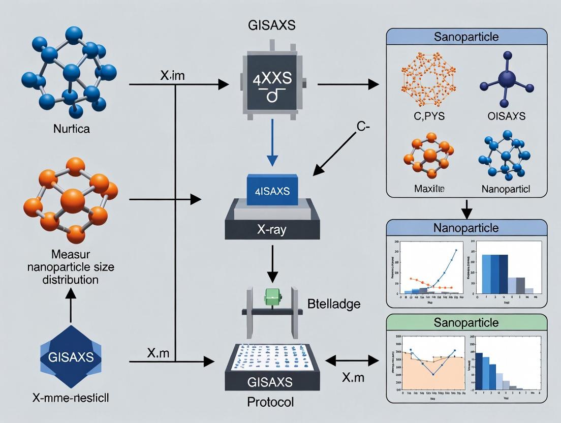
GISAXS Nanoparticle Size Distribution: A Complete Protocol for Biomedical Research
This comprehensive guide details a complete Grazing-Incidence Small-Angle X-ray Scattering (GISAXS) protocol for measuring nanoparticle size distributions, specifically tailored for drug delivery systems and nanomedicine applications.
GISAXS Nanoparticle Size Distribution: A Complete Protocol for Biomedical Research
Abstract
This comprehensive guide details a complete Grazing-Incidence Small-Angle X-ray Scattering (GISAXS) protocol for measuring nanoparticle size distributions, specifically tailored for drug delivery systems and nanomedicine applications. It covers the foundational principles of GISAXS, provides a step-by-step methodological workflow for data acquisition and analysis, addresses common troubleshooting and optimization challenges, and validates the technique against complementary methods like TEM and DLS. Aimed at researchers and drug development professionals, this article serves as a practical resource for reliable nanoscale characterization.
Understanding GISAXS: The Essential Theory for Nanoparticle Characterization
What is GISAXS? Core Principles and Scattering Geometry Explained.
1. Introduction & Thesis Context Within the broader thesis on developing a robust Grazing Incidence Small-Angle X-ray Scattering (GISAXS) protocol for measuring nanoparticle (NP) size distributions in pharmaceutical formulations, a precise understanding of the core principles is foundational. This protocol is critical for researchers and drug development professionals characterizing nanocarriers, liposomes, or virus-like particles immobilized on substrates or at interfaces, where traditional bulk solution SAXS fails.
2. Core Principles GISAXS is an advanced X-ray scattering technique used to investigate the nanoscale structure of thin films, surfaces, and interfaces. Its power lies in combining two main features:
- Grazing Incidence: An X-ray beam strikes the sample surface at a very shallow angle (αi), typically below the critical angle of the material (0.1° to 1°). This results in a large illuminated footprint, high surface sensitivity, and the excitation of an evanescent wave that propagates along the surface, probing structures within the top ~100 nm.
- Small-Angle Scattering: The scattered intensity at small angles (0.1° to 10°) is recorded, providing information on nanoscale electron density fluctuations, corresponding to particle size, shape, spacing, and ordering.
The key outcome is the ability to statistically analyze NP assemblies on a substrate without requiring long-range order, making it ideal for real-world, disordered pharmaceutical formulations.
3. Scattering Geometry Explained The GISAXS geometry defines the coordinate system for data acquisition and interpretation. The following diagram details the critical angles and vectors.
The scattering pattern is analyzed in terms of the momentum transfer vector q = kf - ki, with |k| = 2π/λ. The critical components are:
- qy: Horizontal component, sensitive to in-plane ordering and correlations.
- qz: Vertical component, sensitive to out-of-plane shape, film thickness, and substrate interface.
4. Quantitative Data Summary: GISAXS vs. Related Techniques
Table 1: Comparison of X-ray Scattering Techniques for Nanomaterial Analysis
| Technique | Typical q-range (nm⁻¹) | Probed Length Scale | Sample Environment | Key Strengths for NP Analysis |
|---|---|---|---|---|
| GISAXS | 0.01 – 5 | 1 – 500 nm | Solid/Thin Film, Liquid Interface | Surface/interface specificity, statistical data from NP assemblies on substrates. |
| SAXS (Solution) | 0.1 – 10 | 0.5 – 50 nm | Bulk Solution | Ensemble average size/shape in native state, high-throughput. |
| WAXS | 5 – 50 | 0.1 – 1 nm | Solid or Solution | Atomic/molecular crystal structure, lattice parameters. |
| XRR | 0.01 – 1 | 0.5 – 200 nm | Thin Film/Surface | Precise film thickness, density, and interfacial roughness. |
Table 2: Representative GISAXS Parameters for Pharmaceutical NP Measurement
| Parameter | Typical Range / Value | Protocol Notes for Thesis |
|---|---|---|
| X-ray Wavelength (λ) | ~0.1 nm (12.4 keV) | Synchrotron source preferred for flux and beam collimation. |
| Incident Angle (αi) | 0.1° – 0.5° (near critical angle) | Must be optimized for each substrate/NP system to maximize surface signal. |
| Beam Footprint | 5 – 20 mm (length) | Large footprint ensures statistical sampling of NP ensemble. |
| Detector Distance | 1 – 5 m | Determines q-range resolution; longer distance for smaller q. |
| Exposure Time | 0.1 – 10 s (synchrotron) | Minimize to prevent radiation damage to organic/pharma NPs. |
5. Detailed Experimental Protocol for NP Size Distribution This protocol outlines the key steps for measuring the in-plane radius of spherical NPs.
Protocol Title: GISAXS Measurement of In-Plane Nanoparticle Size Distribution on a Solid Support.
5.1. Sample Preparation
- Materials: Silicon wafer (low roughness), NP solution (e.g., polymeric NPs, liposomes), spin coater, plasma cleaner.
- Procedure:
- Clean substrate via oxygen plasma for 10-15 minutes to ensure hydrophilic surface.
- Deposit a 20-50 µL droplet of NP suspension onto the static wafer.
- Spin-coat at 2000-4000 rpm for 60 s to form a homogeneous, dense monolayer.
- Air-dry sample for 1 hour before loading into the GISAXS chamber.
5.2. Instrument Alignment & Data Collection
- Materials: Synchrotron beamline with GISAXS setup, 2D X-ray detector (e.g., Pilatus), vacuum chamber.
- Procedure:
- Align sample stage to intersect the incident X-ray beam. Precisely set the incident angle (αi) using a laser or X-ray beam viewer.
- Position the 2D detector at the desired sample-to-detector distance (e.g., 2 m).
- Close chamber and evacuate to minimize air scattering.
- Acquire a 2D scattering image with an exposure time of 1-5 seconds. Ensure the direct beam is blocked by a beamstop.
- Acquire a reference image (empty substrate) and a background image (dark current) for subtraction.
5.3. Data Reduction & Analysis
- Software: Igor Pro with Nika or SAXSGUI packages, FitGISAXS, or custom Python scripts.
- Procedure:
- Subtract dark current and background scattering from the sample image.
- Perform geometric corrections (solid angle, polarization).
- Convert the 2D image from detector coordinates (x,y) to reciprocal space coordinates (qy, qz).
- Extract a horizontal line cut at the critical angle position (Yoneda wing) to analyze in-plane scattering.
- Fit the line cut with an appropriate model (e.g., form factor for spheres + structure factor for interactions) using the Distorted Wave Born Approximation (DWBA).
6. The Scientist's Toolkit: Key Research Reagent Solutions & Materials
Table 3: Essential Materials for GISAXS Sample Preparation in Pharmaceutical NP Research
| Item | Function / Relevance | Example Product/Type |
|---|---|---|
| Low-Roughness Substrate | Provides a flat, defined surface for NP deposition; minimizes background scattering. | Single-side polished Silicon (100) wafer. |
| Plasma Cleaner | Creates a chemically clean, hydrophilic surface to ensure uniform NP spreading and adhesion. | Harrick Plasma, Oxygen plasma. |
| Precision Spin Coater | Produces a homogeneous, thin film of NP suspension, crucial for monolayer formation. | Laurell Technologies WS-650. |
| Calibrated Size Standards | Validate GISAXS size measurement protocol against known references. | NIST-traceable polystyrene or silica nanoparticles. |
| Micro-Syringe | Allows precise, reproducible deposition of small volumes of precious NP suspension. | Hamilton Gastight syringe (25-100 µL). |
| X-ray Transparent Windows | For in-situ liquid cell studies of NP assembly at liquid-air or liquid-solid interfaces. | Silicon Nitride (SiN) membranes. |
Why GISAXS for Nanoparticles? Advantages Over Bulk and Solution Techniques
Grazing Incidence Small Angle X-ray Scattering (GISAXS) is a critical technique for characterizing nanoparticles (NPs), especially when deposited on substrates, as in many functional devices. Within the broader thesis on developing robust GISAXS protocols for nanoparticle size distribution measurement, this application note establishes why GISAXS is indispensable compared to bulk and solution-phase techniques. It provides superior, statistically relevant data for supported nanoparticle systems without requiring dispersion, which can alter native states.
Comparative Advantages of GISAXS
Direct Comparison of Characterization Techniques
The table below summarizes the key limitations of common techniques when analyzing substrate-supported nanoparticles, which GISAXS directly addresses.
Table 1: Comparison of Nanoparticle Characterization Techniques
| Technique | Sample Form | Key Limitation for Supported NPs | GISAXS Advantage |
|---|---|---|---|
| Dynamic Light Scattering (DLS) | Solution, dispersed | Requires particle suspension; measures hydrodynamic diameter; insensitive to shape and substrate effects. | Measures particles in situ on substrate; provides shape, size, and spatial correlation data. |
| Transmission Electron Microscopy (TEM) | Dry, on grid (local) | Provides superb local detail but is destructive and offers poor statistical sampling (~100s of particles). | Non-destructive; probes millions of particles over a large area (~mm²), yielding excellent statistics. |
| X-ray Diffraction (XRD) | Powder, thin film | Provides crystal structure and average size via Scherrer analysis but lacks detailed size distribution. | Provides a full size distribution (mean, median, dispersion) alongside structural info from the same measurement. |
| UV-Vis Spectroscopy | Solution, thin film | Provides plasmon resonance (for metals) but gives only indirect, model-dependent size estimates. | Directly measures particle dimensions and interparticle distances, decoupling size from electronic effects. |
| BET Surface Area Analysis | Powder | Provides specific surface area and average particle size but requires a large powder mass. | Non-destructive; works on small sample quantities (e.g., a single catalytic wafer). |
Quantitative Advantages of GISAXS
The following table presents typical quantitative data obtainable from a GISAXS experiment on gold nanoparticles, compared to other methods.
Table 2: Typical Output Metrics from GISAXS vs. Other Techniques
| Metric | GISAXS Output (Example) | TEM (Same Sample) | DLS (Dispersed Sample) |
|---|---|---|---|
| Mean Particle Diameter | 12.3 ± 0.4 nm | 11.8 ± 2.1 nm (from n=200) | 15.6 ± 3.8 nm |
| Size Distribution (σ) | 1.8 nm (narrow log-normal) | Manual fitting required | Polydispersity Index: 0.24 |
| Interparticle Distance | 15.2 ± 2.1 nm | Measurable but labor-intensive | Not Applicable |
| Particle Shape | Truncated spheres | Directly visible | Assumed spherical |
| Statistical Basis | ~10⁹ particles | ~10² particles | ~10¹² particles (in solution) |
Experimental Protocols
Protocol: GISAXS Measurement of Monolayer Nanoparticles on Silicon
This protocol is central to the thesis for establishing a standard operational procedure.
I. Sample Preparation
- Substrate: Use a pristine, single-crystal silicon wafer with a native oxide layer (Si/SiO₂). Clean via successive sonication in acetone and isopropanol for 10 minutes each, followed by oxygen plasma treatment for 5 minutes.
- Nanoparticle Deposition: Deposit nanoparticles via drop-casting, spin-coating, or Langmuir-Blodgett transfer. For citrate-stabilized AuNPs (e.g., 12 nm), use spin-coating at 2000 rpm for 60 seconds from a dilute aqueous solution.
II. GISAXS Data Collection
- Instrument Setup: Utilize a synchrotron beamline or laboratory GISAXS system with a microfocus X-ray source (e.g., Cu Kα, λ = 1.5418 Å).
- Alignment: Mount the sample on a high-precision goniometer. Align the sample surface to the incident X-ray beam using a laser and the direct beam. Set the incident angle (αᵢ) to 0.5°, which is typically above the critical angle of the substrate (~0.2° for Si) but below that of the nanoparticles, to probe the near-surface structure.
- Beline Configuration: Use a 2D pixelated detector (e.g., Pilatus 1M) placed approximately 2.0 - 2.5 meters downstream from the sample. Ensure the beam is attenuated to prevent detector saturation.
- Exposure: Acquire a 2D scattering pattern with an exposure time of 60-300 seconds, depending on source brightness.
III. Data Reduction and Analysis
- Image Processing: Use software (e.g., GIXSGUI, DPDAK, or FitGISAXS) to subtract dark current and correct for detector sensitivity and geometric distortions.
- Slicing: Extract a horizontal line cut (at the Yoneda peak position) or a vertical line cut to analyze in-plane and out-of-plane structures, respectively.
- Modeling: Fit the 1D scattering profile using a form factor (e.g., sphere, cylinder) and a structure factor (e.g., paracrystal, hard-sphere). For spherical particles, use:
- Form Factor P(q): Spherical form factor.
- Structure Factor S(q): Percus-Yevick closure for hard spheres.
- Size Distribution: Assume a log-normal distribution. Fit parameters: mean radius (R), distribution width (σ), and particle volume fraction (η).
- Output: The fit yields the mean particle diameter, polydispersity, and average interparticle distance.
Protocol: Complementary TEM Validation
- Sample Prep: Deposit an identical NP solution onto a TEM grid (e.g., ultrathin carbon on Cu grid). Allow to dry.
- Imaging: Acquire high-resolution TEM images at multiple, random locations (e.g., 5 images at 100kX magnification).
- Analysis: Use image analysis software (e.g., ImageJ) to manually or automatically count and measure the diameter of at least 200 particles.
- Comparison: Compare the mean and distribution from TEM to the GISAXS results to validate the GISAXS model and protocol.
Visualization of Workflows
Diagram 1: GISAXS Protocol for NP Sizing
Diagram 2: Technique Decision Logic
The Scientist's Toolkit
Table 3: Essential Research Reagents & Materials for GISAXS on Nanoparticles
| Item | Function & Specification | Critical Notes |
|---|---|---|
| Single-Crystal Si Wafer | Standard substrate. Provides a smooth, flat, and well-defined surface for NP deposition and scattering. | P-type, ⟨100⟩, with native oxide. Thickness ~500 µm. |
| Citrate-Stabilized AuNPs | Model nanoparticle system for protocol development and validation. | Diameter: 5-50 nm. Low polydispersity recommended. |
| Oxygen Plasma Cleaner | For substrate surface activation. Removes organic contaminants and creates a hydrophilic surface for uniform NP adhesion. | Typical settings: 50-100 W for 1-5 minutes. |
| Precision Spin Coater | For creating uniform, large-area nanoparticle monolayers from colloidal solutions. | Programmable speed (500-3000 rpm) and acceleration. |
| Calibrated Attenuators | Metal foils (e.g., Al) of known thickness. Used to reduce incident beam intensity and prevent detector damage/saturation. | A set with varying transmission factors (e.g., 10%, 1%, 0.1%). |
| Direct Beam Stop | Absorbs the intense specular reflected and direct transmitted beams on the detector. | Usually made of lead or tungsten. Position is calibrated. |
| Standard Sample (Silver Behenate) | Powder with well-known diffraction rings (d-spacing = 58.38 Å). Used for precise calibration of the detector distance and q-scale. | Essential for quantitative analysis. |
| Analysis Software (e.g., FitGISAXS) | Enables modeling and fitting of 2D GISAXS patterns to extract physical parameters. | Requires a theoretical model matching the sample geometry. |
Within the framework of a thesis on developing a robust Grazing-Incidence Small-Angle X-ray Scattering (GISAXS) protocol for measuring nanoparticle size distributions in thin-film drug delivery systems, understanding three core parameters is fundamental. The incidence angle (αi), the critical angle (αc), and their relationship to the scattering vector (q-space) dictate the probing depth, scattering geometry, and data interpretation. For pharmaceutical researchers, precise control of these parameters enables the non-destructive characterization of nanoparticle size, shape, and spatial distribution within polymer matrices, critical for optimizing drug release kinetics and stability.
Core Parameters & Quantitative Data
Definition of Key Parameters
- Incidence Angle (αi): The angle between the incoming X-ray beam and the sample surface. It controls the penetration depth and the effective scattering volume.
- Critical Angle (αc): The angle below which total external reflection occurs for a given material and X-ray wavelength. It is dependent on the material's electron density.
- Q-Space (q): The momentum transfer vector in scattering experiments, defined as q = (4π/λ) sin(θ/2), where λ is the X-ray wavelength and θ is the scattering angle. In GISAXS, it is typically decomposed into components: qz (out-of-plane) and qy (in-plane).
Table 1: Critical Angles for Common Materials in Drug Delivery Films (at Cu Kα, λ = 1.54 Å)
| Material | Electron Density (e⁻/ų) | Critical Angle, αc (degrees) | Primary Function in Film |
|---|---|---|---|
| Silicon (Si) | 0.70 | ~0.22 | Standard substrate |
| Poly(lactic-co-glycolic acid) (PLGA) | ~0.38 | ~0.16 | Biodegradable polymer matrix |
| Polyethylene glycol (PEG) | ~0.33 | ~0.15 | Stabilizer / stealth coating |
| Gold (Au) Nanoparticle | 4.66 | ~0.52 | Drug carrier / contrast agent |
| Water (H₂O) | 0.33 | ~0.15 | Simulant for physiological environment |
Table 2: Incidence Angle Regimes and Their Implications for GISAXS
| Incidence Angle Regime | Condition | Penetration Depth | Information Gained | Application in Drug Delivery Research |
|---|---|---|---|---|
| Total Reflection | αi < αc (film) | ~1-5 nm (evanescent wave) | Surface structure, top-layer nanoparticles | Study of surface segregation or coating uniformity. |
| Shallow Penetration | αi ≈ αc (film/substrate) | ~10-100 nm | Near-surface structure, film-substrate interface | Analysis of nanoparticle distribution at the film-substrate interface. |
| Deep Penetration | αi > αc (film & substrate) | Several microns | Bulk film structure, depth-averaged information | Measurement of bulk nanoparticle size distribution within the polymer matrix. |
Experimental Protocols
Protocol: Determination of Critical Angle via X-ray Reflectivity (XRR)
Objective: To experimentally determine the critical angle of a thin-film sample prior to GISAXS measurement, essential for defining αi. Materials: Thin-film sample on flat substrate, synchrotron or laboratory X-ray source (Cu Kα), goniometer, 2D detector. Procedure:
- Align the sample surface to be coincident with the instrument's rotation axis.
- Set the detector at 2θ = 0° to capture the specularly reflected beam.
- Scan the incidence angle αi from 0° to ~1.0° with fine steps (e.g., 0.005°).
- Record the reflected intensity (I) as a function of αi.
- Plot log(I) vs. αi. The critical angle αc is identified as the angle at which the intensity drops precipitously (typically by ~50%).
- Fit the reflectance curve using a model (e.g., Parratt formalism) to extract precise electron density and film thickness.
Protocol: GISAXS Measurement for Nanoparticle Size Distribution
Objective: To collect GISAXS data for analyzing the size distribution of nanoparticles embedded in a thin film. Materials: Nanoparticle-loaded thin film, synchrotron beamline with grazing-incidence geometry, 2D area detector, beamstop. Procedure:
- Pre-characterization: Perform XRR (Protocol 3.1) on the sample to determine αc.
- Angle Selection: Choose αi based on the region of interest (see Table 2). For bulk statistics, set αi > αc (film). A common choice is αi = 0.2° - 0.5°.
- Alignment: Precisely align the sample using the specular reflection spot. Ensure the beamstop is positioned to block the intense specular and reflected beams.
- Data Acquisition: Expose the sample to the X-ray beam and collect the 2D scattering pattern. Typical exposure times range from 1-10 seconds (synchrotron) to hours (lab source).
- Data Collection Strategy: Collect data at multiple positions on the sample (mapping) to assess homogeneity. Optionally, collect at multiple αi to probe different depths.
- Calibration: Use a standard sample (e.g., silver behenate) to calibrate the q-space scale of the detector.
- Data Processing: Correct the 2D image for detector sensitivity, background scattering, and geometric distortions. Perform sectoral averaging to obtain 1D intensity profiles I(qy) at fixed qz.
Visualizations
Title: GISAXS Protocol Workflow for Nanoparticle Sizing
Title: Incidence Angle vs. Probing Depth in Thin Film
The Scientist's Toolkit: Essential Research Reagents & Materials
Table 3: Key Research Reagent Solutions for GISAXS Sample Preparation
| Item | Function | Example in Drug Delivery Research |
|---|---|---|
| Polymer Matrix Solution | Forms the thin film host for nanoparticles. Properties define αc and degradation kinetics. | PLGA in chloroform or acetone for controlled-release films. |
| Nanoparticle Suspension | The active component to be characterized (drug carrier). | PEGylated gold nanoparticles or polymeric micelles in aqueous buffer. |
| Substrate | Provides a smooth, flat support for film deposition. | Silicon wafer (single-side polished), cleaned via piranha solution. |
| Spin Coater | Creates uniform thin films of reproducible thickness. | Used to deposit polymer/nanoparticle solution at 1000-3000 rpm. |
| Calibration Standard | Enables accurate conversion of detector pixels to q-space. | Silver behenate powder for exact d-spacing calibration. |
| Beamstop | Protects the detector from the intense direct and specularly reflected beam. | Tantalum or lead beamstop on a wire, positioned precisely. |
| Data Analysis Software | Processes 2D images, performs fitting, extracts size distributions. | Igor Pro with Nika & Irena packages, or DAWN Science. |
This document details the protocols and application notes for interpreting Grazing-Incidence Small-Angle X-ray Scattering (GISAXS) data, framed within a thesis focused on measuring nanoparticle size distributions for advanced drug delivery systems.
Core Data Interpretation Principles
GISAXS data analysis transforms a 2D scattering pattern (q-space) into real-space structural parameters, primarily size, shape, and spatial distribution of nano-objects. The key relationship is between the scattering vector q and the real-space dimension d: d = 2π / q. For a distribution of particles, this inverse relationship is applied through modeling.
Table 1: Key GISAXS Parameters and Their Real-Space Correlates
| Scattering Pattern Feature (q-space) | Primary Real-Space Information | Typical Analysis Model |
|---|---|---|
| Position of Yoneda streak / Bragg rods | Inter-particle distance, lattice spacing | Peak fitting (e.g., Gaussian) to find q_xy |
| In-plane (q_xy) intensity modulation | In-plane particle spacing & order | 2D Fast Fourier Transform (FFT) |
| Shape of diffuse scattering halo | Nanoparticle form factor (size, shape) | Local monodisperse approximation (LMA) |
| Vertical (q_z) intensity cut profile | Particle height, substrate correlation length | Distorted Wave Born Approximation (DWBA) |
| Full 2D pattern asymmetry | Particle shape anisotropy (e.g., ellipsoids, cylinders) | Form factor models (Sphere, Core-Shell, etc.) |
Table 2: Common Nanoparticle Form Factors and GISAXS Signatures
| Nanoparticle Type | Primary GISAXS Signature | Key Fitting Parameters |
|---|---|---|
| Isolated Sphere | Semicircular fringes in qz cuts at fixed qxy | Radius (R), Size distribution width (σ) |
| Core-Shell Sphere | Damped fringe pattern with modified periodicity | Core Radius, Shell Thickness |
| Cylinder (standing) | Elongated streaks along q_z | Radius, Height, Orientation |
| Ellipsoid | Asymmetric 2D pattern, elliptical iso-intensity contours | Major Axis, Minor Axis, Aspect Ratio |
Experimental Protocols
Protocol 2.1: GISAXS Measurement for Nanoparticle Size Distribution
- Objective: Acquire a 2D GISAXS pattern suitable for quantitative analysis of nanoparticle size distribution on a substrate.
- Materials: See "The Scientist's Toolkit" (Section 4).
- Procedure:
- Sample Alignment: Mount the nanoparticle-coated substrate on the goniometer. Using a laser guide and the detector, align the sample surface to be parallel to the incident X-ray beam (grazing condition).
- Incident Angle Selection: Perform an incident angle (αi) scan (e.g., 0.1° to 0.5°) using a point detector to locate the critical angle of the substrate (αc) and the Yoneda peak. Set αi slightly above αc (typically 0.2° - 0.3°) to enhance surface sensitivity while penetrating the nanoparticle layer.
- Beamstop Positioning: Precisely position the beamstop to block the intense specular reflected beam and direct beam, preventing detector saturation.
- 2D Exposure: Insert the 2D area detector. Acquire scattering pattern with exposure time sufficient for good signal-to-noise (typically 1-10 seconds for synchrotron, 1+ hour for lab source). Use a beam-defining slit to control footprint.
- Data Calibration: Acquire calibration images: a) Direct beam for q-calibration, b) Background from bare substrate for subtraction.
- Multiple Positions: Raster the sample to 3-5 different spots to check for homogeneity and average results.
Protocol 2.2: Data Reduction and Preliminary Analysis
- Objective: Convert raw detector images into corrected, calibrated intensity maps I(qxy, qz).
- Software: Use packages like GIXSGUI (MATLAB), DPDAK, or SAXSLAB.
- Corrections: Subtract dark current/background image. Apply flat-field correction if necessary.
- Masking: Mask dead pixels and the shadow of the beamstop.
- Geometric Calibration: Using the direct beam position and sample-to-detector distance, transform pixel coordinates (x, y) to scattering vector components (qxy, qz).
- Normalization: Normalize intensity by incident flux, exposure time, and sample footprint.
- Binning/Slicing: Create 1D intensity profiles: a) In-plane cut (I vs. qxy) at the Yoneda peak position (qz ~ 0.1 nm⁻¹), b) Out-of-plane cut (I vs. qz) at a fixed qxy corresponding to the form factor maxima.
Protocol 2.3: Modeling for Size Distribution Extraction
- Objective: Fit models to data to extract mean nanoparticle size and distribution (e.g., polydispersity index, PDI).
- Model: Local Monodisperse Approximation (LMA) coupled with a defined form factor (e.g., sphere) and size distribution model (e.g., Gaussian, Log-normal).
- Software: FitGISAXS, BornAgain, or custom scripts in Igor Pro or Python.
- Initial Guessing: From the position of the first form factor minimum in a qz cut, estimate mean radius: R ≈ π / Δqz.
- Define Model: In the fitting software, define:
- Form Factor: Sphere.
- Structure Factor: Often decoupled approximation (use if particles are non-interacting). For ordered layers, include a 2D paracrystalline lattice factor.
- Size Distribution: Log-normal distribution (characterized by mean radius R0 and distribution width σ).
- DWBA: Ensure the model uses the DWBA for correct accounting of reflection/refraction at the substrate.
- Fitting: Perform a least-squares fit of the simulated 2D pattern to the calibrated data. The primary fitting parameters are R0, σ, and particle surface density.
- Validation: Check fit residual map (data - model) for random noise, indicating a good fit. Extract PDI = (σ/R0)².
Visualization of Workflows
Diagram 1: GISAXS Data Analysis Workflow (82 chars)
Diagram 2: GISAXS Data Interpretation Logic (74 chars)
The Scientist's Toolkit
Table 3: Essential Research Reagent Solutions & Materials
| Item | Function in GISAXS Protocol | Key Specifications / Notes |
|---|---|---|
| Nanoparticle Suspension | The sample of interest, deposited on a substrate. | For drug delivery: Lipid NPs, polymeric micelles, inorganic carriers. Well-characterized in solution prior to deposition. |
| Ultra-Smooth Substrate | Provides a flat, low-background surface for NP deposition and X-ray reflection. | Single-crystal silicon wafer (P/Boron doped), < 5 Å roughness. Thermally oxidized Si wafers for hydrophilic surface. |
| Sample Mounting Tape | Securely attaches the fragile substrate to the metallic sample holder without damaging it. | Double-sided carbon tape or copper tape. Must be non-outgassing in vacuum. |
| Calibration Standard | Used for precise q-space calibration of the detector. | Silver behenate (for small-angle) or silicon powder (for wide-angle). Known lattice spacing. |
| Beam-Defining Slits | Shapes the incident X-ray beam, defining its size and divergence on the sample. | Typically four independent tantalum or tungsten carbide blades. |
| X-ray Transparent Window | Seals the sample environment (e.g., vacuum chamber) while allowing the beam to pass. | Polyimide (Kapton) film or beryllium. Low scattering background is critical. |
| Area Detector | Captures the 2D scattering pattern. | Key parameters: Pixel size, point spread function, dynamic range, sensitivity (e.g., Eiger2 1M, Pilatus3). |
| Data Analysis Software Suite | For data reduction, modeling, and fitting. | GIXSGUI (MATLAB, DWBA modeling), BornAgain (Monte Carlo fitting), DPDAK (Python-based reduction), Igor Pro with Nika macros. |
Application Notes
This application note provides a comparative analysis of synchrotron and laboratory X-ray sources for Grazing-Incidence Small-Angle X-ray Scattering (GISAXS) within a thesis focused on protocol standardization for nanoparticle size distribution (PSD) measurement in drug delivery system characterization.
Core Performance Parameter Comparison
The choice of source fundamentally dictates experimental throughput, resolution, and sample scope.
Table 1: Synchrotron vs. Laboratory X-ray Source Specifications for GISAXS
| Parameter | Synchrotron Source | Laboratory Source (Metal Anode, e.g., Cu) |
|---|---|---|
| Photon Flux | 10¹² - 10¹⁵ ph/s | 10⁸ - 10⁹ ph/s |
| Beam Divergence | < 0.1 mrad | ~ 1-5 mrad |
| Beam Size (FWHM) | 10-100 µm (easily tunable) | 100-500 µm |
| Wavelength | Tunable (0.5-2.0 Å typical) | Fixed (Cu Kα = 1.5418 Å) |
| Typical Exposure Time | 0.01 - 1 second | 10 minutes - several hours |
| Energy Resolution (ΔE/E) | ~ 10⁻⁴ | ~ 10⁻³ |
| Anisotropic/Complex Samples | Excellent (fast raster mapping) | Limited (long exposures problematic) |
| Operational Accessibility | Limited (beamtime proposals) | High (in-lab, on-demand) |
| Primary Advantage | Ultra-high flux, tunability, coherence | Accessibility, cost, dedicated instrument time |
Table 2: Suitability Assessment for PSD Measurement Tasks
| Research Task | Optimal Source | Rationale |
|---|---|---|
| High-Throughput Screening of formulations | Laboratory | On-demand use supports rapid iteration. |
| Kinetic Studies (e.g., film drying, NP self-assembly) | Synchrotron | Millisecond temporal resolution captures dynamics. |
| Weak Scatterers (e.g., polymeric NPs, low contrast) | Synchrotron | High flux provides sufficient signal-to-noise. |
| Mapping lateral inhomogeneity on a substrate | Synchrotron | Micro/nano-beam allows spatially resolved GISAXS. |
| Routine QA/QC of batch consistency | Laboratory | Cost-effective and readily available for standardized tests. |
| Anomalous GISAXS near absorption edges | Synchrotron | Requires tunable X-ray energy. |
Experimental Protocols
Protocol 1: Laboratory-Based GISAXS for Routine Nanoparticle Film Characterization
Objective: To determine the mean size and size distribution of gold nanoparticles deposited on a silicon wafer using a laboratory Cu Kα source.
Materials & Pre-Measurement:
- Sample: Gold nanoparticle colloidal dispersion spin-coated onto a clean Si wafer.
- Alignment: Pre-align the diffractometer's direct beam center and sample stage height using a standard (e.g., Ag behenate or Si powder).
- Safety: Ensure all interlocks are functional. Use beamstop and guard to minimize stray radiation.
Procedure:
- Mounting: Secure the sample on the vacuum-compatible stage using a small piece of adhesive tape at the substrate's edge.
- Incidence Angle Alignment:
- Perform an incident angle (αᵢ) scan (e.g., 0.0° to 0.5°) while monitoring the Yoneda streak intensity on the 2D detector.
- Set αᵢ to the critical angle of the substrate (αc,Si ≈ 0.18°) for maximum surface sensitivity and to minimize substrate penetration/background.
- Beam Definition: Insert motorized slits to define beam size (e.g., 0.2 x 0.2 mm²).
- Acquisition:
- Close the X-ray shutter. Set the detector distance (typically 1-2 m).
- Configure acquisition software (e.g., with a PILATUS detector). Set exposure time to 1800 seconds (30 minutes). Use a high-voltage setting of 50 kV and a current of 1 mA for the Cu source.
- Evacuate the flight tube to minimize air scattering.
- Open the shutter and begin acquisition.
- Data Saving: Save the raw 2D image in a standard format (e.g., .tiff, .h5).
Data Analysis:
- Use SAXS analysis software (e.g., GIXSGUI, DPDAK, Fit2D) to perform radial integration around the direct beam, converting the 2D pattern to a 1D intensity I(q) vs. scattering vector q profile.
- Fit the 1D profile with a model (e.g., a form factor for spheres combined with a log-normal size distribution) to extract mean radius and distribution width.
Protocol 2: Synchrotron-Based GISAXS forIn-SituKinetic Measurement
Objective: To monitor the self-assembly kinetics of polymer nanoparticles during solvent evaporation in real-time.
Materials: A droplet of nanoparticle solution placed in a sealed, X-ray transparent cell with controlled atmosphere.
Procedure:
- Beamline Setup:
- Select X-ray energy (e.g., 12.4 keV, λ = 1.0 Å) for optimal flux and detector efficiency.
- Set up a high-speed 2D detector (e.g., EIGER 4M) in a vacuum chamber.
- Define a micro-beam (e.g., 50 x 50 µm²) using KB mirrors or slits.
- Rapid Alignment: Utilize the high flux to quickly find the sample edge and set αᵢ just above the substrate critical angle using a fast ion chamber or diode.
- Kinetic Experiment Scripting:
- Program the beamline control software for a time-resolved series.
- Parameters: 1000 frames, with an exposure time of 0.05 seconds per frame and a 0.01 second dead time between frames. Total experiment time: ~60 seconds.
- Trigger & Acquire: Initiate the acquisition sequence simultaneously with the start of controlled solvent evaporation (e.g., by opening a valve to a dry gas stream).
- Data Streaming: Stream the frame-by-frame 2D data directly to high-performance storage.
Data Analysis:
- Process the image stack using a batch-processing macro.
- Extract a key parameter (e.g., integrated intensity of a specific Bragg peak or correlation ring) from each frame.
- Plot this parameter versus time to reveal the kinetics of the ordering process.
Visualizations
Decision Workflow for X-ray Source Selection
Laboratory GISAXS Measurement Protocol
The Scientist's Toolkit: Key Research Reagent Solutions
Table 3: Essential Materials for GISAXS Sample Preparation & Calibration
| Item | Function | Example/Notes |
|---|---|---|
| High-Purity Silicon Wafers | Standard substrate for GISAXS due to low roughness, well-defined critical angle, and compatibility with spin-coating. | Single-side polished, P/B doped, with native oxide layer. |
| Silver Behenate Powder | Primary calibration standard for q-range. Provides sharp Bragg peaks for precise detector distance and geometry calibration. | [CH₃(CH₂)₂₀COOAg], d-spacing = 58.38 Å. |
| Colloidal Silica/Nanosphere Standards | Secondary size calibration standard. Validates the entire PSD analysis pipeline from measurement to fitting. | Polystyrene or silica spheres with certified mean diameter and low polydispersity (e.g., NIST RM 8011-8013). |
| Low-Background Sample Holders | Securely mounts fragile wafer samples without adding parasitic scattering. | Vacuum-compatible holders with precision masks to define sample area. |
| X-ray Transparent Windows | For in-situ cells (liquid, humidity, temperature control). Allows the beam to enter/exit the sample environment. | Kapton or graphene films for lab sources; diamond for high-power synchrotrons. |
| Precision Syringe & Pipettes | For reproducible deposition of nanoparticle dispersions onto substrates for film formation. | Critical for consistent film thickness and morphology. |
Step-by-Step GISAXS Protocol: From Sample Prep to Size Distribution
Sample Preparation Protocols for Supported Nanoparticle Films and Layers
This document provides standardized protocols for preparing supported nanoparticle (NP) films and layers, a critical preparatory step for accurate nanoparticle size distribution analysis using Grazing-Incidence Small-Angle X-ray Scattering (GISAXS). Reproducible, uniform, and non-aggregated samples are paramount for extracting reliable size, shape, and spatial correlation data from GISAXS patterns. These application notes detail methodologies to achieve optimal substrates for subsequent structural characterization.
Key Research Reagent Solutions & Essential Materials
| Item Name | Function & Brief Explanation |
|---|---|
| Ultra-Flat Silicon Wafers (SiO₂/Si) | Primary substrate. The native oxide layer provides a hydrophilic, chemically uniform, and atomically smooth surface for NP deposition. |
| Piranha Solution (3:1 H₂SO₄:H₂O₂) | CAUTION: Extremely hazardous. Used for deep cleaning and hydroxylation of Si surfaces, rendering them highly hydrophilic and contaminant-free. |
| Oxygen Plasma Cleaner | Alternative to piranha. Removes organic contaminants and activates the substrate surface by introducing polar functional groups. |
| Poly(diallyldimethylammonium chloride) (PDDA) | Cationic polyelectrolyte used in Layer-by-Layer (LbL) assembly to create a charged surface for electrostatic NP adsorption. |
| (3-Aminopropyl)triethoxysilane (APTES) | Silane coupling agent used to functionalize oxide surfaces with terminal amine (-NH₂) groups for covalent or electrostatic NP attachment. |
| Toluene (Anhydrous) | Common solvent for silanization reactions and for dispersing hydrophobic nanoparticles (e.g., oleylamine-capped NPs). |
| Ethanol & Acetone (HPLC Grade) | Solvents for ultrasonic cleaning and rinsing of substrates to remove particulate and organic matter. |
| Polymer Capping Agents (e.g., PVP, PEG) | Stabilize nanoparticles in solution and can prevent aggregation during deposition. May be removed post-deposition via calcination. |
Detailed Experimental Protocols
Protocol 1: Substrate Pre-Cleaning (Piranha Treatment)
Objective: To achieve a perfectly clean, hydrophilic silicon substrate. Materials: Single-side polished Si wafers, concentrated sulfuric acid (H₂SO₄), 30% hydrogen peroxide (H₂O₂), Teflon wafer holders, DI water. Procedure:
- Cut & Handle: Cut wafer into desired pieces (~1.5 x 1.5 cm) using a diamond scribe. Handle only with tweezers.
- Solvent Clean: Sonicate substrates sequentially in acetone and ethanol for 10 minutes each. Dry under a stream of nitrogen.
- Piranha Etch: In a fume hood, slowly add 75 mL of H₂SO₄ to 25 mL of H₂O₂ in a clean glass beaker. Always add acid to peroxide.
- Immerse: Immediately immerse the solvent-cleaned substrates in the fresh piranha solution for 15-30 minutes.
- Rinse: Remove substrates and rinse extensively with copious amounts of DI water (> 200 mL per substrate).
- Dry & Store: Dry under N₂ stream. Use immediately or store in DI water for up to 24 hours.
Protocol 2: Spin-Coating of Nanoparticle Monolayers
Objective: To deposit a uniform, close-packed monolayer of nanoparticles. Materials: Piranha-cleaned Si wafer, NP dispersion (e.g., 15 nm Au NPs in toluene, ~2 mg/mL), spin coater, micropipette. Procedure:
- Dispersion Prep: Sonicate the NP dispersion for 30 minutes to ensure no aggregates.
- Substrate Mount: Fix the clean, dry substrate on the spin coater chuck via vacuum.
- Deposit & Spread: While static, pipette 50-100 µL of dispersion onto the center of the substrate. Wait 10 seconds for initial spread.
- Spin Program: Execute a two-step program: (1) 500 rpm for 10 s (low-speed spread), (2) 2000-4000 rpm for 30-60 s (thinning and drying). Optimize speed for desired coverage.
- Post-treatment: Anneal on a hotplate at 150°C for 5 minutes to improve adhesion, if compatible with NPs.
Protocol 3: Layer-by-Layer (LbL) Electrostatic Assembly
Objective: To build uniform, controlled multilayer NP films with precise thickness. Materials: Piranha-cleaned substrate, PDDA solution (1% w/w in 0.5 M NaCl), polyelectrolyte (e.g., PSS), NP dispersion (oppositely charged to final layer), DI water rinse baths. Procedure:
- Prime Surface: Immerse substrate in PDDA solution for 20 min. Rinse with DI water (3 x 1 min) and dry with N₂.
- Alternate Adsorption: For each NP layer: a. Immerse the charged substrate into the well-dispersed, oppositely charged NP solution for a set time (e.g., 30 min). b. Rinse thoroughly in two consecutive DI water baths (2 min each) to remove loosely bound NPs. c. Dry gently with N₂. d. To add another layer, reintroduce to the oppositely charged polyelectrolyte solution (e.g., PSS) for 10 min, rinse, dry, and repeat from step a.
- Final Rinse: After the final desired layer, perform a final rinse and dry. The film is now ready for GISAXS.
Protocol 4: Functionalization with APTES for Covalent Attachment
Objective: To create an amine-terminated surface for bonding to functionalized NPs. Materials: O₂ plasma-cleaned Si wafer, anhydrous toluene, APTES, nitrogen glovebox (optional). Procedure:
- Plasma Clean: Treat substrate with O₂ plasma for 5 minutes to activate surface.
- Solution Prep: In a dry vessel, prepare a 2% v/v solution of APTES in anhydrous toluene.
- Silanization: Immediately immerse plasma-treated substrates in the APTES solution. Incubate for 2 hours under inert atmosphere or in a sealed container.
- Rinse: Remove substrates and rinse sequentially with toluene, ethanol, and DI water to remove physisorbed silane.
- Cure: Bake substrates at 110°C for 10-15 minutes to complete the condensation reaction.
Table 1: Key Parameters for Spin-Coating Protocols
| Nanoparticle Type | Solvent | Concentration (mg/mL) | Spin Speed (rpm) | Resultant Film Characteristics (Typical) |
|---|---|---|---|---|
| Au Citrate (15 nm) | Water | 0.5 | 3000 | Sub-monolayer, isolated particles |
| Au Oleylamine (10 nm) | Toluene | 2.0 | 2000 | Dense monolayer, hexagonal packing |
| SiO₂ (30 nm) | Ethanol | 5.0 | 1500 | Multilayer, uniform coverage |
| Fe₃O₄ (12 nm) | Hexane | 1.5 | 2500 | Discontinuous monolayer |
Table 2: LbL Assembly Build-Up Metrics
| Bilayer # | Adsorption Time (NP layer) | Estimated Layer Thickness (nm) | Surface Roughness (RMS, nm) | GISAXS Suitability |
|---|---|---|---|---|
| 1 | 30 min | ~15 nm | 2.1 | Excellent for in-plane order |
| 3 | 20 min | ~42 nm | 3.5 | Good for vertical structure |
| 5 | 15 min | ~68 nm | 5.8 | Moderate (increased scattering) |
| 10 | 10 min | ~135 nm | 12.3 | Challenging (multiple scattering) |
Experimental Workflow Diagrams
Title: Overall Sample Preparation Workflow
Title: Layer-by-Layer Assembly Process
Within the broader thesis on establishing a robust, high-throughput GISAXS (Grazing-Incidence Small-Angle X-ray Scattering) protocol for measuring nanoparticle size distributions in drug delivery formulations, precise beamline setup is the foundational step. This document details the application notes and protocols for optimizing beam position and detector distance, which are critical for achieving sufficient reciprocal space resolution, minimizing parasitic scattering, and ensuring accurate, reproducible quantitative analysis.
Core Principles & Quantitative Targets
Optimal setup is defined by the experimental goals: measuring nanoparticle sizes typically between 1 nm and 100 nm. The key parameters and their target values are summarized below.
Table 1: Key GISAXS Parameters for Nanoparticle Sizing
| Parameter | Symbol | Typical Target Value/Range | Rationale for Nanoparticle Sizing |
|---|---|---|---|
| Incidence Angle | αᵢ | 0.1° - 0.5° (above critical angle) | Ensures surface sensitivity while maximizing scattering volume from nanoparticles on substrate or in thin film. |
| Beam Energy / Wavelength | E / λ | 10-15 keV / 0.083-0.124 nm (e.g., Cu Kα: 8.05 keV) | Shorter λ increases q-range; standard lab sources often used for protocol development. |
| Beam Size at Sample | - | 50 μm x 200 μm (V x H) | Balances intensity and spatial resolution for heterogeneous samples. |
| Sample-Detector Distance | SDD | 1.0 m - 4.0 m | Determines q-range and angular resolution. Longer SDD provides higher resolution at low q. |
| Q-range (Vertical) | qz | 0.01 - 2 nm⁻¹ | Must cover form factor oscillations of target nanoparticle size distribution. |
| Q-range (Horizontal) | qy | 0.01 - 1 nm⁻¹ | Sensitive to in-plane ordering and shape. |
Table 2: Detector Distance vs. Accessible Q-min for λ=0.1 nm
| Sample-Detector Distance (m) | Pixel Size (μm) | Minimum Accessible q (nm⁻¹)* | Suitable Nanoparticle Radius |
|---|---|---|---|
| 1.0 | 75 | ~0.075 | < 15 nm |
| 2.0 | 75 | ~0.0375 | < 30 nm |
| 3.0 | 75 | ~0.025 | < 40 nm |
| 4.0 | 75 | ~0.0188 | < 50 nm |
*Approximation for direct beam at Yoneda wing, qmin ≈ (1/SDD) * (pixelsize) / (λ/2π).
Detailed Experimental Protocols
Protocol 3.1: Initial Beam Position and Profile Characterization
Objective: To locate and define the direct beam position and profile before the sample. Materials: Beamstop, knife-edge (e.g., Si wafer), X-ray sensitive beam profile monitor or high-dynamic-range detector. Steps:
- Beam Blocking: Insert a beamstop in the direct path. Ensure no beam hits the detector directly.
- Knife-Edge Scan: a. Mount a sharp, X-ray absorbing edge (knife-edge) on a motorized stage at the sample position. b. Perform a fine scan (step size ~1 μm) with the edge moving perpendicularly through the beam. c. Record the transmitted intensity on a downstream diode.
- Data Analysis: The derivative of the transmission vs. position curve gives the beam intensity profile. Fit to an error function to determine beam center and full width at half maximum (FWHM).
- Beam Visualization (Optional): Use a beam viewing screen or a short exposure on the main detector (with heavy attenuation) to visually confirm position and shape.
Protocol 3.2: Sample Alignment and Incident Angle Calibration
Objective: To precisely set the sample surface to the desired grazing incidence angle (αᵢ). Materials: Flat reference substrate (e.g., pristine Si wafer), laser aligner, sample stage with high-precision goniometry. Steps:
- Coarse Laser Alignment: Use an optical laser co-aligned with the X-ray beam to roughly align the sample surface.
- X-ray Reflectivity (XRR) Rocking Curve: a. Replace sample with the reference Si wafer. b. Set detector at 0° (in the plane of reflection) with a point detector or pixel detector. c. Scan the sample ω (theta) angle through 0° with a very fine step (~0.001°). d. The specular reflection will appear as a sharp peak. The maximum intensity defines ω = 0°.
- Angle Zeroing: Set the ω motor position at the peak maximum as the new zero.
- Set Incidence Angle: Offset the ω stage by the desired αᵢ (e.g., 0.2°).
Protocol 3.3: Detector Distance Optimization and Q-Space Calibration
Objective: To select the optimal detector distance and calibrate the scattering pattern into reciprocal space (q). Materials: Calibration standard (e.g., Ag behenate, Si grating), tape for attenuation. Steps:
- Distance Selection: Based on Table 2, choose a starting SDD. For unknown polydisperse samples, start at ~2m.
- Beamstop Alignment: Precisely center the beamstop to block the direct and specularly reflected beam.
- Q-Calibration:
a. Mount a standard with known d-spacing (e.g., Ag behenate, d=5.838 nm).
b. Acquire a transmission SAXS pattern at normal incidence.
c. Fit the ring positions (in pixels) to the equation:
q = (4π/λ) * sin(0.5 * arctan(r / SDD)), where r is the ring radius. d. Generate a pixel-to-q conversion matrix. - GISAXS Pattern Check: Acquire a pattern from a known nanoparticle sample. Verify that the expected form factor oscillations are resolved and within the detector's dynamic range.
Visualized Workflows and Relationships
Title: GISAXS Beamline Setup Sequential Protocol
Title: Key Parameters Affecting Measurable Size Range
The Scientist's Toolkit: Research Reagent Solutions
Table 3: Essential Materials for GISAXS Alignment & Calibration
| Item | Function in Setup & Alignment | Specific Example/Notes |
|---|---|---|
| Reference Silicon Wafer | Provides an atomically flat, clean surface for precise incident angle determination via X-ray reflectivity rocking curves. | Single-side polished, P/B doped, native oxide layer acceptable. |
| Knife-Edge | Used for beam profiling to determine precise beam center, size, and shape at the sample position. | Tantalum or tungsten foil with a laser-cut sharp edge. |
| Attenuation Filters | Prevents detector saturation during alignment and direct beam checks, especially with high-flux synchrotron beams. | Sets of Al or Cu foils of varying thickness (e.g., 50 μm to 1 mm). |
| Q-Calibration Standard | Allows conversion of pixel coordinates on detector to reciprocal space vector q (nm⁻¹). | Silver behenate (AgBh) powder, catalase, or gratings with known periodicity. |
| Beamstop | Protects the detector from damage by the intense direct and specularly reflected beams. Must be precisely centered. | Lead or tungsten core, often on a motorized stage for alignment. |
| Sample Leveling Stage | Provides precise control over sample tilt (ω) and rotation (φ) to set grazing incidence angle. | Goniometer stage with < 0.001° resolution. |
| Beam Position Monitor | A non-intrusive tool to monitor beam stability and position upstream of the sample. | Diamond or Si CVD blade with photodiode. |
| Alignment Laser | Co-aligned with the X-ray beam path for safe and quick initial sample and optical component alignment. | Red diode laser, mounted on beamline optics hutch. |
This application note details the critical parameters for Grazing Incidence Small-Angle X-ray Scattering (GISAXS) experiments within a comprehensive thesis on measuring nanoparticle size distributions. Accurate data acquisition is paramount for deriving reliable structural and statistical information, particularly in pharmaceutical nanoparticle characterization for drug development. The strategy revolves around optimizing exposure time, angular ranges (incident and exit angles), and instrumental resolution to maximize signal-to-noise while minimizing radiation damage and measurement artifacts.
The optimal settings are interdependent and depend on sample type, beamline geometry, and detector specifications. The following table synthesizes current recommendations from recent synchrotron and laboratory-source studies.
Table 1: Quantitative Data Acquisition Parameters for GISAXS on Nanoparticles
| Parameter | Typical Range | Recommended for Au/SiO2 NPs (50-200 nm) | Recommended for Polymer NPs (20-80 nm) | Rationale & Impact on Resolution (Δq) |
|---|---|---|---|---|
| Incident Angle (αᵢ) | 0.1° - 1.0° | 0.2° - 0.5° (above critical angle) | 0.15° - 0.3° (near critical angle) | Defines penetration depth, footprint, and surface sensitivity. Must be > critical angle for bulk scattering. |
| Angular Range (Exit, 2θ) | 0° - 5° | 0° - 3° | 0° - 5° | Captures the relevant q-range for target NP sizes. Limited by detector size and sample-detector distance. |
| Exposure Time (Synchrotron) | 0.1 - 10 s | 1 - 3 s | 0.5 - 2 s | Balances photon count (SNR) with sample stability. Vital for radiation-sensitive soft materials. |
| Exposure Time (Lab Source) | 600 - 3600 s | 1200 - 1800 s | 1800 - 3600 s | Requires long integration due to lower flux. Check for detector linearity over long exposures. |
| Beam Size (H x V) | 50x50 μm² to 500x500 μm² | 100x200 μm² | 200x300 μm² | Smaller size improves in-plane resolution but reduces scattered intensity. |
| Sample-Detector Distance (SDD) | 1.0 - 4.0 m | 2.0 - 2.5 m | 1.5 - 2.0 m | Longer SDD improves angular resolution (Δq ∝ 1/SDD) but reduces intensity. |
| Target q-range (q = 4πsinθ/λ) | 0.01 - 1.0 nm⁻¹ | 0.02 - 0.5 nm⁻¹ | 0.05 - 1.0 nm⁻¹ | q ≈ 2π / D, where D is nanoparticle diameter. |
Detailed Experimental Protocols
Protocol 1: Preliminary Calibration and Angle Optimization
Objective: To determine the critical angle and optimal incident angle for the sample.
- Sample Preparation: Spin-coat a thin film of the nanoparticle suspension (e.g., polystyrene or gold NPs in aqueous buffer) onto a clean silicon wafer. Dry under inert atmosphere.
- X-ray Reflectivity (XRR) Scan: Prior to GISAXS, perform a quick XRR scan near αᵢ = 0° to 1.0°. Identify the critical angle (α_c) from the steep drop in reflected intensity.
- Incident Angle Selection: Set the GISAXS incident angle to a value 0.05° - 0.1° above αc for surface-sensitive measurements, or 0.3° - 0.5° above αc to probe the entire film and substrate interface.
- Beline Calibration: Use a silver behenate or similar standard to calibrate the q-scale and detector geometry (pixel position vs. scattering angle). Record at αᵢ = 0.5° for 1 second.
Protocol 2: Main GISAXS Data Acquisition for Size Distribution
Objective: To acquire statistically robust 2D GISAXS patterns for analysis.
- Alignment: Precisely align the sample surface to the incident beam using the laser guide and stage goniometer. Ensure the beam footprint fully illuminates the sample without overspill.
- Pilot Exposure: Take a short exposure (e.g., 0.5 s synchrotron, 60 s lab source) to check for intense specular reflection and detector saturation. Adjust beamstop position if necessary.
- Primary Data Collection: Acquire the main GISAXS image with the parameters defined in Table 1. For a lab source, collect multiple frames (e.g., 30 x 60s) to monitor beam stability and allow for outlier removal.
- Background Subtraction: Immediately collect an identical exposure from a clean, empty spot on the substrate (or a pure solvent-cast film for solution cells). This is the background/scattering from the cell and substrate.
- Redundancy: Move the sample to a fresh, unexposed spot and repeat steps 2-4. Collect data from at least 3 distinct spots to assess sample homogeneity and improve statistical counting.
Protocol 3: Resolution and smearing Check via Standard Sample
Objective: To characterize the instrumental resolution function.
- Standard Measurement: Use a monodisperse nanoparticle standard (e.g., NIST-traceable 50 nm Au nanoparticles). Acquire GISAXS data using the standard protocol.
- Line Shape Analysis: Perform an azimuthal integration of the 2D pattern to obtain the 1D intensity I(q) vs. q profile.
- Fitting: Fit the first-order Bragg peak or form factor minima with a Gaussian or pseudo-Voigt function. The full width at half maximum (FWHM) of this peak, Δq, defines the effective instrumental resolution. This value dictates the minimum detectable size difference between nanoparticles.
Workflow and Relationships
Diagram Title: GISAXS Data Acquisition Strategy Workflow
The Scientist's Toolkit: Key Research Reagent Solutions & Materials
Table 2: Essential Materials for GISAXS Nanoparticle Experiments
| Item | Function | Example/Details |
|---|---|---|
| Ultra-Flat Single Crystal Substrate | Provides a low-roughness, low-background scattering surface for film deposition. | Silicon wafers (with native oxide), Fused silica, Mica sheets. |
| Nanoparticle Size Standard | Calibrates the q-range and characterizes instrumental resolution function. | NIST-traceable Au nanoparticles (e.g., 30 nm, 50 nm, 100 nm). |
| Calibration Standard | Precise determination of sample-to-detector distance and detector tilt. | Silver behenate (d-spacing = 5.838 nm), Rat tail collagen. |
| Precision Sample Cell (Liquid) | Enables GISAXS measurement of nanoparticles in solution or under controlled environment. | Kapton or quartz capillaries, Humidity-controlled cells. |
| Spin Coater | Produces uniform, thin films of nanoparticle suspensions for solid-state measurements. | Programmable spin coater with vacuum chuck. |
| Low-Scattering Adhesive/Glue | Secures samples and standards in holders without adding parasitic scattering. | Vacuum grease, double-sided carbon tape. |
| Precision Goniometer Stage | Allows micron-level positioning and precise control of incident and exit angles. | Multi-axis (x,y,z, θ, χ) goniometer. |
| X-ray Detector | Records the 2D scattering pattern with high dynamic range and low noise. | Hybrid Pixel Detector (e.g., Pilatus, Eiger), CCD-based detector. |
Within the broader thesis on establishing a robust Grazing-Incidence Small-Angle X-ray Scattering (GISAXS) protocol for nanoparticle size distribution measurement in drug delivery systems, this document details critical application notes for data reduction. Accurate extraction of size distributions from GISAXS patterns necessitates meticulous correction for parasitic background scattering and instrumental effects prior to modeling.
Core Correction Workflow
Logical Correction Sequence
Diagram Title: GISAXS Data Correction Workflow for Nanoparticle Sizing
Quantitative Impact of Corrections on Key Parameters
Table 1: Effect of Sequential Corrections on Derived Nanoparticle Parameters (Simulated Data for 20 nm Gold Nanoparticles on Si Substrate)
| Correction Step | Apparent Mean Radius (nm) | Polydispersity (σ/R) | Peak Intensity I(0) (a.u.) | Notes |
|---|---|---|---|---|
| Raw Data | 18.7 ± 4.1 | 0.31 | 1.00 | Uncorrected data shows bias and high error. |
| After Dark Current Subtraction | 19.2 ± 3.8 | 0.28 | 0.92 | Reduces low-q noise floor. |
| After Pixel Sensitivity/Flat Field | 19.8 ± 2.9 | 0.22 | 0.95 | Corrects detector inhomogeneities. |
| After Parasitic Background Subtraction | 20.1 ± 1.9 | 0.11 | 0.41 | Most critical step; removes substrate/air scattering. |
| After Geometric (Footprint) Correction | 20.0 ± 1.8 | 0.10 | 0.40 | Accounts for illuminated sample area. |
| Fully Corrected Data | 20.0 ± 1.8 | 0.10 | 0.40 | Ready for accurate model fitting. |
Detailed Experimental Protocols
Protocol A: Measurement of Parasitic Background Scattering
Objective: To acquire the background scattering profile of the substrate and solvent/support film devoid of nanoparticles. Materials: See Scientist's Toolkit. Procedure:
- Sample Preparation: Prepare an identical substrate (e.g., silicon wafer) using the same cleaning protocol (e.g., piranha etch, UV-Ozone) as used for nanoparticle deposition.
- Solvent Deposition: If nanoparticles are deposited from a solvent (e.g., toluene, water), deposit an identical volume of the pure solvent onto the substrate and allow it to dry under identical conditions.
- Beamline Alignment: Mount the background sample in the GISAXS holder. Align the incident X-ray beam to the same grazing angle (αi) used for the nanoparticle sample (typically 0.1° - 0.5° above the critical angle).
- Data Acquisition: Acquire a 2D scattering image with an exposure time equal to or greater than that used for the nanoparticle sample. Repeat for 2-3 different spots on the substrate to check for homogeneity.
- Data Storage: Save the image in a standard format (e.g., .tiff, .h5) with metadata noting αi, exposure time, and sample details.
Protocol B: Dark Current and Detector Flat-Field Correction
Objective: To correct for detector-specific electronic noise and pixel-to-pixel sensitivity variations. Procedure:
- Dark Current Image: With the detector shutter closed, acquire multiple images (e.g., 10) using the exact same exposure time and readout settings as the sample measurement. Average these images to create a master
dark_image. - Flat-Field Image: Using a homogeneous, weakly scattering source (e.g., a fluorescent screen with direct beam, severely attenuated to avoid detector damage), acquire an image to map pixel sensitivity. Ensure the intensity is within the linear response range of the detector. Average multiple exposures to create a master
flat_field_image. - Application: Correct each raw frame (
raw_image) using the formula:corrected_image = (raw_image - dark_image) / (flat_field_image - dark_image)Perform this operation before any other analysis.
Protocol C: Geometric and Intensity Corrections
Objective: To account for variations in irradiated sample volume and beam decay. Procedure:
- Footprint Correction: Calculate the beam footprint on the sample:
Footprint = Beam_Size / sin(αi). The scattering intensity must be normalized by this length, as it varies with αi. - Transmission Correction: Measure the incident beam intensity (I0) using an upstream monitor (e.g., ion chamber) for both sample and background. Normalize scattered intensities by I0. If I0 is not directly available, use the intensity of the direct beam attenuated through a pinhole as a reference.
- Beam Decay: If using synchrotron radiation, monitor I0 over time and correct for beam current decay, especially during long exposures.
The Scientist's Toolkit
Table 2: Essential Research Reagent Solutions & Materials for GISAXS Sample Preparation and Background Correction
| Item | Function/Description | Example Product/Catalog |
|---|---|---|
| High-Purity Silicon Wafers | Standard, low-roughness substrate for GISAXS. Provides a consistent parasitic background. | Single-side polished, ⟨100⟩, 1x1 cm², 1-10 Ω·cm resistivity. |
| Piranha Solution | CAUTION: Highly corrosive. Used to clean substrates, removing organic residue to minimize background. | Freshly mixed H₂SO₄ (96%) : H₂O₂ (30%) in a 3:1 ratio. |
| UV-Ozone Cleaner | Alternative to piranha for substrate cleaning; oxidizes organic contaminants. | Benchtop UV-Ozone system (e.g., 185 nm & 254 nm lamps). |
| Anhydrous Toluene | Common solvent for dispersing hydrophobic nanoparticles (e.g., oleylamine-capped AuNPs). Minimizes water-related scattering. | Sigma-Aldrich, 99.8%, inhibitor-free. |
| Milli-Q Water | Solvent for hydrophilic nanoparticles. Must be filtered (0.2 µm) to remove dust. | 18.2 MΩ·cm resistivity, < 5 ppb TOC. |
| Attenuator Set | Calibrated X-ray attenuators (e.g., Al foils) to reduce beam intensity for direct beam/flat-field measurements. | Set with varying thicknesses (e.g., 50 µm to 1 mm Al). |
| Direct Beam Stop | Prevents damage to the detector from the intense specularly reflected and direct beams. | Lead, tantalum, or compound material on a thin Kapton film. |
| Calibration Standard | Known scatterer for q-range calibration (e.g., silver behenate, polystyrene beads). | Silver behenate powder, d-spacing = 58.38 Å. |
Data Integration and Modeling Pathway
Diagram Title: GISAXS Modeling Pathway for Size Distribution
This document constitutes a core chapter in a broader thesis on establishing a standardized Grazing-Incidence Small-Angle X-ray Scattering (GISAXS) protocol for determining the size, shape, and distribution of nanoparticles (NPs). Accurate fitting of GISAXS patterns is paramount for extracting quantitative structural parameters. The Distorted Wave Born Approximation (DWBA) is the essential theoretical framework for analyzing GISAXS data from NPs on substrates, as it correctly accounts for the reflection and refraction effects at the substrate interface that the simple Born approximation neglects. This application note details the implementation of DWBA-based fitting models for nanoparticle systems.
Theoretical Foundation: DWBA for Nanoparticles
In GISAXS, an X-ray beam impinges on a sample at a grazing angle (α~i~) near the critical angle of the substrate (α~c~). The DWBA treats the scattering as a perturbation of the ideal reflected wave (the "distorted wave"). For nanoparticles on a surface, the scattering cross-section is calculated by considering four scattering processes: (1) incident wave scattered by particle, (2) incident wave reflected then scattered, (3) incident wave scattered then reflected, and (4) incident wave reflected, scattered, and reflected again.
The intensity I(q) for an ensemble of NPs is: [ I(\mathbf{q}) \propto \left| \int d\mathbf{r} e^{i\mathbf{q}\cdot\mathbf{r}} \Delta\eta(\mathbf{r}) [e^{i qz z} + R(\alphai)e^{-i qz z}] [e^{i qz' z} + R(\alphaf)e^{-i qz' z}] \right|^2 ] where Δη is the scattering length density difference, q is the scattering wavevector, and R(α) is the Fresnel reflection coefficient.
Key Research Reagent Solutions & Materials
| Item | Function in DWBA-GISAXS Experiment |
|---|---|
| Monodisperse Nanoparticle Standards (e.g., Au, SiO₂, PS) | Calibrate the GISAXS setup and validate the DWBA fitting model parameters. Provide known size/shape for model benchmarking. |
| Low-Roughness Single-Crystal Substrates (Si, SiO~x~/Si, Quartz) | Provide a flat, well-defined interface with known critical angle and refractive index for precise DWBA calculations. |
| Precision Goniometer | Enables accurate control of incident and exit angles (α~i~, α~f~, 2θ~f~) which are critical inputs for the DWBA formalism. |
| High-Brilliance Synchrotron X-ray Source | Provides the high-intensity, monochromatic, and collimated beam required for collecting statistically robust 2D GISAXS patterns in short exposures. |
| 2D Pixel Detector (Pilatus, Eiger) | Captures the full 2D scattering pattern, essential for analyzing anisotropic structures and separating Yoneda from Bragg peaks. |
| DWBA-Fitting Software (IsGISAXS, BornAgain, HipGISAXS) | Implements the DWBA theory for various particle shapes (sphere, cylinder, cube, etc.) and includes necessary corrections (roughness, size dispersion). |
Experimental Protocol for DWBA-Based GISAXS Measurement
Sample Preparation
- Substrate Cleaning: Sonicate substrate (e.g., silicon wafer) sequentially in acetone, isopropanol, and deionized water for 10 minutes each. Dry under nitrogen stream.
- Nanoparticle Deposition: Deposit nanoparticle solution (e.g., colloidal Au NPs) via spin-coating (typical: 3000 rpm for 30 s) or drop-casting onto the prepared substrate.
- Sample Characterization: Prior to GISAXS, characterize sample with SEM or AFM to obtain preliminary size/distribution data and confirm particle integrity.
GISAXS Data Collection at Synchrotron Beamline
- Beam Alignment: Align the sample surface to the X-ray beam with micrometer precision. Determine the exact incident angle (α~i~) using a direct beam scan or substrate reflectivity.
- Angle Selection: Set α~i~ to be at or slightly above the critical angle of the substrate (α~c~ for Si is ~0.22° at 10 keV) to enhance surface sensitivity and the Yoneda band signal.
- Exposure: Acquire 2D scattering pattern using a pixel detector placed ~1-5 m from the sample. Typical exposure times range from 0.1 to 10 seconds at a synchrotron.
- Calibration: Collect scattering pattern from a known standard (e.g., silver behenate) for q-calibration of the detector.
Data Reduction & DWBA Fitting Workflow
- Image Processing: Correct raw 2D image for detector dark current, flat field, and spatial distortions. Mask bad pixels and beam stop shadow.
- q-Space Conversion: Convert pixel coordinates to scattering vector components (q~y~, q~z~) using calibrated sample-detector distance and beam center.
- Model Selection: Choose an appropriate form factor (e.g., sphere, cylinder) and interference function (e.g., decoupling approximation, local monodisperse approximation) in the DWBA-fitting software.
- Parameter Fitting: Fit the 2D pattern or 1D cuts (e.g., horizontal at Yoneda peak) by varying parameters like particle radius, height, center-to-center distance, and size distribution width (σ). Constrain parameters using prior knowledge from SEM/AFM.
- Validation: Assess fit quality via residual maps and χ² values. Cross-validate extracted size distribution with TEM/AFM results.
Quantitative Data from DWBA Fitting of Nanoparticle Systems
Table 1: Representative Fitted Parameters for Different Nanoparticle Systems Using DWBA
| Nanoparticle System (Substrate) | Form Factor Model | Fitted Radius (nm) | Size Dispersion (σ, nm) | Inter-particle Distance (nm) | Key Reference |
|---|---|---|---|---|---|
| Colloidal Au NPs (Si/SiO₂) | Sphere (DWBA) | 7.2 ± 0.3 | 0.8 | 45 ± 10 | Renaud et al., Science (2003) |
| PS-b-PMMA Polymer NPs (Si) | Cylinder (DWBA) | 12.5 (Radius) | 1.2 | 35 (Center-to-center) | Busch et al., Macromolecules (2007) |
| Self-Assembled Iron Oxide NPs (Si) | Truncated Sphere (DWBA) | 5.0 ± 0.4 | 0.5 | 11 ± 2 | Lazzari et al., J. Appl. Cryst. (2006) |
| Sputtered Pt NPs (Glass) | Parallelepiped (DWBA) | 3.1 (Height) | 0.7 (Ht. Disp.) | N/A (Random) | Chushkin et al., J. Appl. Cryst. (2014) |
Table 2: Comparison of Key Outputs from Simple Born Approximation vs. DWBA Fitting
| Fitting Aspect | Born Approximation | Distorted Wave Born Approximation (DWBA) | Impact on NP Characterization |
|---|---|---|---|
| Angular Dependence | Ignores reflection/refraction | Explicitly includes angle-dependent Fresnel coefficients | Correctly models intensity near α~c~; essential for accurate size. |
| Yoneda Peak | Cannot reproduce it | Accurately models the diffuse scattering peak at α~f~ = α~c~ | Provides a strong intensity feature for precise fitting. |
| Substrate Effect | Neglected | Fully incorporated via distorted waves | Critical for NPs on or near an interface; prevents systematic error. |
| Computational Load | Low | High (4 scattering terms) | Requires specialized software and more fitting time. |
Visualization of Workflows and Relationships
Title: DWBA-Based GISAXS Data Analysis Workflow
Title: The Four DWBA Scattering Processes for a Nanoparticle
This protocol is a core chapter of a thesis focused on establishing a robust, standardized Grazing-Incidence Small-Angle X-ray Scattering (GISAXS) workflow for characterizing nanoparticle (NP) ensembles in pharmaceutical formulations. The precise extraction of size distributions from GISAXS data is critical, as NP size directly influences drug loading, release kinetics, and cellular uptake. Moving beyond simple monodisperse models, this document details the quantitative fitting of experimental data to theoretical form factors for spheres, cylinders, and other shapes to recover polydisperse size distributions, essential for Quality-by-Design in drug development.
Theoretical Framework & Data Fitting Principles
The scattered intensity I(q) in GISAXS is proportional to the product of a form factor P(q), describing the shape and size of the NP, and a structure factor S(q), describing inter-particle interactions. For dilute systems, S(q) ≈ 1. The form factor for an ensemble with a size distribution D(R) is calculated by integration:
I(q) ∝ ∫ P(q, R) D(R) dR
Fitting involves minimizing the difference between this modeled intensity and the experimental 1D GISAXS profile (obtained by sector averaging). Key distribution models include:
- Monodisperse: Single size.
- Gaussian/Normal: Symmetric dispersion around a mean.
- Log-Normal: Asymmetric, naturally constrains sizes to positive values, commonly used for nanoparticles.
- Schulz-Zimm: Useful for polymer and particle dispersity.
Diagram Title: GISAXS Size Distribution Extraction Workflow
Application Notes: Form Factors and Fitting Tables
Table 1: Common Form Factors for Nanoparticle Characterization
| Form Factor Model | Key Shape Parameters | Typical Fitting Parameters | Pharmaceutical Relevance |
|---|---|---|---|
| Sphere | Radius (R) | Mean Radius, σ (dist. width), Scale, Bkg | Solid Lipid NPs, Polymeric NPs, Virus-like particles. |
| Cylinder (Height) | Radius (R), Height (H) | Mean R, Mean H, σR, σH, Scale, Bkg | Nanorods, certain fibrous structures, elongated micelles. |
| Cylinder (Length) | Radius (R), Length (L) | Mean R, Mean L, σR, σL, Scale, Bkg | Carbon nanotubes, rod-shaped viruses. |
| Core-Shell Sphere | Core Radius (R_c), Shell Thickness (t) | Mean R_c, Mean t, σRc, σt, Scale, Bkg | Polymeric NPs with PEG corona, liposomes, nanocapsules. |
| Ellipsoid | Semi-axes (a, b) or Radius & Aspect Ratio | Mean Radius, Aspect Ratio, σ, Scale, Bkg | Non-spherical protein aggregates, some metal NPs. |
Table 2: Size Distribution Models & Metrics
| Distribution Model | Probability Density Function D(R) | Fitted Parameters | Polydispersity Index (PDI) / Dispersity |
|---|---|---|---|
| Log-Normal | [1/(√(2π) σ R)] exp( - (ln R - μ)²/(2σ²) ) | μ (log mean), σ (log width) | PDI = exp(σ²) |
| Gaussian | (1/(σ√(2π)) exp( - (R - R₀)²/(2σ²) ) | R₀ (mean), σ (width) | PDI = (σ/R₀)² |
| Schulz-Zimm | [ (z+1)^(z+1) R^z / (R₀^(z+1) Γ(z+1)) ] exp(-(z+1)R/R₀) | R₀ (mean), z (width parameter) | Đ = 1/(z+1) |
Detailed Experimental Protocol: GISAXS Data Acquisition & Fitting
A. Sample Preparation & Measurement (Preceding the Fit)
- Substrate: Use a pristine silicon wafer. Clean via piranha solution (Caution: Highly corrosive) or oxygen plasma treatment for 5 minutes.
- Deposition: Spin-coat or drop-cast the nanoparticle suspension (e.g., 50 µL) onto the substrate at a controlled speed (e.g., 2000-4000 rpm) to form a thin, homogeneous film. Air-dry.
- GISAXS Alignment: Mount the sample on the goniometer. Align the substrate surface to the X-ray beam using a laser and the goniometer's translation/rotation stages. The incident angle (α_i) should be set slightly above the critical angle of the substrate (typically ~0.2° for Si) to enhance surface sensitivity while probing the NPs.
- Data Collection: Use a synchrotron beamline or lab-source GISAXS instrument with a 2D detector (e.g., Pilatus or Eiger). Typical settings: Beam energy ~10-15 keV (λ ~0.1 nm), exposure time 1-60 seconds, sample-detector distance 1-3 m. Ensure the beam is attenuated to prevent detector saturation.
B. Data Reduction Protocol (Pre-Fitting)
- Beam Center & Masking: Determine the direct beam position on the 2D image. Mask the beam stop and any defective detector pixels.
- Sector Averaging: Using software (e.g., Igor Pro with Nika package, DAWN Science, or FitGISAXS), define a narrow horizontal sector (Δαf ≈ 0.1°) just above the Yoneda band to extract the *qy* ~ 0 cut. This approximates a standard SAXS pattern (I vs. q_z, where q = 4π sin(θ)/λ).
- Background Subtraction: Subtract the scattering profile from an identically prepared bare substrate.
C. Fitting Protocol for Size Distribution This example uses a Log-Normal distribution of Spheres.
- Software Setup: Operate within a fitting environment (e.g., SASView, Igor Pro, custom Python script using
lmfit/scipy). - Define Model: Construct the intensity function:
I_model(q) = Scale * ∫ [P_sphere(q, R)]² * D_LogNormal(R; μ, σ) dR + Incoherent_BackgroundwhereP_sphere(q,R) = 3 * [sin(qR) - qR cos(qR)] / (qR)³. - Initial Parameters: Estimate initial values: R from q at the first intensity minimum (~4.49/q). Set initial Scale to match intensity, Bkg from high-q tail, and σ (~0.1-0.3 for moderate polydispersity).
- Fitting Execution: Perform a non-linear least squares minimization (e.g., Levenberg-Marquardt algorithm). Constrain R and σ to positive values.
- Validation: Assess fit quality via reduced chi-squared (χ²_ν ~1), visual agreement, and randomness of residuals. Use error analysis on parameters (standard error from covariance matrix).
The Scientist's Toolkit: Key Research Reagents & Materials
Table 3: Essential Materials for GISAXS Sample Prep & Analysis
| Item / Reagent | Function / Purpose |
|---|---|
| High-Purity Silicon Wafer | Standard, flat, low-roughness substrate with known critical angle for precise GISAXS alignment. |
| Piranha Solution (H₂SO₄:H₂O₂) | Extreme Caution. Used for ultra-cleaning Si wafers to remove organic residue and ensure hydrophilic surface. |
| Oxygen Plasma Cleaner | Alternative to piranha for substrate cleaning and surface activation to improve NP suspension wetting. |
| Anhydrous Toluene or Chloroform | Common solvents for dispersing hydrophobic nanoparticles (e.g., PLGA NPs) prior to spin-coating. |
| PBS Buffer (pH 7.4) | Aqueous medium for dispersing biocompatible or protein-conjugated NPs to mimic physiological conditions. |
| Poly-L-lysine Solution (0.1% w/v) | Substrate coating agent to enhance adhesion of negatively charged nanoparticles via electrostatic interaction. |
| Spin Coater | Instrument to create uniform, thin films of NP suspensions, minimizing coffee-ring effects and aggregate formation. |
| Calibration Standard (Silver Behenate) | Powder standard used to calibrate the q-range and detector distance of the GISAXS/SAXS instrument. |
| SASView / Irena (Igor Pro) Software | Primary software packages for modeling form factors, polydispersity, and fitting GISAXS/SAXS data. |
Solving Common GISAXS Challenges: Troubleshooting and Data Quality Optimization
Within the broader thesis on developing robust Grazing-Incidence Small-Angle X-ray Scattering (GISAXS) protocols for nanoparticle size distribution measurement, beam-induced damage presents a critical challenge. Sensitive samples, including polymer nanocomposites, lipid nanoparticles, and proteinaceous drug delivery systems, undergo morphological and chemical alterations under X-ray irradiation, skewing size distribution data. This document outlines current strategies and detailed protocols to mitigate these effects, ensuring data fidelity.
Mechanisms and Quantification of Beam Damage
Beam damage arises primarily through radiolysis (for soft materials in solution/ humidity) and heating. The key metrics are the critical dose (Dc) and dose-rate. Current literature indicates significant variation in tolerable doses.
Table 1: Representative Critical Doses for Sensitive Materials
| Material Class | Sample Form | Typical Critical Dose (kGy) | Primary Damage Manifestation |
|---|---|---|---|
| Polymers (e.g., PS, PMMA) | Thin Film | 1 - 10 | Chain scission, cross-linking, loss of GISAXS ordering |
| Lipid Bilayers / Vesicles | Hydrated Film | 0.1 - 1 | Loss of lamellar order, vesicle fusion |
| Proteins / Enzymes | Solution or Crystal | 0.01 - 0.1 | Loss of tertiary structure, aggregation |
| DNA-based Nanostructures | Aqueous Buffer | < 0.05 | Strand breakage, loss of shape |
| Block Copolymer Thin Films | Self-assembled | 5 - 20 | Order-disorder transition, pattern fading |
Core Mitigation Strategies & Protocols
Strategy 1: Dose Management
Protocol A: Dose Fractionation & Low-Dose GISAXS Acquisition
- Pre-calculation: Use the formula Dose (Gy) = (Flux * Exposure time * Energy Absorption Coefficient) / Area to estimate dose. Utilize online tools like RADDOSE-3D for complex geometries.
- Setup: At the synchrotron beamline, employ a high-efficiency, fast-readout detector (e.g., Pilatus/Eiger).
- Acquisition: Collect a large series of very short exposures (e.g., 0.01s each) rather than one long exposure.
- Processing: Align and sum frames offline using software like SAXSutilities or DPDAK. Monitor radial/ azimuthal integration for changes in peak position/intensity over frame sequence. Discard frames where damage signatures (e.g., peak broadening) appear.
- Validation: Compare the summed "low-dose" scattering pattern with an early-frame subset. Key GISAXS features (Yoneda, Bragg rods) should remain consistent.
Strategy 2: Cryogenic Cooling
Protocol B: Vitrification of Hydrated Samples for GISAXS
- Sample Preparation: Apply sample (e.g., lipid nanoparticle suspension) onto a cleaned silicon wafer. Use a spin-coater for thin, even films.
- Vitrification: Rapidly plunge the wafer into liquid ethane cooled by liquid nitrogen using a manual plunger or vitrification robot.
- Transfer & Mount: Under continuous liquid nitrogen, transfer the vitrified wafer to a pre-cooled cryo-stage in the GISAXS instrument (typically ≤ 100 K).
- Data Collection: Perform GISAXS in a vacuum or dry nitrogen atmosphere to prevent frost. Use a beam-defining slit to minimize exposed area.
- Note: This suppresses radiolysis-derived reactive species diffusion, increasing Dc by ~10-100x for hydrated samples.
Strategy 3: Radical Scavengers
Protocol C: Incorporating Scavengers for Solution GISAXS For in-situ GISAXS of nanoparticles in solution using a flow cell or capillary.
- Scavenger Preparation: Prepare stock solutions of effective radical scavengers:
- 100 mM Sodium Ascorbate (aqueous, pH ~7)
- 50 mM Cysteine
- 10-20% (v/v) DMSO for some systems
- Sample Mixing: Mix the nanoparticle suspension (e.g., protein-based therapeutic) with scavenger stock to achieve final recommended concentrations (e.g., 10-50 mM ascorbate).
- Control Experiment: Prepare an identical sample without scavenger.
- GISAXS Run: Acquire data on both samples under identical flux and exposure conditions. Monitor the time evolution of the Guinier region or the pair-distance distribution function.
- Analysis: The scavenger-containing sample should show stable scattering profiles over a longer time period compared to the control.
Strategy 4: Computational Correction
Protocol D: Modeling and Subtracting Damage Effects
- Time-Series Acquisition: Collect a GISAXS image series at constant flux, ensuring the final frames show clear damage.
- Feature Tracking: Isolate a specific GISAXS feature (e.g., a Bragg peak intensity, Iq at a specific q).
- Model Fitting: Fit the decay of this feature over time (frame number) to an exponential decay or multi-step model.
- Extrapolation: Use the model to extrapolate the undamaged intensity (It=0) back from the initial, non-linear decay regime.
- Generate Corrected Pattern: Reconstruct a "damage-corrected" 2D GISAXS pattern using the extrapolated scaling factors. This is most effective for uniform dose deposition.
Integrated Experimental Workflow
Title: Integrated Beam Damage Mitigation Workflow for GISAXS
The Scientist's Toolkit: Research Reagent & Material Solutions
Table 2: Essential Materials for Beam Damage Mitigation
| Item | Function & Rationale |
|---|---|
| Silicon Nitrace (SiN) Membranes | Low-X-ray-absorption windows for liquid cells; enable transmission GISAXS/SAXS on sensitive solutions with minimal dose. |
| Cryo Plunger (e.g., Vitrobot) | For reproducible vitrification of hydrated samples, trapping amorphous ice to suppress radiolysis. |
| Liquid Nitrogen Cryo-Stage | Maintains sample at cryogenic temperatures (≤100 K) during GISAXS measurement. |
| Radical Scavengers (Ascorbate, Cysteine) | Competitive scavengers of diffusive hydroxyl and secondary radicals generated by water radiolysis. |
| Fast-readout 2D X-ray Detector (Eiger2) | Enables dose fractionation; allows collection of many short frames to monitor damage onset. |
| Precision Beam-Defining Slits | Reduce illuminated sample volume, limiting total dose and damage footprint. |
| In-vacuum Sample Chamber | Removes oxygen and water vapor, reducing formation of reactive species for dry polymer films. |
| Flow-through Capillary Cell | Allows continuous renewal of sample volume, providing fresh material to the beam. |
Integrating these strategies into the standard GISAXS protocol for nanoparticle sizing is non-optional for sensitive materials. A tiered approach—starting with pre-experiment dose calculation, employing cryo-cooling or radical scavengers where applicable, utilizing low-dose acquisition protocols, and applying computational corrections—forms a robust defense against beam damage, ensuring the extracted size distributions are accurate and representative of the native state.
Correcting for Substrate Roughness and Background Scattering Artifacts
Within the broader thesis on establishing robust, standardized Grazing-Incidence Small-Angle X-ray Scattering (GISAXS) protocols for nanoparticle (NP) size distribution measurement, this document addresses a critical experimental challenge. Accurate quantification of in-situ NP dispersions, essential for pharmaceutical nanocrystal and liposomal drug delivery system characterization, is confounded by scattering from rough substrates and inherent background signals. These Application Notes provide detailed protocols and correction methodologies to isolate the true nanoparticle scattering contribution, thereby increasing data fidelity for research and development.
GISAXS is a powerful technique for statistically analyzing nanoscale structures on surfaces and in thin films. For drug development, it enables non-destructive sizing of therapeutic nanoparticles adsorbed at interfaces. However, the scattered intensity I(q) is a superposition:
I_total(q) = I_NP(q) + I_substrate(q) + I_background(q)
where I_NP is the signal of interest, I_substrate arises from substrate roughness and density fluctuations, and I_background includes diffuse scattering, air scattering, and detector noise. Uncorrected, these artifacts lead to significant errors in derived size distributions, particularly for polydisperse systems or small NPs (<20 nm).
Key Artifacts and Quantitative Impact
Table 1: Primary Artifacts and Their Effect on GISAXS Analysis
| Artifact Source | Typical q-range affected | Impact on NP Size Distribution | Magnitude of Error (Example) |
|---|---|---|---|
| Substrate Root-Mean-Square Roughness (σ > 2 nm) | Low q (< 0.1 nm⁻¹) | Overestimation of NP radius, false detection of large aggregates | Can inflate R_avg by 20-40% |
| Substrate Correlated (Lateral) Roughness | Medium q (0.1 - 1 nm⁻¹) | Broadening of distribution, introduction of spurious peaks | Polydispersity (σ) error up to 15% |
| Thermal/Detector Background (Dark Current) | All q | Increased intensity floor, reduces signal-to-noise ratio (SNR) | Can obscure weak scattering from low-concentration species |
| Specular Reflection Streak (Yoneda Band) | Along q_z | Masks scattering in critical regions, complicates data reduction | Requires masking in 2D analysis |
| Incident Beam Flux Fluctuations | All q | Introduces noise, compromises absolute intensity calibration | Normalization errors of 5-10% |
Experimental Protocols for Artifact Correction
Protocol 3.1: Preparation and Characterization of Reference Substrates
Aim: To obtain a background scattering profile I_substrate(q) for subtraction.
Materials: Silicon wafers (P/Boron, <100>), Piranha solution (3:1 H₂SO₄:H₂O₂), RCA-1 cleaning standard, plasma cleaner.
Procedure:
- Cleaning: Treat Si wafer in fresh Piranha solution (Caution: Highly exothermic and oxidizing) for 30 minutes. Rinse extensively with Milli-Q water (18.2 MΩ·cm).
- Surface Activation: Perform oxygen plasma treatment (100 W, 0.2 mbar, 2 minutes) to ensure a hydrophilic, clean surface.
- Roughness Measurement: Characterize the substrate via Atomic Force Microscopy (AFM) in tapping mode (5x5 μm² area). Calculate RMS roughness (σ). Acceptable σ < 1 nm for NP sizing.
- GISAXS Reference Measurement: Mount the clean, dry substrate in the GISAXS chamber. Acquire scattering pattern using identical beam parameters (energy, incidence angle α_i, exposure time) as for subsequent NP sample measurements. This is the substrate reference file.
Protocol 3.2: GISAXS Data Acquisition with Background Subtraction
Aim: To collect NP sample data with matched background. Materials: Nanoparticle dispersion (e.g., PEGylated liposomes, nanocrystals), calibrated micropipette, sample stage with vacuum chuck. Procedure:
- Incidence Angle Selection: Set αi to 0.5° above the critical angle of the substrate (αc for Si ~ 0.22° at 10 keV) to enhance surface sensitivity while minimizing penetration.
- Background Collection:
a. Acquire dark image (beam shutter closed) with exposure time
t_expto capture detector dark current. b. Acquire direct beam image (attenuated by order of 10⁶) for precise beam center and solid-angle calibration. c. Acquire empty cell/bare substrate image as per Protocol 3.1. - Sample Measurement: a. Deposit 50 μL of NP suspension on the characterized substrate, allowing gentle spin-coating to form a monolayer. b. Immediately mount sample and acquire GISAXS pattern with identical settings as background. c. Measure two additional sample spots to check for spatial heterogeneity.
Protocol 3.3: Data Reduction and Correction Workflow
Aim: To computationally isolate I_NP(q).
Software: Custom Python (using libraries: numpy, scipy, pyFAI, matplotlib) or specialized SAXS reduction packages.
Algorithm:
- Primary Subtraction:
I_corrected_1 = I_sample - I_dark - Substrate Subtraction:
I_corrected_2 = I_corrected_1 - (I_substrate - I_dark) - Intensity Normalization: Normalize
I_corrected_2by incident beam flux (from ion chamber), exposure time, and sample transmission factor. - Geometric Corrections: Apply solid angle correction and mask pixels affected by beam stop, specular streak, and detector gaps.
- Azimuthal Integration: Convert corrected 2D image to 1D intensity profile
I(q)vs scattering vectorq.
Visualization of Workflows
Diagram Title: GISAXS Artifact Correction Experimental Workflow
The Scientist's Toolkit
Table 2: Essential Research Reagent Solutions & Materials
| Item | Function & Rationale | Example Product/Chemical |
|---|---|---|
| High-Flatness Substrates | Minimizes I_substrate. Single-crystal Si wafers provide atomically smooth, reproducible reference surfaces. |
Silicon Wafer, P-type/Boron, <100>, 1-side polished (UniversityWafer) |
| Piranha Solution | Removes organic contaminants via aggressive oxidation, ensuring a clean starting substrate. | 3:1 v/v Sulfuric Acid (H₂SO₄, 96%) : Hydrogen Peroxide (H₂O₂, 30%) |
| Plasma Cleaner | Creates a hydrophilic, chemically active surface for uniform NP adhesion and removes final trace organics. | Harrick Plasma, PDC-32G |
| Calibrated Attenuators | Allows direct beam measurement for absolute intensity calibration without detector saturation. | Ta foil set of varying thicknesses |
| Certified Nanoparticle Standards | Validate the correction protocol and instrument performance with known size distributions. | NIST Traceable Gold Nanoparticles (e.g., 30 nm ± 1.5 nm) |
| Precision Syringe/ Pipette | Enables reproducible deposition of nanoliter-to-microliter volumes for monolayer formation. | Eppendorf Research plus, 10-100 μL |
| Low-Background Sample Holder | Minimizes extraneous scattering from stages and mounts. | Custom-made, polished aluminum or carbon-fiber pin |
| Data Reduction Software | Implements correction algorithms, azimuthal integration, and fitting routines. | Python with pyFAI, GSAS-II, Irena package for Igor Pro |
Managing Interparticle Interference and Assessing Sample Monolayer Quality
Within the broader thesis on establishing a robust Grazing-Incidence Small-Angle X-ray Scattering (GISAXS) protocol for nanoparticle size distribution measurement, two critical challenges emerge: managing interparticle interference effects and quantitatively assessing sample monolayer quality. Interparticle interference, if unaccounted for, skews size distribution analysis by altering the scattering profile. Concurrently, achieving a homogeneous, non-aggregated monolayer is paramount for accurate GISAXS measurement. These Application Notes provide detailed protocols to address these challenges, ensuring data fidelity for researchers in nanomaterial science and drug development.
Core Concepts & Data
Quantifying Interparticle Interference Effects
Interparticle interference becomes significant when the average interparticle distance is less than ~3 times the particle radius. The table below summarizes key metrics and their impact on GISAXS analysis.
Table 1: Metrics for Assessing Interparticle Interference
| Metric | Formula/Description | Critical Threshold | Impact on GISAXS Profile |
|---|---|---|---|
| Volume Fraction (φ) | φ = (4/3)πR³ * (N/A) | φ > 0.05 (5%) | Pronounced structure factor peak near q=0; distorts form factor. |
| Average Interparticle Distance (d) | d ≈ (A/N)^(1/2) for 2D | d < 6R | Interference fringes appear in the Yoneda region. |
| Correlation Length (ξ) | From decay of g(r) | ξ >> R | Indicates ordered domains, causes sharp, Bragg-like peaks. |
| Structure Factor S(q) Magnitude | S(q) = Itotal(q) / Iform(q) | Deviation from 1 > 10% | Requires explicit fitting with form factor * structure factor models. |
Monolayer Quality Assessment Parameters
Table 2: Quantitative Parameters for Monolayer Assessment
| Assessment Method | Parameter Measured | Ideal Value for GISAXS | Measurement Technique |
|---|---|---|---|
| GISAXS Line Shape | FWHM of Yoneda peak | < 0.02 Å⁻¹ (q_y) | Direct from GISAXS detector image. |
| Atomic Force Microscopy | Coverage (%) | > 90% | Image analysis of 5+ 5µm x 5µm areas. |
| RMS Roughness (Rq) | < Particle Radius | ||
| Particle Density Variation | Coefficient of Variation < 15% | ||
| SEM/TEM Analysis | Nearest Neighbor Distance Std. Dev. | < 20% of mean distance | Statistical analysis on >200 particles. |
| Contact Angle | Water Contact Angle | Consistent ±3° across substrate | Goniometry. |
Experimental Protocols
Protocol 1: GISAXS Measurement with In-Situ Dilution for Interference Management
Objective: To record scattering data at multiple surface concentrations to isolate and model the structure factor. Materials: See "Scientist's Toolkit" (Section 6). Procedure:
- Initial Dense Monolayer Preparation: Deposit nanoparticles via Langmuir-Blodgett or spin-coating to achieve high coverage (~80%).
- GISAXS Alignment: Align the sample at the critical angle of the substrate (typically ~0.1-0.2° for Si). Set the detector distance for desired q-range (e.g., 0.005 - 0.5 Å⁻¹).
- Data Collection - Series 1: Acquire a 2D GISAXS pattern of the dense monolayer (Exposure: 1-10s).
- In-Situ Dilution: Introduce a controlled flow of pure solvent (e.g., hexane for hydrophobic particles) over the substrate surface for 60s to induce partial desorption/dilution.
- Drying: Gently dry under inert gas (N₂) flow for 30s.
- Data Collection - Series 2: Acquire a 2D GISAXS pattern at the reduced density. Repeat steps 4-6 to obtain 3-5 datasets of progressively lower density.
- Data Processing:
- Use SAXSGUI or BornAgain software to perform horizontal integration (along q_xy) near the Yoneda region.
- For each dilution, fit the low-q (< 0.02 Å⁻¹) data with a simple model (e.g., sphere form factor + hard-sphere structure factor).
- Plot the extracted structure factor S(q) amplitude vs. calculated surface coverage. Extrapolate to zero coverage to obtain the "interference-free" form factor.
Protocol 2: Pre-GISAXS Monolayer Quality Assessment via Optical Microscopy & Image Analysis
Objective: To pre-screen monolayer homogeneity and coverage rapidly and non-destructively. Procedure:
- Substrate Priming: Clean substrate (e.g., Si wafer) with piranha solution (Caution: Highly corrosive), rinse with Milli-Q water, and dry under N₂. Treat with appropriate self-assembled monolayer (e.g., OTS for hydrophobicity).
- Monolayer Deposition: Apply nanoparticle solution via drop-casting or spin-coating. Optimize concentration and spin speed to produce a large-area film.
- Dark-Field Optical Microscopy:
- Place sample on microscope stage.
- Use a dark-field condenser to illuminate at a high angle. Use a 20x-50x objective.
- Capture images from at least 10 random, non-overlapping locations across the substrate.
- Image Analysis (Using ImageJ/FIJI):
- Convert images to 8-bit. Apply "Subtract Background" (rolling ball radius 50 pixels).
- Adjust threshold to highlight nanoparticles. Use "Analyze Particles" function.
- Record for each image: Area Coverage (%), Particle Count, and Interparticle Distance (using "Voronoi" plugin).
- Calculate the mean and coefficient of variation (CV = Std. Dev./Mean) for coverage across all images. A CV < 15% indicates good spatial homogeneity.
- Decision Point: If coverage is between 40-90% and CV < 20%, proceed to GISAXS. If not, adjust deposition parameters.
Data Analysis Workflow
Diagram Title: GISAXS Data Analysis Decision Workflow for Interference
Monolayer Preparation & Screening Protocol
Diagram Title: Monolayer Preparation and Quality Screening Workflow
The Scientist's Toolkit
Table 3: Key Research Reagent Solutions & Materials
| Item | Function/Description | Example Product/Chemical |
|---|---|---|
| High-Purity Silicon Wafers | Primary substrate for GISAXS due to low roughness and well-defined critical angle. | Si(100), p-type, native oxide. |
| Piranha Solution | Extreme cleaning agent to remove organic residue from substrates. CAUTION: Highly exothermic and corrosive. | 3:1 v/v conc. H₂SO₄ : 30% H₂O₂. |
| Octadecyltrichlorosilane (OTS) | Common self-assembled monolayer (SAM) agent to create a hydrophobic surface for nanoparticle self-assembly. | OTS in toluene (1-2 mM). |
| Anhydrous Toluene | Solvent for SAM preparation and nanoparticle dispersion; anhydrous to prevent SAM hydrolysis. | Sigma-Aldrich, 99.8%, sealed. |
| Size-Standard Nanoparticles | Calibrated nanoparticles for validating the GISAXS protocol and instrument alignment. | Au nanoparticles (e.g., 20nm ± 1nm NIST-traceable). |
| Langmuir-Blodgett Trough | For producing highly uniform, compressible nanoparticle monolayers at the air-liquid interface. | KSV NIMA or equivalent. |
| Polydimethylsiloxane (PDMS) Stamps | Used for contact-printing methods to create patterned nanoparticle monolayers. | Sylgard 184 Kit. |
| GISAXS Simulation Software | Essential for modeling and fitting complex scattering patterns. | BornAgain, IsGISAXS, SASfit. |
| Image Analysis Software | For quantitative assessment of monolayer microscopy images. | FIJI/ImageJ with custom macros. |
Optimizing Signal-to-Noise Ratio for Low-Concentration or Small Nanoparticles
Within the broader thesis on establishing robust Grazing-Incidence Small-Angle X-ray Scattering (GISAXS) protocols for nanoparticle size distribution analysis, a central challenge is the accurate characterization of systems with inherently weak scattering signals. This includes nanoparticles at low concentrations (e.g., below 0.1 wt%) or with small dimensions (e.g., sub-10 nm). The signal-to-noise ratio (SNR) in such experiments is critically low, obscuring the subtle scattering features necessary for reliable size distribution determination. This application note details specialized methodologies for optimizing SNR, thereby extending the practical applicability and precision of GISAXS in pharmaceutical nanomaterial research.
Key Factors Influencing SNR in GISAXS
Quantitative Impact of Experimental Parameters
The following table summarizes the primary factors affecting SNR, their typical operational range, and their quantitative impact on the detected scattering intensity (I), where I ∝ SNR.
Table 1: Key Parameters Affecting GISAXS SNR for Low-Signal Samples
| Parameter | Typical Range for Low-SNR Samples | Effect on Scattering Intensity (I) | Primary Mechanism |
|---|---|---|---|
| X-ray Flux | > 10¹² ph/s (Synchrotron) | I ∝ Flux | Direct increase in incident photons. |
| Beam Size (at sample) | 50 x 200 μm² to 500 x 500 μm² | I ∝ 1/Area (for constant flux) | Smaller area increases flux density but reduces illuminated sample volume. |
| Incidence Angle (α_i) | 0.1° - 0.5° (near critical angle) | I ∝ V_eff (Effective Illuminated Volume) | Maximizes scattering volume within the evanescent wave. |
| Exposure Time | 1 - 60 seconds (Synchrotron); 1+ hours (Lab) | I ∝ Time | Integrates more scattering events. |
| Detector Distance | 1 - 5 m | I ∝ 1/Distance² | Lower solid angle of detection; reduces spatial overlap. |
| Nanoparticle Concentration | 0.01 - 0.1 wt% | I ∝ Concentration | Direct proportionality to number of scatterers. |
| Background Scattering | Minimized via substrate choice & chamber | SNR ∝ Isample / √(Ibackground) | Reduces parasitic scattering noise. |
Detailed Experimental Protocols
Protocol A: Substrate Preparation for Minimal Background
Objective: Fabricate an ultra-smooth, low-scattering substrate to minimize parasitic background.
- Materials: Prime-grade silicon wafer (e.g., Si<100>), Piranha solution (3:1 H₂SO₄:30% H₂O₂), high-purity water (18.2 MΩ·cm), spectroscopic-grade toluene.
- Procedure: a. Cleaning: Immerse wafer in fresh Piranha solution for 30 minutes at 90°C. CAUTION: Extremely corrosive. b. Rinsing: Rinse copiously with high-purity water for 5 minutes. c. Drying: Dry under a stream of dry nitrogen gas. d. Optional Hydrophobic Coating: For non-aqueous samples, vapor-phase silanization with hexamethyldisilazane (HMDS) for 1 hour.
- Validation: Characterize substrate via atomic force microscopy (AFM) to confirm RMS roughness < 0.5 nm.
Protocol B: Sample Deposition for Optimal Surface Density
Objective: Achieve a monolayer or sub-monolayer coverage of nanoparticles without aggregation.
- Materials: Prepared substrate, nanoparticle suspension, spin coater, precision micropipette.
- Procedure for Spin-Coating: a. Pipette 50-100 µL of nanoparticle suspension onto the static substrate center. b. Initiate spinning in two stages: 500 rpm for 5 s (spread), then immediately ramp to 2000-4000 rpm for 30 s (thin). c. Optimize speed to achieve a non-close-packed monolayer, avoiding "coffee-ring" effects.
- Alternative: Drop-Casting with Controlled Drying: Place substrate in sealed container with a small volatile solvent reservoir to slow, uniform drying.
Protocol C: GISAXS Measurement with SNR Optimization
Objective: Acquire scattering data with maximized SNR for a given beamline configuration.
- Alignment: a. Pre-align the substrate surface to the beam center using a laser or diode. b. Precisely set the incidence angle (α_i) using a high-resolution goniometer. For silicon, start at ~0.2° (near the critical angle of Si).
- Beam Definition: Use upstream slits to define a beam size that balances flux density and sample area coverage (e.g., 100 µm vertical x 500 µm horizontal).
- Beamstop Alignment: Precisely align the beamstop to block the specular reflected beam and direct beam without obscuring the Yoneda streak region.
- Detector Configuration: Set detector distance (typically 2-5 m) to resolve the desired q-range. Use a vacuum flight tube if available to minimize air scattering.
- Exposure: Acquire multiple frames (e.g., 10 x 10 s) rather than one long exposure to monitor for radiation damage. For lab sources, single exposures of several hours may be necessary.
- Background Subtraction: Immediately measure an identical, blank substrate under identical conditions for background subtraction.
Protocol D: Data Reduction and Analysis
Objective: Extract the nanoparticle form factor from noisy 2D GISAXS patterns.
- Pre-processing: Use SAXS software (e.g., DAWN, SAXSGUI, Fit2D). Subtract dark current and background substrate image. Apply solid angle and polarization corrections.
- Horizontal Line Cut (at Yoneda Peak): Extract intensity I(q_xy) along the detector horizon at the vertical position of the substrate's Yoneda peak. This integrates scattering from nanoparticles at the surface.
- Form Factor Fitting: Fit the I(q_xy) data to a model (e.g., sphere, cylinder). For polydisperse systems, use a distribution model (e.g., Schulz, log-normal). I(q) ∝ |Δρ|² * V² * P(q) * S(q) + Background Where P(q) is the form factor and S(q) the structure factor (~1 for dilute monolayers).
The Scientist's Toolkit
Table 2: Essential Research Reagent Solutions & Materials
| Item | Function/Application | Key Consideration |
|---|---|---|
| Prime-Grade Silicon Wafers | Ultra-smooth, low-scattering substrate. | <100> orientation, native oxide provides hydrophilic surface. |
| Piranha Solution | Removes organic contaminants from substrate. | Highly dangerous. Must be used with appropriate PPE and protocol. |
| HMDS (Hexamethyldisilazane) | Creates a hydrophobic substrate surface via vapor-phase silanization. | Reduces capillary forces during drying to prevent aggregation. |
| Size-Standard Gold Nanoparticles (e.g., 5, 10, 20 nm) | Calibration of instrument resolution and data analysis pipeline. | NIST-traceable standards ensure validation. |
| Precision Micro-pipettes | Accurate deposition of low-volume nanoparticle suspensions. | Critical for reproducible sample preparation. |
| Vacuum Desiccator | Controlled environment for slow, uniform drying of drop-cast samples. | Minimizes "coffee-ring" effect and aggregation. |
| Pilatus or Eiger 2D Detector | Low-noise, high-dynamic-range X-ray photon counting. | High sensitivity and fast readout for synchrotron applications. |
| SAXS Data Analysis Software (e.g., SASfit, Irena, DAWN) | Model fitting and size distribution extraction from 1D scattering profiles. | Essential for quantitative analysis post-measurement. |
Visualization: GISAXS SNR Optimization Workflow
Diagram Title: Workflow and Factors for GISAXS SNR Optimization
Diagram Title: Data Analysis Path for Low SNR GISAXS
Dealing with Polydispersity and Non-Spherical Particle Shapes in Analysis.
Application Note: GISAXS Protocol Refinement for Complex Nanoparticle Dispersions
Within the broader thesis research on developing robust GISAXS (Grazing-Incidence Small-Angle X-ray Scattering) protocols for nanoparticle (NP) size distribution measurement, the primary analytical challenge is the deconvolution of signals from polydisperse and non-spherical particle ensembles. Standard spherical model fitting fails, introducing significant error in calculated size parameters. This note details refined methodologies for handling these complexities.
Core Challenges in Quantitative Analysis:
- Polydispersity: A distribution of particle sizes broadens and dampens the characteristic scattering oscillations, making unique structural determination difficult.
- Non-Spherical Shapes (e.g., rods, plates, cubes): Shape anisotropy adds orientational parameters, producing a scattering pattern that is a superposition of contributions from all possible particle orientations relative to the X-ray beam.
Protocol: GISAXS Measurement and Analysis for Complex NP Ensembles
1. Sample Preparation & Deposition (Substrate-Matched Films)
- Objective: Create a homogeneous, non-aggregated sub-monolayer of nanoparticles on a flat substrate (e.g., silicon wafer).
- Protocol: Spin-coating from a dilute, optimized solvent solution is preferred. For ligand-stabilized NPs, a solvent with low surface tension (e.g., hexane) is used. The concentration and spin speed are tuned to achieve isolated particles. Sample uniformity is verified by optical microscopy prior to synchrotron measurement.
2. GISAXS Data Acquisition
- Objective: Collect statistically significant 2D scattering data over a wide q-range.
- Protocol: Measurements are performed at a synchrotron beamline. The incident angle (α_i) is set slightly above the critical angle of the substrate (typically 0.1° - 0.5°) to enhance surface sensitivity. A 2D detector captures the scattering pattern. Multiple exposures at different sample positions are averaged to improve statistics. A beamstop blocks the specular reflected beam.
3. Data Pre-processing and Reduction
- Objective: Obtain a 1D scattering profile I(q) suitable for modeling.
- Protocol: The 2D image is corrected for detector dark current, spatial distortion, and normalized by incident flux. A geometric transformation is applied to convert pixel coordinates to reciprocal space coordinates (qy, qz). For isotropic in-plane orientation distributions, the data is radially averaged over the qy axis (parallel to the surface) to produce an effective 1D intensity profile I(qxy).
4. Advanced Modeling and Fitting Strategy
- Objective: Extract size, shape, and dispersity parameters by fitting the scattering data.
- Protocol: Use a form factor P(q) for the hypothesized particle shape (e.g., cylinder, cube, core-shell) combined with a structure factor S(q)≈1 for dilute systems. The model is numerically calculated and fitted to the experimental I(q) using a least-squares algorithm.
- For Polydispersity: Implement a size distribution function (e.g., Gaussian, Log-normal, Schulz) within the model. The fitting parameters become the mean size and the distribution's standard deviation.
- For Non-Spherical Shapes: A shape-specific form factor is mandatory. For example, for nanorods, the form factor depends on the radius R and length L. The model must account for the orientational distribution of particles relative to the substrate (often assumed to be isotropic in-plane but with a defined out-of-plane tilt).
Table 1: Impact of Polydispersity and Shape on GISAXS Fitting Parameters
| Particle Characteristic | Primary Fitting Model | Key Fitting Parameters | Consequence of Using Incorrect Model |
|---|---|---|---|
| Monodisperse Spheres | Sphere Form Factor | Radius (R) | N/A (Ideal case) |
| Polydisperse Spheres | Sphere Form Factor + Size Distribution (e.g., Log-normal) | Mean Radius (μ), Std. Dev. (σ) | Underestimated mean size, poor fit quality at high q. |
| Monodisperse Nanorods | Cylinder Form Factor | Radius (R), Length (L) | Physically meaningless "size" parameters, systematic fitting errors. |
| Polydisperse Nanorods | Cylinder Form Factor + Distribution on R and/or L | μR, σR, μL, σL | Highly complex, often requires prior knowledge to constrain parameters. |
GISAXS Analysis Workflow for Complex NPs
The Scientist's Toolkit: Key Research Reagents & Materials
| Item | Function in Protocol |
|---|---|
| High-Purity Silicon Wafer (P-type, <100>) | Atomically flat, low-roughness substrate for NP deposition; provides a well-defined background for GISAXS. |
| Anhydrous, Spectroscopic-Grade Solvents (Hexane, Toluene) | Used to prepare dilute, stable NP dispersions for spin-coating; minimizes residual contamination. |
| Polymer Stabilizers (e.g., PS-b-PMMA, PVP) | Used in some protocols to control NP dispersion and prevent aggregation on the substrate during film formation. |
| Plasma Cleaner (O₂/Ar) | For rigorous substrate cleaning to ensure uniform wettability and remove organic contaminants before NP deposition. |
| Precision Spin Coater | Enables reproducible creation of sub-monolayer NP films with controlled density and homogeneity. |
| GISAXS Simulation Software (e.g., IsGISAXS, BornAgain, SASfit) | Essential for calculating the theoretical scattering pattern of a given NP model (shape, size, distribution) for fitting. |
| Non-Linear Fitting Suite (e.g., in Igor Pro, MATLAB, SciPy) | Used to iteratively adjust model parameters to minimize the difference between simulation and experimental I(q) data. |
Logical Flow for Data Interpretation
Validating GISAXS Results: Comparison with TEM, DLS, and Other Techniques
Within the broader thesis on developing a robust Grazing-Incidence Small-Angle X-ray Scattering (GISAXS) protocol for nanoparticle size distribution measurement, it is critical to contrast this statistical, ensemble-averaging technique with the direct, particle-by-particle imaging capability of Transmission Electron Microscopy (TEM). This comparison is foundational for researchers and drug development professionals selecting the optimal characterization tool for nanotherapeutics, where size distribution critically influences biodistribution, efficacy, and safety.
Core Comparison: Ensemble Statistics vs. Direct Imaging
Table 1: Fundamental Comparison of GISAXS and TEM
| Feature | GISAXS | TEM |
|---|---|---|
| Primary Output | Statistical size distribution from an ensemble (~10⁹ particles) | Direct images of individual particles |
| Measurement Type | Indirect, reciprocal space scattering pattern | Direct, real-space imaging |
| Sample State | Typically in-situ, can be in liquid cell or dried film | Ex-situ, high vacuum (typically) |
| Sample Volume Analyzed | Large area (mm²), bulk-sensitive | Extremely small area (µm²), surface-sensitive |
| Statistical Relevance | Very High (Excellent for polydispersity) | Lower (Requires counting many images) |
| Resolution Range | 1 nm to ~200 nm | <0.1 nm to micron scale |
| Throughput & Automation | High (Rapid data collection, automated analysis possible) | Low (Manual imaging and particle counting) |
| Key Artifacts | Paracrystal distortions, beam damage | Sample preparation artifacts, aggregation on grid |
Table 2: Quantitative Performance Metrics for Nanoparticle Sizing
| Metric | GISAXS Protocol | TEM Protocol |
|---|---|---|
| Typical Measurement Time | 1-10 minutes per sample position | 30 mins - several hours (for statistically valid count) |
| Number of Particles Analyzed | ~10⁹ - 10¹² | ~100 - 1000 (for manual analysis) |
| Size Precision (Monodisperse Sample) | ± 0.2 nm (with good modeling) | ± 0.5 nm (limited by pixel size, staining) |
| Polydispersity (PDI) Accuracy | Excellent (inherently measured) | Good, but limited by particle count |
| Primary Data Analysis Method | Fitting to scattering models (e.g., DWBA, form factor) | Digital image analysis (e.g., ImageJ) |
Detailed Experimental Protocols
Protocol 1: GISAXS for Nanoparticle Size Distribution on a Solid Support
This protocol is central to the thesis, designed for characterizing spray-dried or spin-coated nanoparticle films relevant to inhaled or implantable drug formulations.
Research Reagent Solutions & Essential Materials:
| Item | Function |
|---|---|
| Synchrotron Beamtime | Provides high-flux, monochromatic X-ray beam required for GISAXS. |
| 2D X-ray Detector (e.g., Pilatus) | Captures the scattered intensity pattern in reciprocal space. |
| Precision Goniometer | Allows fine control of the sample's incident angle (αi) near the critical angle. |
| Silicon Wafer Substrate | Atomically flat, low-roughness substrate for depositing nanoparticle films. |
| Spin Coater or Spray Dryer | For creating a uniform, non-aggregated film of nanoparticles on the substrate. |
| GISAXS Analysis Software (e.g., Irena, IsGISAXS) | For model fitting (e.g., sphere form factor, paracrystal distortion) to extract size, shape, and spacing parameters. |
Methodology:
- Sample Preparation: A colloidal suspension of nanoparticles (e.g., polymeric micelles, liposomes) is spin-coated onto a clean silicon wafer at 3000 rpm for 60 seconds to create a dry, planar film.
- Beamline Alignment: The sample is mounted on the goniometer in the synchrotron hutch. The incident X-ray beam energy is set (e.g., 10 keV, λ=1.24 Å). Using a diode, the direct beam position is precisely marked on the detector.
- Angle Calibration: The sample stage is adjusted to find the substrate's critical angle (typically ~0.2° for Si at 10 keV) by monitoring the Yoneda band in the scattering pattern.
- Data Acquisition: The incident angle (αi) is set slightly above the critical angle (e.g., 0.3°) to enhance scattering from nanoparticles while penetrating the film. A 2D scattering pattern is collected for 1-5 seconds, ensuring the detector is not saturated.
- Data Reduction: The 2D image is corrected for detector sensitivity, beam polarization, and background scattering from air and the substrate.
- Model Fitting: A horizontal line cut (qy) at the Yoneda peak position is extracted. This 1D intensity profile vs. qz is fitted using the Distorted Wave Born Approximation (DWBA) and a form factor model (e.g., sphere, cylinder) to extract the mean radius, standard deviation (polydispersity), and inter-particle distance.
Protocol 2: TEM for Direct Nanoparticle Imaging and Sizing
This protocol is the standard for direct visualization and is used to validate the statistical results from GISAXS.
Research Reagent Solutions & Essential Materials:
| Item | Function |
|---|---|
| TEM Grid (e.g., Carbon-coated Copper, 400 mesh) | Provides a thin, electron-transparent support for the sample. |
| Negative Stain (e.g., 2% Uranyl Acetate) | Enhances contrast by embedding around particles, outlining their shape. |
| Plasma Cleaner | Makes the grid hydrophilic for even sample spreading. |
| High-Resolution TEM | Provides the electron beam and lenses for imaging at atomic resolution. |
| Digital CCD Camera | Captures the electron micrograph. |
| Image Analysis Software (e.g., ImageJ/Fiji) | For manual or semi-automated particle counting and sizing. |
Methodology:
- Grid Preparation: A carbon-coated TEM grid is plasma-cleaned for 30 seconds to create a hydrophilic surface.
- Sample Application: 5 µL of diluted nanoparticle suspension is pipetted onto the grid. After 1 minute, excess liquid is wicked away with filter paper.
- Negative Staining: Immediately, 5 µL of 2% uranyl acetate is applied for 30 seconds, then wicked away. The grid is air-dried completely.
- TEM Imaging: The grid is loaded into the TEM holder. At an accelerating voltage of 80-120 kV, low-magnification images are taken to assess distribution, followed by higher-magnification images (e.g., 50,000x-100,000x) of multiple, non-overlapping regions.
- Particle Analysis: Images are imported into ImageJ. After scale calibration, particles are thresholded and analyzed using the "Analyze Particles" function to obtain Feret's diameter or area-based diameter for at least 300 particles across multiple images.
- Statistical Reporting: The mean diameter, standard deviation, and a histogram of the size distribution are generated.
Visualization of Workflows and Logical Relationships
Title: GISAXS Protocol Workflow for Nanoparticle Sizing
Title: TEM Protocol Workflow for Nanoparticle Sizing
Title: Decision Logic: GISAXS or TEM for Nanoparticle Analysis?
This application note, developed within the broader thesis research on GISAXS Protocol for Nanoparticle Size Distribution Measurement, directly addresses the critical choice between in-situ solution-state analysis and ex-situ dried-film characterization. DLS and GISAXS are complementary techniques whose selection is dictated by the sample state (dispersion vs. solid film) and the required information (hydrodynamic diameter vs. in-plane/out-of-plane structure). This document provides protocols and comparative data to guide researchers in pharmaceutical development, where understanding nanoparticle properties in both final dosage forms (often films) and during formulation (in solution) is paramount.
Comparative Technique Analysis
Table 1: Core Comparison of GISAXS and DLS
| Parameter | Grazing-Incidence Small-Angle X-ray Scattering (GISAXS) | Dynamic Light Scattering (DLS) |
|---|---|---|
| Primary Sample State | Solid, dried films on a substrate (e.g., silicon wafer). | Liquid dispersion (solution, suspension). |
| Measured Size | Particle radius of gyration (Rg), shape, and spatial arrangement (lateral & vertical). | Hydrodynamic diameter (Dh) via diffusion coefficient. |
| Size Range | ~1 nm to >200 nm. | ~0.3 nm to ~10 μm. |
| Key Output | Size distribution, particle shape, ordering, and correlation distances in the film. | Intensity-weighted size distribution (Z-average), PDI, stability (zeta potential). |
| Sample Preparation | Requires film formation on a flat, smooth substrate. Non-destructive. | Requires dilution to avoid multiple scattering. Minimal preparation. |
| Information Depth | Probes entire film thickness (nanometer to micrometer scale). | Probes bulk of the cuvette volume. |
| Thesis Relevance | Core protocol for final, dried pharmaceutical film formulations (e.g., coatings, implants, printed arrays). | Benchmarking for initial nanoparticle synthesis and stability in solution prior to film casting. |
Table 2: Quantitative Data Comparison for Polystyrene Nanoparticle Standards
| Sample (Nominal 50 nm PS) | Technique | Reported Size (Mean ± SD) | Polydispersity Index (PDI) / Dispersion | State Measured |
|---|---|---|---|---|
| Batch A | DLS | 52.3 ± 0.8 nm (Z-Avg) | 0.04 | Dilute aqueous solution |
| GISAXS | 49.1 ± 5.2 nm (Rg) | Isotropic dispersion in film | Spin-coated dried film | |
| Batch B (Aggregated) | DLS | 128.4 ± 25.1 nm (Z-Avg) | 0.31 | Dilute aqueous solution |
| GISAXS | 55.7 ± 8.1 nm (Primary particle); Correlation peak at ~220 nm | Evidence of aggregate ordering | Spin-coated dried film |
Experimental Protocols
Protocol 1: DLS for Pre-Film Characterization (Thesis Benchmarking Step)
Title: Pre-fabrication Nanoparticle Solution Characterization by DLS. Purpose: To determine the hydrodynamic size and stability of nanoparticle dispersions prior to film casting for GISAXS analysis. Materials: See "The Scientist's Toolkit" below. Procedure:
- Sample Preparation: Dilute the nanoparticle stock suspension in the appropriate filtered solvent (e.g., deionized water, PBS) to achieve a final concentration that yields an optimal scattering intensity. A general guideline is to aim for a count rate between 200-500 kcps. Filter the diluted sample using a 0.22 μm or 0.45 μm syringe filter (non-protein samples) to remove dust.
- Instrument Setup: Power on the DLS instrument and laser, allowing a 15-30 minute warm-up period. Rinse a clean, dust-free cuvette with filtered solvent. Load the prepared sample into the cuvette, ensuring no bubbles are present.
- Measurement Parameters: Set the equilibration temperature to 25.0 °C (or as required). Allow 2 minutes for temperature stabilization. Set the measurement angle (commonly 173° for backscatter or 90°). Configure the software for automatic measurement duration determination or set to 10-15 runs of 10 seconds each.
- Data Acquisition & Analysis: Perform a minimum of three consecutive measurements. The software will calculate the intensity-weighted size distribution, the Z-average hydrodynamic diameter, and the Polydispersity Index (PDI). For a robust thesis dataset, repeat measurements on at least three independently prepared dilutions.
- Quality Control: Acceptable data should have a PDI < 0.2 for monodisperse systems. Inspect the correlation function decay; it should be smooth and single-phase for monodisperse samples.
Protocol 2: GISAXS for Dried Film Analysis (Core Thesis Methodology)
Title: GISAXS Analysis of Nanoparticle Size Distribution in Solid Films. Purpose: To quantitatively determine the nanoparticle size, shape, and spatial distribution within a dried film relevant to a final drug product. Materials: See "The Scientist's Toolkit" below. Procedure:
- Substrate Preparation: Clean a single-crystal silicon wafer (or other low-roughness substrate) using piranha solution (Caution: Extremely corrosive) or sequential sonication in acetone and isopropanol. Dry under a stream of nitrogen or argon.
- Film Deposition: Deposit nanoparticles onto the substrate using a controlled method. Spin-coating is standard: Apply 50-100 μL of the nanoparticle dispersion (pre-characterized by DLS, Protocol 1) to the wafer center. Spin at a predetermined speed (e.g., 2000-4000 rpm for 30-60 seconds) to form a uniform, dry film. Optimize concentration and speed for monolayer/bilayer coverage.
- GISAXS Alignment: Mount the sample on the goniometer in the synchrotron beamline or lab-source instrument. Align the sample surface to the incident X-ray beam using a laser and/or theodolite. Set the grazing-incidence angle (αi) to a value between 0.1° and 0.5°, typically just above the critical angle of the film material to enhance scattering volume and signal.
- Data Collection: Open the beam shutter and collect the 2D scattering pattern on a 2D detector (e.g., Pilatus). Typical exposure times range from 1-10 seconds at a synchrotron to several hours with a lab source. Collect data at a sample-to-detector distance calibrated with a known standard (e.g., silver behenate).
- Data Reduction & Analysis: Use software (e.g., GIXSGUI, Irena, FitGISAXS) to correct the 2D image for detector geometry, flat field, and background. Perform a horizontal line cut (at the Yoneda wing) to obtain the 1D scattering profile I(qy). Model the data using the Distorted Wave Born Approximation (DWBA) and appropriate form factors (e.g., sphere, cylinder) to extract the radius of gyration (Rg) and size distribution.
The Scientist's Toolkit
| Research Reagent / Material | Function in Experiment |
|---|---|
| Single-Crystal Silicon Wafer | Ultra-smooth, flat substrate for film deposition, providing a well-defined interface for GISAXS. |
| Piranha Solution (H₂SO₄/H₂O₂) | Powerful oxidizing cleaner for silicon wafers to remove organic residues and ensure a hydrophilic surface. (Extreme Hazard). |
| Anodisc or Syringe Filter (0.22 μm) | Removes dust particles from nanoparticle dispersions prior to DLS measurement, critical for accurate results. |
| Disposable Cuvette (DLS-grade) | Holds liquid sample for DLS measurement; low dust and specified for the instrument's optical geometry. |
| Precision Micropipettes (2-100 μL) | For accurate handling and dilution of small volumes of nanoparticle suspensions. |
| Spin Coater | Creates uniform, thin films of nanoparticles on substrates by centrifugal force for GISAXS analysis. |
| Silver Behenate Powder | Calibration standard for GISAXS detector distance and q-range determination. |
| Standard Latex Nanoparticles (e.g., 50 nm) | Used for routine verification and calibration of both DLS and GISAXS instrument performance. |
Visualized Workflows
Title: DLS Protocol for Solution Nanoparticle Sizing
Title: GISAXS Protocol for Dried Nanoparticle Film Analysis
Title: Thesis Workflow Integrating DLS and GISAXS
Correlating GISAXS with Atomic Force Microscopy (AFM) for Height and Shape
This application note details a critical protocol for correlating Grazing-Incidence Small-Angle X-ray Scattering (GISAXS) with Atomic Force Microscopy (AFM) to extract comprehensive morphological data of nanoparticle ensembles. Within the broader thesis on establishing robust GISAXS protocols for nanoparticle size distribution measurement, this correlation is essential. While GISAXS provides statistically superior, volume-averaged data on in-plane and out-of-plane dimensions from large sample areas, it requires models for data fitting which can introduce ambiguities. AFM provides direct, real-space topographic measurements of height and shape for individual particles but is limited to surface analysis and small scan areas. Their combination offers a powerful validation tool, cross-verifying GISAXS-derived parameters (e.g., radius, height, inter-particle distance) with direct AFM observations, thereby refining scattering models and increasing confidence in the final nanoparticle size and shape distribution analysis critical for drug development platforms.
Key Comparative Data: GISAXS vs. AFM
Table 1: Core Capabilities and Outputs of GISAXS and AFM Techniques
| Aspect | GISAXS | Atomic Force Microscopy (AFM) |
|---|---|---|
| Measurement Type | Reciprocal-space, statistical scattering. | Real-space, direct imaging. |
| Primary Height/Shape Output | Mean particle height and form factor from Yoneda wing/rocking curve analysis. | Topographic profile; Z-height measurement per particle. |
| Lateral Information | Mean in-plane radius, correlation length (inter-particle distance). | Individual particle width (convolution with tip). |
| Probed Area | Large (mm²), excellent ensemble averaging. | Small (typically up to 100x100 µm²). |
| Depth Sensitivity | Sub-surface and surface (tunable via angle). | Topmost surface only. |
| Throughput | Fast data acquisition (minutes). | Slow single image acquisition (minutes to hours). |
| Sample Environment | Vacuum/Air, possible in-situ cells. | Ambient air/Liquid. |
| Model Dependence | High (requires fitting model). | Low (direct measurement). |
Table 2: Example Correlation Data from a Gold Nanoparticle Study
| Parameter | GISAXS Result (Mean ± Std) | AFM Result (Mean ± Std) | Correlation Notes |
|---|---|---|---|
| Nanoparticle Height (nm) | 15.2 ± 1.8 | 15.8 ± 2.1 | Excellent agreement validates GISAXS form factor model. |
| In-plane Radius (nm) | 24.5 ± 3.2 | 28.1 ± 3.5* | AFM value larger due to tip convolution; GISAXS value is more accurate. |
| Inter-particle Distance (nm) | 52.0 ± 5.0 | 50.5 ± 7.0 | Good agreement confirms GISAXS correlation peak analysis. |
| Shape Assignment | Truncated sphere model best fit. | Observed hemispherical/truncated cap morphology. | Consistent observation supports model choice. |
*AFM lateral dimensions are overestimated without deconvolution.
Experimental Protocols
Protocol 1: Sample Preparation for Correlative GISAXS-AFM Analysis
Objective: Prepare a clean, stable substrate with deposited nanoparticles suitable for both techniques. Materials: Silicon wafer (with native oxide), Piranha solution (3:1 H₂SO₄:H₂O₂ CAUTION), nanoparticle suspension (e.g., citrate-stabilized Au NPs), spin coater, plasma cleaner. Procedure:
- Substrate Cleaning: Cut silicon wafer into appropriate chips (~1x1 cm²). Immerse in piranha solution for 30 minutes at 90°C. Rinse thoroughly with deionized water (18.2 MΩ·cm) and dry under N₂ stream. Perform in a fume hood with appropriate PPE.
- Surface Hydrophilization: Treat cleaned substrate with oxygen plasma for 2 minutes to create a hydrophilic, uniformly OH-terminated surface.
- Nanoparticle Deposition: Dilute nanoparticle suspension to optimal concentration. Pipette 50-100 µL onto the substrate center. Spin coat at 1500-3000 rpm for 60 seconds to form a sub-monolayer. Allow to dry in a clean environment.
- Sample Mapping: Using an optical microscope, create a coordinate map of regions of interest (ROIs) with suitable nanoparticle density. Mark the substrate edges for re-location.
Protocol 2: GISAXS Measurement for Morphological Parameters
Objective: Acquire GISAXS data to extract ensemble parameters of height, shape, and spatial correlation. Equipment: Synchrotron beamline or lab-source GISAXS instrument, 2D X-ray detector, sample stage with goniometry. Procedure:
- Alignment: Mount the sample on the vacuum-compatible stage. Align the sample surface to the incident X-ray beam using a laser or direct beam. Set the grazing-incidence angle (αᵢ) to ~0.2-0.5°, typically above the critical angle of the substrate but below that of the nanoparticles to enhance surface sensitivity.
- Beam Definition: Use slits to define a beam footprint of ~0.1 x 5 mm (V x H). Select X-ray energy (e.g., 10-15 keV).
- Data Acquisition: Acquire a 2D scattering pattern with an exposure time of 1-60 seconds, ensuring no detector saturation. Optionally, perform a rocking curve (ω-scan) around the critical angle for enhanced height sensitivity.
- Data Reduction: Correct the 2D image for detector geometry, flat field, and background scattering (subtract pattern from a clean substrate).
Protocol 3: AFM Measurement on Correlated ROIs
Objective: Obtain topographical images of the exact regions measured by GISAXS. Equipment: Atomic Force Microscope (preferably with large-range stage), tapping mode probes (e.g., RTESPA-300). Procedure:
- Relocation: Using the optical microscope of the AFM and the sample map from Protocol 1, locate the marked ROI as precisely as possible.
- Scan Parameters: Engage a sharp tapping mode probe. Set a scan size encompassing at least 50-100 nanoparticles (e.g., 5x5 µm²). Use a resolution of 512x512 pixels. Optimize scan rate (0.5-1 Hz) and setpoint to minimize tip-sample interaction force.
- Image Acquisition: Acquire height and amplitude images simultaneously. Perform scans in multiple ROIs within the larger GISAXS footprint to assess heterogeneity.
- Image Processing: Apply first-order flattening to correct for sample tilt. No additional filtering should be applied before critical dimension analysis.
Protocol 4: Data Correlation and Analysis Workflow
Objective: Correlate parameters from GISAXS fitting and AFM image analysis. Software: GISAXS analysis package (e.g., GIXSGUI, HipGISAXS), AFM analysis software (e.g., Gwyddion, NanoScope Analysis), data plotting tool. Procedure:
- GISAXS Modeling: Fit the calibrated 2D GISAXS pattern using the Distorted Wave Born Approximation (DWBA). Use a model form factor (e.g., sphere, cylinder, truncated sphere) and a structure factor (e.g., hard sphere, paracrystal). Extract parameters: mean particle radius (R), height (H), inter-particle distance (D), and size distribution σ.
- AFM Particle Analysis: Use particle analysis toolbox. Identify particles by thresholding the height image. For each particle, measure: a) Maximum height (Zmax). b) Equivalent circular diameter from the projected area. Note: Lateral size is broadened by tip convolution.
- Direct Comparison: Create scatter plots and histograms of AFM-derived heights (Zmax). Compare the mean and distribution directly to the GISAXS-derived height (H). Validate the in-plane GISAXS radius (R) by deconvolving the AFM lateral size using known tip geometry or by comparing AFM-derived center-to-center distances with GISAXS D-values.
- Model Refinement: If discrepancies exist, iterate the GISAXS fitting model (e.g., change form factor from sphere to hemisphere) and re-compare until statistical agreement is reached within experimental uncertainty.
Visualization of the Correlation Workflow
Title: GISAXS-AFM Correlation Workflow
Title: Iterative GISAXS Model Validation via AFM
The Scientist's Toolkit: Key Research Reagent Solutions & Materials
Table 3: Essential Materials for GISAXS-AFM Correlation Studies
| Item Name | Function/Description | Critical Notes |
|---|---|---|
| High-Purity Silicon Wafers | Standard substrate with low roughness, well-defined critical angle for X-rays, and excellent AFM compatibility. | Use with native oxide (~1.5 nm) or thermally grown oxide for consistency. |
| Piranha Solution | Ultra-cleaning solution for removing organic contaminants from silicon substrates. | Extreme Hazard. Use with concentrated acids/peroxides only in dedicated fume hoods with full PPE. |
| Oxygen Plasma Cleaner | Creates a uniformly hydrophilic, chemically clean surface for reproducible nanoparticle adhesion. | Essential for removing trace organics and controlling surface energy before deposition. |
| Certified Nanoparticle Suspensions | Provide monodisperse nanoparticles (e.g., NIST-traceable Au NPs) for method calibration and validation. | Crucial for establishing baseline accuracy of the correlated technique. |
| Tapping Mode AFM Probes | Sharp silicon tips with high resonance frequency for high-resolution topographic imaging with minimal sample damage. | e.g., BudgetSensors Tap300GD series; tip radius <10 nm for accurate particle delineation. |
| Spin Coater | Creates uniform sub-monolayers of nanoparticles over large areas compatible with GISAXS footprint. | Optimize speed and concentration to prevent aggregation and achieve desired coverage. |
| Calibration Gratings (AFM) | Grids with known pitch and height (e.g., TGZ1, PG) for verifying AFM lateral and vertical dimensional accuracy. | Use before sample measurement to confirm instrument performance. |
| GISAXS Test Sample | A periodic nanostructure (e.g., PS-b-PMMA block copolymer film) with known morphology to align and validate GISAXS instrument. | Ensures proper beam alignment and q-calibration before measuring unknown samples. |
This application note details experimental protocols for validating nanoparticle drug delivery systems (liposomes and polymeric NPs). The work is framed within a broader thesis developing robust Grazing-Incidence Small-Angle X-ray Scattering (GISAXS) protocols for in situ and ex situ nanoparticle size distribution measurement. These validation methods provide complementary, orthogonal data to correlate with GISAXS structural analysis, ensuring comprehensive characterization of Critical Quality Attributes (CQAs).
Effective validation requires a multi-parametric approach. The following table summarizes core CQAs and typical target ranges for pre-clinical formulations.
Table 1: Summary of Key CQAs and Measurement Techniques
| Critical Quality Attribute (CQA) | Preferred Measurement Technique(s) | Typical Target Range (Pre-clinical) | Relevance to GISAXS Correlation |
|---|---|---|---|
| Hydrodynamic Diameter & PDI | Dynamic Light Scattering (DLS) | 50-200 nm; PDI < 0.2 | Benchmarks overall size; GISAXS provides core size & distribution in dry/film state. |
| Zeta Potential (Surface Charge) | Electrophoretic Light Scattering | ±30 mV for high stability | Indicates colloidal stability; informs GISAXS sample prep to avoid aggregation. |
| Particle Concentration | Nanoparticle Tracking Analysis (NTA), UV-Vis | 1e12 - 1e14 particles/mL | Essential for dosing and in vitro studies. |
| Drug Loading & Encapsulation Efficiency | HPLC/UV-Vis after separation (e.g., dialysis) | > 90% EE, 5-20% w/w DL | Core therapeutic metric. |
| In Vitro Drug Release Kinetics | Dialysis in sink conditions, HPLC sampling | Sustained release over hours-days | Functional performance; GISAXS can monitor structural changes during release. |
| Morphology & Lamellarity (Liposomes) | Cryo-Transmission Electron Microscopy | Spherical, unilamellar vesicles | Gold-standard visualization; directly validates GISAXS structural models. |
| Sterility & Endotoxin | Microbial culture, LAL assay | Sterile, < 0.25 EU/mL | Clinical transition requirement. |
Experimental Protocols
Protocol 3.1: Determination of Encapsulation Efficiency (EE%) and Drug Loading (DL%)
Principle: Separate unencapsulated/free drug from nanoparticles via centrifugation or size-exclusion chromatography, then quantify drug.
Materials:
- Purified nanoparticle formulation
- Free drug standard
- Release medium (e.g., PBS, pH 7.4)
- Centrifugal filter units (MWCO 10-50 kDa) or Sephadex G-50 columns
- HPLC system or plate reader for quantification
Procedure:
- Total Drug: Dilute an aliquot of the formulation 1:100 in a suitable organic solvent (e.g., 10% Triton X-100 for liposomes, DMSO for polymeric NPs) to disrupt all particles. Vortex vigorously for 15 minutes. Analyze drug content (C_total).
- Free Drug: Place 500 µL of formulation into a centrifugal filter device. Centrifuge at 2,000 x g for 30 min. Collect the filtrate containing unencapsulated drug. Analyze drug content (C_free).
- Calculation:
- Encapsulation Efficiency (%) = [(Ctotal – Cfree) / C_total] x 100
- Drug Loading (%) = [Mass of encapsulated drug / Total mass of nanoparticle (lipid+polymer+drug)] x 100
Protocol 3.2: In Vitro Drug Release under Sink Conditions
Principle: Use dialysis to physically separate nanoparticles from release medium, allowing continuous sampling of released drug.
Materials:
- Dialysis tubing (MWCO > 10x drug MW)
- Release buffer (e.g., PBS, pH 7.4, with 0.5% w/v Tween 80 to maintain sink)
- Water bath/shaker maintaining 37°C
- HPLC for time-point analysis
Procedure:
- Dialyze 1 mL of nanoparticle formulation against 200 mL of pre-warmed release buffer.
- At predetermined time points (e.g., 0.5, 1, 2, 4, 8, 12, 24, 48 h), remove 1 mL from the external buffer and replace with fresh pre-warmed buffer.
- Quantify drug concentration in each sample via HPLC.
- Plot cumulative drug release (%) vs. time. Fit data to release models (e.g., Higuchi, Korsmeyer-Peppas).
Protocol 3.3: Sample Preparation for Complementary GISAXS Analysis
Principle: Create uniform, thin films of nanoparticles on ultra-smooth substrates (e.g., silicon wafers) for GISAXS measurement.
Materials:
- P-type, prime grade Silicon wafers
- Piranha solution (3:1 H2SO4:H2O2) CAUTION: Highly corrosive
- Spin coater
- Nitrogen gas stream
Procedure:
- Substrate Cleaning: Cut wafer into ~1 cm² pieces. Clean in piranha solution for 30 min. Rinse extensively with Milli-Q water and dry under N2 stream.
- Film Deposition: Pipette 20-50 µL of concentrated nanoparticle suspension onto the clean wafer. Spin-coat at 2000-5000 rpm for 60 sec.
- Drying: Allow the film to dry under ambient conditions in a laminar flow hood.
- GISAXS Measurement: Mount sample on GISAXS stage. Align at grazing incidence (typically 0.1° - 0.5°). The scattering pattern will provide information on nanoparticle size, shape, and ordering in the dried state.
Visualization of Workflows
Diagram Title: Integrated NP Validation & GISAXS Workflow
Diagram Title: Drug Release Mechanisms from Liposomes vs Polymeric NPs
The Scientist's Toolkit: Essential Research Reagent Solutions
Table 2: Key Reagents and Materials for NP Validation
| Item | Function/Application | Example/Note |
|---|---|---|
| Size-Exclusion Chromatography (SEC) Columns | Purification of NPs from unencapsulated drug/impurities. | Sephadex G-50, Sepharose CL-4B for liposomes; Sephacryl S-500 for larger polymeric NPs. |
| Dialysis Membranes/Tubing | In vitro release studies, buffer exchange. | Regenerated cellulose membranes with appropriate MWCO (e.g., 10-100 kDa). |
| Centrifugal Filter Units | Rapid separation of free drug for EE% determination. | Amicon Ultra units (MWCO 10-100 kDa). Compatible with various solvents. |
| HPLC Columns & Standards | Quantification of drug (encapsulated, free, released). | C18 reversed-phase columns. Use certified reference standards for calibration. |
| Cryo-TEM Grids & Vitrobot | Sample preparation for high-resolution morphological analysis. | Quantifoil or Lacey carbon grids. Vitrobot for automated plunge-freezing. |
| Ultra-Smooth Substrates | Sample support for GISAXS and AFM. | Prime-grade Silicon wafers, freshly cleaved mica sheets. |
| Stable Reference Materials | Calibration of DLS, NTA, and other instruments. | Polystyrene latex beads of known size (e.g., 100 nm ± 3 nm). |
| Endotoxin Detection Kit | Ensuring lack of pyrogenic contamination for in vivo studies. | Limulus Amebocyte Lysate (LAL) chromogenic or gel-clot assay kits. |
1. Introduction Within the broader thesis on developing robust GISAXS protocols for measuring nanoparticle (NP) size distributions in pharmaceutical formulations, a critical assessment of its limitations is essential. This application note details scenarios where alternative techniques are superior, ensuring researchers select the optimal tool.
2. Quantitative Comparison of Size Analysis Techniques The table below summarizes key parameters, highlighting GISAXS limitations in specific regimes.
Table 1: Comparative Analysis of Nanoparticle Sizing Techniques
| Technique | Optimal Size Range | Resolution | Measurement Environment | Key Limitation for Drug Formulations |
|---|---|---|---|---|
| GISAXS | 1 – 200 nm | ~1-2 nm (lateral size) | In-situ, dried films, liquid cells | Polydisperse (>20%) samples yield ambiguous data. |
| Dynamic Light Scattering (DLS) | 0.3 nm – 10 μm | Low (hydrodynamic diameter) | Native solution state | Poor resolution for polydisperse or aggregated samples. |
| Transmission Electron Microscopy (TEM) | 0.5 nm – 1 μm | Atomic to ~1 nm | High vacuum, dried grid | Sample preparation can alter native state; statistics limited. |
| Nano Tracking Analysis (NTA) | 10 nm – 2 μm | Moderate (single-particle) | Native solution state | Lower concentration limit (~10^6 particles/mL). |
| Analytical Ultracentrifugation (AUC) | 0.1 nm – 5 μm | High (sedimentation coefficient) | Native solution state | Long experiment time; complex data analysis. |
3. Experimental Protocols for Cited Key Experiments
Protocol 3.1: DLS for Polydisperse Protein Aggregates (Alternative to GISAXS)
- Objective: Determine hydrodynamic size distribution of a polydisperse monoclonal antibody (mAb) solution.
- Materials: Malvern Zetasizer Ultra, disposable microcuvettes, 0.22 µm filtered PBS, mAb sample.
- Procedure:
- Equilibrate instrument and sample to 25°C.
- Filter PBS into a clean cuvette as background.
- Load mAb sample at 1 mg/mL concentration.
- Set measurement angle to 173° (backscatter).
- Perform a minimum of 12 sequential 10-second runs.
- Analyze correlation function using the "Multiple Narrow Modes" algorithm.
- Data Interpretation: Report Z-average diameter and polydispersity index (PdI). A PdI >0.7 indicates high polydispersity, invalidating the intensity-based distribution for precise sizing; use volume distribution with caution.
Protocol 3.2: TEM with Statistical Analysis for Sub-5 nm Gold NPs
- Objective: Obtain primary particle size distribution for ultrasmall, monometallic NPs.
- Materials: TEM (e.g., JEOL JEM-1400Plus), carbon-coated copper grids, NP solution.
- Procedure:
- Glow-discharge grid to increase hydrophilicity.
- Apply 5 µL of diluted NP solution for 60 seconds.
- Wick away excess with filter paper. Air dry.
- Image at 80-120 kV at multiple, random grid squares (≥50 images).
- Using ImageJ/Fiji, measure Feret's diameter of ≥500 individual particles.
- Fit histogram data to a log-normal function to obtain mean size and geometric standard deviation (GSD).
- Data Interpretation: GSD >1.25 indicates significant polydispersity. GISAXS analysis of such a sample would be challenging due to weak, diffuse scattering.
4. Visualization of Decision Logic
Title: Decision Workflow for GISAXS Applicability
5. The Scientist's Toolkit: Research Reagent Solutions
Table 2: Essential Materials for Complementary Size Analysis
| Item | Function & Relevance to Limitation |
|---|---|
| Zetasizer Ultra (DLS/NIBS) | Provides hydrodynamic size and PdI in solution. Critical for initial assessment of polydispersity, which disqualifies GISAXS. |
| Carbon-coated TEM Grids | Support film for high-resolution imaging of primary NP size/shape when GISAXS data is ambiguous. |
| NanoSight NS300 (NTA) | Visualizes and sizes particles in low-concentration suspensions (e.g., viral vectors), a weak point for GISAXS. |
| Analytical Ultracentrifuge | Resolves complex mixtures by mass/shape in native state, overcoming GISAXS's model-fitting limitations for polydisperse systems. |
| Size Exclusion Chromatography (SEC) Columns | Pre-fractionates polydisperse protein samples prior to analysis, enabling cleaner GISAXS or DLS measurement. |
Conclusion
GISAXS emerges as a powerful, statistically robust tool for determining nanoparticle size distributions in thin-film or supported configurations, directly relevant to coatings, sensors, and implantable drug delivery systems. By mastering the foundational theory, adhering to a meticulous protocol, proactively troubleshooting data quality issues, and validating results against complementary techniques, researchers can obtain highly reliable nanoscale metrics. The future of GISAXS in biomedical research lies in its integration with in-situ and in-operando studies, enabling real-time monitoring of nanoparticle behavior under physiological conditions, which will be crucial for advancing intelligent therapeutic platforms and personalized nanomedicine.