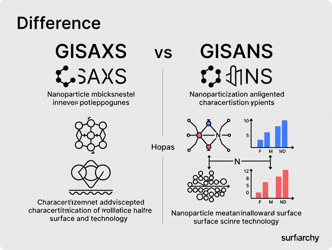
GISAXS vs GISANS: A Comprehensive Guide to Nanoparticle Characterization Techniques
This article provides an in-depth comparative analysis of Grazing Incidence Small-Angle X-ray Scattering (GISAXS) and Grazing Incidence Small-Angle Neutron Scattering (GISANS) for nanoparticle characterization.
GISAXS vs GISANS: A Comprehensive Guide to Nanoparticle Characterization Techniques
Abstract
This article provides an in-depth comparative analysis of Grazing Incidence Small-Angle X-ray Scattering (GISAXS) and Grazing Incidence Small-Angle Neutron Scattering (GISANS) for nanoparticle characterization. Targeted at researchers, scientists, and drug development professionals, it explores the fundamental principles, methodological applications, common troubleshooting strategies, and validation approaches for each technique. The guide explains how GISAXS excels in probing electron density contrasts and nanoscale morphology, while GISANS leverages neutron scattering length density to reveal isotopic and magnetic information, particularly valuable for complex core-shell or lipid-based nanoparticle systems in biomedical research. By synthesizing current methodologies and comparative insights, this resource aims to empower users in selecting and optimizing the appropriate scattering technique for their specific nanoparticle characterization challenges.
Core Principles Explained: Understanding GISAXS and GISANS from the Ground Up
Within the field of nanomaterial and thin film characterization, GISAXS and GISANS are essential, non-destructive grazing incidence scattering techniques. They provide statistically averaged, in-depth information about the morphology, ordering, and size distribution of nanostructures at surfaces and interfaces. This whitepaper frames these techniques within the context of a broader thesis on their distinct roles and complementary nature in advanced nanoparticle characterization research, particularly for applications in drug delivery system development.
Core Principles and Comparative Foundation
Both techniques utilize a grazing incidence beam geometry, where the incident angle (α_i) is typically between 0.1° and 1.0°, slightly above the critical angle for total external reflection of the substrate. This configuration maximizes the beam footprint on the sample, enhances surface sensitivity by limiting penetration depth, and probes the near-surface and interfacial nanostructures.
The fundamental difference lies in the probe: GISAXS uses X-rays (photons), while GISANS uses neutrons. This distinction leads to profound implications for scattering contrast, penetration, and sample environment.
- GISAXS: Scattering contrast arises from electron density differences. It is highly sensitive to heavy elements and provides excellent resolution for size and shape.
- GISANS: Scattering contrast arises from nuclear scattering length density (SLD) differences and is particularly sensitive to light elements (e.g., H, D, Li). The strong isotope effect (e.g., H vs. D) allows for contrast variation by isotopic labeling, making it unparalleled for studying organic, polymeric, and biological nanostructures in complex matrices.
Quantitative Comparison of Technique Parameters
Table 1: Core Technical Specifications of GISAXS vs. GISANS
| Parameter | GISAXS | GISANS |
|---|---|---|
| Probe Particle | X-ray Photon | Neutron |
| Typical Source | Synchrotron, Laboratory rotating anode (Cu Kα, λ≈1.54 Å) | Reactor or Spallation Source (e.g., λ=4-20 Å) |
| Interaction | With electron density | With atomic nuclei |
| Contrast Origin | Electron density difference (Δρ_e) | Scattering Length Density difference (ΔSLD) |
| Key Sensitivity | Heavy elements, inorganic materials, size/shape | Light elements (H, D), isotopes, magnetism |
| Penetration Depth | Microns (tunable via α_i) | Centimeters (highly penetrating) |
| Beam Footprint | ~10-50 mm (long, thin ellipse) | ~10-50 mm (long, thin ellipse) |
| Typical Q-range | 0.01 - 5 nm⁻¹ | 0.01 - 2 nm⁻¹ |
| Resolution (ΔQ/Q) | ~0.01 | ~0.05 |
| Sample Environment | Vacuum/air, limited by X-ray windows | Flexible (high-pressure cells, complex in-situ setups) |
| Primary Data | 2D scattering pattern on area detector | 2D scattering pattern on He-3 or scintillator detector |
Table 2: Application-Oriented Comparison in Nanoparticle Research
| Research Aspect | GISAXS Strengths | GISANS Strengths |
|---|---|---|
| Inorganic NP Characterization | Excellent for size, shape, and superlattice ordering of metals, oxides. | Limited direct contrast; useful for coated NPs or in polymeric matrices. |
| Polymer/NP Composites | Good for large NPs; weak contrast for polymer matrix. | Excellent. Can match SLD of NP to see polymer structure, or vice versa. |
| Lipid & Polymer Nanoparticles (Drug Delivery) | Maps outer structure, core-shell morphology if heavy core. | Ideal. Distinguishes lipid bilayers, polymeric corona, internal aqueous core via H/D contrast. |
| Protein Adsorption & Bio-nano Interfaces | Requires heavy labeling or high concentrations. | Superior. Can study in-situ protein corona formation on NPs in physiological buffers. |
| Buried Interfaces & Depth Profiling | Limited to ~microns; uses angle-dependent fringes. | Excellent penetration allows study of NPs at deeply buried liquid-solid interfaces. |
| Magnetic Nanoparticle Assembly | Insensitive to magnetism. | Unique. Can separate nuclear and magnetic scattering to map magnetic morphology. |
Detailed Experimental Protocols
Generic GISAXS/GISANS Experiment Workflow
Diagram Title: GISAXS/GISANS Experimental Workflow
Protocol A: Characterizing Nanoparticle Superlattice on a Substrate (GISAXS)
- Sample Preparation: Deposit colloidal nanoparticle solution (e.g., 20 nm Au NPs) onto a pristine silicon wafer via spin-coating or drop-casting. Anneal if necessary to promote ordering.
- Alignment: Mount sample on high-precision goniometer. Use a photodiode or ion chamber to find the direct beam position. Align the sample surface to the beam axis (ω = 0).
- Critical Angle Determination: Perform an αi scan (reflectivity-like) to find the critical angle (αc) of the substrate (Si: ~0.22° for Cu Kα).
- Measurement: Set αi to a value slightly above αc (e.g., 0.25°). This ensures total external reflection and an evanescent wave, confining the probe to the surface. Open detector shutter for a calibrated exposure time (synchrotron: 0.1-10s; lab source: >1hr).
- Data Collection: Collect 2D scattering pattern. A beam-stop blocks the intense specularly reflected and directly transmitted beams.
- Primary Analysis: Integrate the 2D pattern along the Qy (lateral) direction to obtain the in-plane structure factor, revealing superlattice peaks. Integrate along Qz (vertical) for information about particle height and substrate correlation.
Protocol B: Probing Lipid Nanoparticle (LNP) Internal Structure (GISANS)
- Contrast Design: Prepare two identical batches of LNPs: one dispersed in H₂O buffer and one in D₂O buffer. The different SLD of the solvent will highlight different components.
- Sample Cell: Load LNP dispersion into a demountable liquid cell with quartz windows and a precise path length (e.g., 1-2 mm).
- Alignment: Mount cell on spectrometer. Use direct beam to align. The critical angle is less critical for deeply penetrating neutrons in liquid samples, but α_i is kept low (~0.2-0.5°) for surface/interface sensitivity if needed.
- Measurement: For each solvent condition, measure the 2D GISANS pattern at a chosen α_i. Use a neutron wavelength of ~5-8 Å. Count times range from 30 minutes to several hours.
- Contrast Matching Analysis: By subtracting or comparing patterns in H₂O and D₂O, the scattering from the lipid bilayer (whose SLD can be matched to either solvent) can be isolated, allowing precise measurement of bilayer thickness and internal core structure.
The Scientist's Toolkit: Essential Research Reagents & Materials
Table 3: Key Materials and Reagents for GISAXS/GISANS Experiments
| Item | Function & Importance |
|---|---|
| High-Purity Silicon Wafers | Standard substrate due to ultra-smooth surface, well-defined critical angle, and low roughness/background scattering. |
| Deuterated Solvents (D₂O, Toluene-d₈) | Crucial for GISANS. Provides strong contrast variation against hydrogenated materials (polymers, lipids, surfactants) for selective component highlighting. |
| Precision Liquid Cells (Quartz/ Sapphire windows) | Allows in-situ and in-operando studies of nanomaterials in liquid environments, essential for drug delivery research. |
| Standard Calibration Samples | Silver behenate or similar for GISAXS; colloidal silica or polymer films for GISANS. Used for precise Q-calibration of the detector. |
| Beam-Stop (Movable) | Blocks the intense specular reflection and direct beam to prevent detector saturation and allow measurement of the weak diffuse scattering signal. |
| High-Vacuum Compatible Sample Holders | For studies requiring control of atmosphere or prevention of air scattering, especially for very low-angle scattering. |
| Contrast-Matched Polymer Blends | Polymer mixtures with matched electron density (GISAXS) or SLD (GISANS) to "mask" one component and isolate the structure of the other (e.g., NP or pore). |
Diagram Title: Decision Logic for Choosing GISAXS or GISANS
GISAXS and GISANS are not competing techniques but powerful partners in the nanoscale characterization toolkit. GISAXS offers high-resolution, readily accessible structural data on inorganic and hard-matter systems. GISANS, with its unique sensitivity to isotopes and light elements and its superior penetration, is indispensable for soft matter, biological composites, and buried interface studies—areas directly relevant to next-generation drug delivery systems. The choice between them is dictated by the specific scientific question, with the most powerful insights often arising from their combined, complementary use.
Within the research paradigm investigating the structural characterization of nanoparticles and thin films, Grazing Incidence Small-Angle Scattering (GISAXS) and Grazing Incidence Small-Angle Neutron Scattering (GISANS) are indispensable techniques. The fundamental difference between them, and the basis for their complementary use, stems from the distinct physical interaction mechanisms of X-rays and neutrons with matter. This whitepaper delineates these core contrast mechanisms, providing the foundational physics necessary to interpret GISAXS and GISANS data within nanoparticle research for advanced materials and drug delivery systems.
Core Interaction Physics and Scattering Formalism
Scattering intensity, I(Q), where Q is the scattering vector, is governed by the interaction potential and the spatial distribution of scattering centers. The differential cross-section for a collection of N particles is: [ \frac{d\Sigma}{d\Omega}(Q) = \left\langle \left| \sum{i=1}^{N} bi \exp(i\mathbf{Q} \cdot \mathbf{r}i) \right|^2 \right\rangle ] where ( bi ) is the scattering length and ( \mathbf{r}_i ) is the position of scatterer i.
2.1 X-ray Scattering (GISAXS) X-rays interact with the electron cloud of an atom. The scattering length for X-rays is proportional to the atomic number (Z) and the complex atomic form factor, ( f(\mathbf{Q}, E) = f0(\mathbf{Q}) + f'(E) + i f''(E) ). The latter two terms are energy-dependent resonant (anomalous) corrections near absorption edges. The scattering contrast is directly proportional to the electron density difference ((\Delta\rhoe)) between the nanoparticle and the surrounding matrix: [ b{X-ray} \propto re f(\mathbf{Q}, E), \quad \text{Contrast} \propto (\rho{e, particle} - \rho{e, matrix}) ] where ( r_e ) is the classical electron radius.
2.2 Neutron Scattering (GISANS) Neutrons interact with the atomic nucleus via the strong nuclear force and with unpaired electron spins via magnetic dipole interaction. The nuclear scattering length, ( bn ), is isotope-specific, varies irregularly across the periodic table, and can be positive or negative. The key parameter is the scattering length density (SLD), ( \rho{SLD} = \sumi ni b{n,i} ), where ( ni ) is the number density of nucleus i. Neutron contrast arises from the SLD difference: [ \text{Contrast} \propto (\rho{SLD, particle} - \rho{SLD, matrix}) ] Critically, the scattering length varies between isotopes (e.g., ( ^1\text{H} ) and ( ^2\text{H} ) (D)), enabling contrast variation/v matching by tuning the H₂O/D₂O ratio in solvents or matrices.
Quantitative Comparison of Core Mechanisms
Table 1: Fundamental Scattering Properties
| Property | X-ray Scattering (GISAXS) | Neutron Scattering (GISANS) |
|---|---|---|
| Probe | Photons (electromagnetic wave) | Neutrons (particle with spin ½) |
| Interaction | With electron density | With atomic nuclei & magnetic moments |
| Scattering Length | ( \propto Z ), ~10⁻¹⁵ m | Isotope-dependent, irregular, ~10⁻¹⁵ m |
| Key Contrast Parameter | Electron Density, (\rho_e) (e⁻/ų) | Scattering Length Density, SLD (Å⁻²) |
| Element Sensitivity | Strong for high Z | Non-monotonic; can distinguish isotopes |
| Penetration Depth | Microns to mm (material dependent) | cm scale for most non-absorbing materials |
| Beam Typical Source | Synchrotron or Laboratory X-ray Tube | Nuclear Reactor or Spallation Source |
Table 2: Implications for Nanoparticle Characterization (GISAXS vs. GISANS)
| Application Aspect | GISAXS Advantage | GISANS Advantage |
|---|---|---|
| Core-Shell Structure | Excellent for heavy metal cores in light organic shells. | Ideal for organic/organic or soft matter interfaces; can highlight shell via contrast matching. |
| Buried Interfaces | Limited for deeply buried structures in solid matrices. | Superior penetration allows probing deeply buried nanostructures in polymers, lipids, or matrices. |
| Biological/Lipid Systems | Weak contrast in aqueous media; radiation damage can be high. | Excellent contrast in H₂O/D₂O; minimal radiation damage to soft matter. |
| Magnetic Nanostructures | Indirect via anomalous scattering near edges. | Direct probe of magnetic structure via spin-dependent scattering. |
| Kinetics/In-situ | Fast data collection at synchrotrons. | Slower, but enables unique in-situ contrast variation experiments in fluid cells. |
Experimental Protocols for GISAXS and GISANS
Protocol 1: Standard GISAXS Experiment on Nanoparticle Thin Films
- Sample Preparation: Spin-coat or Langmuir-Blodgett deposit nanoparticle (e.g., gold, iron oxide, polymer) dispersion onto a silicon wafer. Measure film thickness via ellipsometry.
- Alignment: Mount sample on a high-precision goniometer in a vacuum chamber to minimize air scattering. Using a direct beam stop, align the sample surface to the incident X-ray beam (( \alpha_i )) with micrometric precision.
- Data Acquisition (Synchrotron): Set ( \alphai ) to a value slightly above the critical angle of the substrate (typically 0.1° - 0.5°). Use a 2D area detector (e.g., Pilatus) placed ~1-5 m downstream. Acquire scattering patterns at fixed incidence or perform a rocking scan (scan ( \alphai )) around the critical angle to enhance surface sensitivity.
- Data Reduction: Correct raw images for detector dark current, spatial distortion, and solid angle. Normalize by incident flux and exposure time. Subtract background scattering from bare substrate.
Protocol 2: Contrast Variation GISANS Experiment on Lipid Nanoparticles
- Sample Preparation: Prepare identical batches of lipid nanoparticles (LNPs) in buffers with varying D₂O/H₂O ratios (e.g., 0%, 50%, 100% D₂O). Load into temperature-controlled, quartz liquid cells with precise path length (1-2 mm).
- Alignment & Contrast Match Point: Mount cell on goniometer. Use a cold neutron beam (wavelength, ( \lambda ), typically 4-10 Å). Align grazing incidence geometry similarly to GISAXS. First, measure scattering from pure buffer solvents to determine the SLD value that gives minimal scattering (the "match point").
- Data Acquisition: Acquire 2D GISANS patterns for each LNP sample at a fixed ( \alpha_i ) above the critical angle of the liquid-substrate interface. Use a 2D He-3 or scintillator detector. Count times are long, typically several hours per sample, due to lower neutron flux.
- Data Analysis: Perform radial averaging to obtain 1D intensity vs. Q profiles. Analyze using models (e.g., core-shell form factor, paracrystal lattice factor) where the SLD of each component is defined based on the known buffer SLD. The variation of the forward scattering I(Q→0) with buffer SLD directly yields the nanoparticle's mean SLD.
Visualizing the Scattering Process and Analysis Workflow
Title: Fundamental Scattering Contrast Pathway
Title: GISAXS vs GISANS Experiment Decision Flow
The Scientist's Toolkit: Essential Research Reagents & Materials
Table 3: Key Materials for Scattering Experiments on Nanoparticles
| Item | Function in Experiment | GISAXS-specific / GISANS-specific / Common |
|---|---|---|
| High-Purity Silicon Wafers | Atomically flat, low-scattering substrate for thin film deposition. | Common |
| Deuterated Solvents (D₂O, Toluene-d₈) | To modulate SLD of the matrix for contrast variation and matching. | GISANS-critical |
| Precision Liquid Cells (Quartz) | Holds liquid samples with defined path length for in-situ studies. | Common (GISANS use more frequent) |
| Micrometric Goniometer | Provides precise angular control for grazing incidence alignment. | Common |
| Beam Stop (Direct & Specular) | Blocks intense direct and specularly reflected beam to protect detector. | Common |
| Calibration Standards | Silver behenate (d-spacing) for X-rays; polymer blends for neutron Q-calibration. | Common (standard-dependent) |
| Ion-Exchange Columns | For precise preparation of buffer mixtures with specific H₂O/D₂O ratios. | GISANS-critical |
| Radiation-Sensitive Detectors | Pilatus/Eiger (X-ray), ³He Tube/Scintillator (Neutron) for 2D pattern capture. | Technique-specific |
Understanding nanoparticle surfaces and interfaces is critical for applications in catalysis, drug delivery, and advanced materials. This in-depth guide examines the core physical interactions of X-rays and neutrons with nanoscale matter, contextualized within a broader thesis comparing Grazing-Incidence Small-Angle X-ray Scattering (GISAXS) and Grazing-Incidence Small-Angle Neutron Scattering (GISANS) for nanoparticle characterization. These complementary techniques leverage the distinct scattering mechanisms of photons and neutrons to probe structure, composition, and morphology at interfaces.
Fundamental Interaction Mechanisms
X-ray Interactions
X-rays interact primarily with the electron cloud of atoms. The key interactions are:
- Elastic Scattering (Thomson Scattering): Coherent, without energy loss, providing structural information.
- Inelastic Scattering (Compton Scattering): Incoherent, with energy loss.
- Photoelectric Absorption: Complete transfer of energy, ejecting a core electron.
- Fluorescence and Auger Emission: Secondary processes following absorption.
The scattering length density (SLD) for X-rays, ( \rho{x-ray} ), is proportional to the electron density and the atomic form factor, ( f ): [ \rho{x-ray} = \frac{re \lambda^2}{\pi} \sumi ni fi ] where ( re ) is the classical electron radius, ( \lambda ) is the wavelength, and ( ni ) is the number density of atom ( i ).
Neutron Interactions
Neutrons interact with atomic nuclei via the strong nuclear force and with unpaired electron spins via magnetic dipole interactions. The interactions are:
- Nuclear Scattering: Can be coherent (structure-sensitive) or incoherent (e.g., from hydrogen-1).
- Magnetic Scattering: From interaction with magnetic moments.
- Absorption: Generally weak, but high for isotopes like ( ^{10}B ), ( ^{3}He ), ( ^{113}Cd ).
The neutron scattering length ( b ) varies irregularly across the periodic table and between isotopes. The SLD for neutrons is: [ \rho{neutron} = \sumi ni bi ]
Table 1: Comparison of Core Interaction Properties
| Property | X-rays | Neutrons (Nuclear) |
|---|---|---|
| Primary Interaction | With electron density | With atomic nuclei |
| Scattering Length | Proportional to atomic number (Z) | No monotonic dependence on Z |
| Isotope Sensitivity | None | High (e.g., ( ^1H ) vs ( ^2H )) |
| Magnetic Sensitivity | Very weak (via polarization) | Direct (with magnetic moments) |
| Penetration Depth | Medium (µm to mm) | Very High (cm scale) |
| Sample Environment | Vacuum/Air typical | Often requires containment |
| Typical Flux | ( 10^{12} - 10^{15} ) ph/s | ( 10^7 - 10^{10} ) n/cm²/s |
Interaction with Nanoparticle Surfaces and Interfaces
At grazing incidence, the beam penetrates only a few nanometers into the substrate, creating an evanescent wave that selectively probes nanoparticles at the interface. The reflected and scattered intensity depends on the angle of incidence relative to the critical angle of the substrate.
Refraction and Reflection
The index of refraction for both probes is slightly less than 1: ( n = 1 - \delta + i\beta ).
- For X-rays, ( \delta \propto \rho_{x-ray} ) and ( \beta ) accounts for absorption.
- For neutrons, ( \delta \propto \rho_{neutron} ) and ( \beta ) is typically very small.
Below the critical angle ( \alpha_c = \sqrt{2\delta} ), total external reflection occurs, confining the probe to the surface region.
Scattering from Surface Nanoparticles
The differential scattering cross-section for particles at an interface incorporates:
- Form Factor (P): Describes scattering from the nanoparticle shape/size.
- Structure Factor (S): Describes inter-particle interference/ordering.
- Distorted Wave Born Approximation (DWBA): Essential for grazing incidence, accounts for multiple reflections/refractions of the incident and scattered waves at the interface.
The intensity in a GISAXS/GISANS pattern is: [ I(q{xy}, qz) \propto |T(\alphai)|^2 |T(\alphaf)|^2 \cdot |F(\vec{q})|^2 \cdot S(\vec{q}) ] where ( T ) are the transmission functions, ( F ) is the form factor amplitude, and ( q{xy}, qz ) are the momentum transfer components parallel and perpendicular to the surface.
Table 2: Signal Sensitivity to Nanoparticle Properties
| Nanoparticle Property | GISAXS Sensitivity | GISANS Sensitivity |
|---|---|---|
| Size & Shape | High (via form factor) | High (via form factor) |
| Surface Coverage | High (via intensity) | High (via intensity) |
| Lateral Ordering | High (via ( q_{xy} ) peaks) | High (via ( q_{xy} ) peaks) |
| Core Composition | Moderate (electron density) | Very High (via SLD contrast) |
| Ligand Shell Density | Low (weak contrast) | Very High (H/D isotope labeling) |
| Buried Interface Structure | Low (penetration) | Very High (deep penetration) |
| Magnetic Structure | None | Very High (spin polarization) |
Experimental Protocols for GISAXS and GISANS
Sample Preparation Protocol
- Substrate: Single crystal silicon wafers (SiO₂ native oxide) are standard for their low roughness and well-defined critical angle.
- Nanoparticle Deposition: Use spin-coating, drop-casting, or Langmuir-Blodgett techniques to achieve monolayer coverage. Critical to avoid multilayer formation for clear interface signals.
- Contrast Matching (GISANS): Prepare nanoparticles with deuterated ligands or disperse in deuterated solvents to match the SLD of specific components, isolating scattering from desired interfaces.
Beamline Setup & Data Acquisition
- Alignment: Pre-align the sample stage to the beam center and rotation axis (omega) with a laser or direct beam.
- Incident Angle Determination: Perform a specular reflectivity scan to find the substrate's critical angle (( \alphac )). Set the measurement angle (( \alphai )) slightly above ( \alpha_c ) for surface sensitivity or below for total external reflection.
- Beam Definition: Use slits to define beam size (typically 50 x 200 µm) to illuminate a ~1 cm long footprint.
- Detector Positioning: Place a 2D area detector (CCD for GISAXS, ( ^3He ) tube or scintillator for GISANS) several meters downstream to capture small-angle scattering.
- Exposure: Typical exposure times range from 0.1-10 seconds for synchrotron GISAXS and 10 minutes to several hours for reactor-source GISANS.
- Background Subtraction: Measure and subtract scattering from a clean, identical substrate.
Data Reduction and Analysis Workflow
- Correction: Apply flat-field, dark-current, and geometric corrections to the 2D image.
- Binning: Bin pixels to improve statistics if necessary.
- Coordinate Transformation: Convert detector pixel coordinates to reciprocal space coordinates (( q{xy}, qz )).
- Sector Cuts: Extract 1D profiles (e.g., horizontal cuts at constant ( q_z ) for in-plane structure, vertical cuts for out-of-plane shape).
- Model Fitting: Fit profiles using DWBA-based models for form factor (sphere, cylinder, core-shell) and structure factor (hard sphere, paracrystal) to extract parameters like radius, spacing, and ordering.
Diagram Title: GISAXS/GISANS Data Analysis Workflow
The Scientist's Toolkit: Key Research Reagent Solutions
Table 3: Essential Materials for Nanoparticle Interface Probing
| Item | Function & Relevance |
|---|---|
| High-Purity Silicon Wafers | Atomically flat, low-roughness substrate with well-defined X-ray/neutron optical properties. |
| Deuterated Solvents (e.g., Toluene-d8, D2O) | For contrast variation in GISANS; matching SLD to hide specific components. |
| Deuterated Ligands (e.g., Thiols, Polymers) | Coating nanoparticles with deuterated molecules to manipulate SLD and highlight core-shell interfaces. |
| Precision Goniometer Stage | Provides micron-resolution control of sample angle (omega, phi, tilt) for precise grazing incidence alignment. |
| Beam-Defining Slits (Tungsten/ Cadmium) | Define beam footprint; cadmium is essential for neutron beam shaping due to high absorption. |
| 2D Area Detector (Pixel/CCD for X-ray, ³He for Neutrons) | Captures the scattered intensity pattern. Fast readout is crucial for time-resolved GISAXS. |
| Vacuum Chamber/Beam Containment | Required for neutron experiments due to beam divergence and biological shielding. Optional for hard X-ray GISAXS. |
| Standard Reference Samples (e.g., PS Latex on Si) | Used for beamline alignment, detector calibration, and resolution verification. |
Comparative Thesis: GISAXS vs. GISANS in Research
The choice between GISAXS and GISANS hinges on the specific scientific question, driven by their fundamental interaction differences.
Table 4: Strategic Selection for Research Objectives
| Research Objective | Preferred Technique | Rationale |
|---|---|---|
| High-throughput morphology screening | GISAXS | Superior flux enables rapid measurement of size, shape, and ordering. |
| Probing ligand shell density & conformation | GISANS | Unmatched contrast via H/D labeling of organic components. |
| Studying nanoparticles at buried solid/liquid interfaces | GISANS | Deep neutron penetration through sample environments (e.g., flow cells). |
| In-situ monitoring of self-assembly kinetics | GISAXS | Time-resolution down to milliseconds at synchrotrons. |
| Characterizing magnetic nanoparticle arrays | GISANS | Direct sensitivity to magnetic moment arrangement via spin polarization. |
| Resolving composition of complex core-shell structures | GISANS | Independent tuning of SLD for core, shell, and matrix via isotopic labeling. |
Diagram Title: Decision Flow: GISAXS vs GISANS Selection
The interactions of X-rays and neutrons with nanoparticle surfaces are fundamentally different, giving rise to the complementary strengths of GISAXS and GISANS. GISAXS, with its high flux and rapid data collection, is the premier tool for morphological and kinetic studies. GISANS, through its nuclear and isotope sensitivity, is unparalleled for resolving compositional, organic, and magnetic structures at interfaces, even when buried. A complete thesis on nanoparticle characterization must leverage both techniques to construct a holistic, multi-contrast picture of nanostructure at interfaces, driving advances in nanomaterials design for catalysis, medicine, and nanotechnology.
This technical guide elucidates the principles and applications of grazing incidence geometries, primarily focusing on X-ray and neutron scattering techniques. Framed within ongoing research differentiating GISAXS (Grazing Incidence Small-Angle X-ray Scattering) and GISANS (Grazing Incidence Small-Angle Neutron Scattering) for nanoparticle characterization, this document details the critical role of geometry in probing surface and interfacial structures. The ultra-shallow penetration afforded by angles below the critical angle enables unparalleled sensitivity to thin films, nanostructured surfaces, and buried interfaces, which is paramount for advanced materials science and pharmaceutical development.
In a grazing incidence setup, a collimated beam of X-rays or neutrons strikes a flat sample surface at an incident angle (α_i) typically ranging from 0.1° to 1.0°. This angle is often chosen to be at or below the critical angle for total external reflection (typically 0.1°-0.5° for X-rays on solid materials). At this condition, the beam propagates as an evanescent wave, confining the probe intensity to the near-surface region (tens of nanometers) and drastically reducing background scattering from the substrate. This geometry is the foundational element for a suite of surface-sensitive techniques.
Core Physical Principles
Critical Angle and Evanescent Wave
The critical angle (αc) is defined by the refractive index of the material, n = 1 - δ + iβ, where δ is related to scattering density. For X-rays: [ αc ≈ \sqrt{2δ} ] Below αc, total external reflection occurs, creating an evanescent wave with a penetration depth (Λ) given by: [ Λ = \frac{λ}{2π\sqrt{αc^2 - α_i^2}} ] where λ is the wavelength. This confines the probe to the surface, enabling exquisite sensitivity to thin films and nanoparticles at interfaces.
The q-Vector Resolution
The scattering vector q is central to all scattering techniques. In grazing incidence, it has distinct components:
- q_z: Perpendicular to the surface, sensitive to film thickness, density profile, and vertical particle correlation.
- q_y: Parallel to the surface (in-plane), sensitive to lateral nanostructure ordering and in-plane correlation lengths.
Precise control of αi and the exit angle (αf) and scattering angle (2θ) in the detector plane allows mapping of the reciprocal space (qy, qz).
GISAXS vs. GISANS: Geometry in Context of Nanoparticle Characterization
While the geometric setup is nearly identical, the probe particle—X-ray vs. neutron—fundamentally differentiates GISAXS and GISANS, leading to complementary information crucial for advanced research.
Comparative Table: Core Parameters
| Parameter | GISAXS (X-rays) | GISANS (Neutrons) |
|---|---|---|
| Typical Wavelength (λ) | 0.5 - 1.5 Å (Synchrotron) | 4 - 20 Å (Reactor/Spallation) |
| Critical Angle (α_c)* | ~0.1° - 0.3° | ~0.2° - 0.6° |
| Primary Contrast Source | Electron density difference | Nuclear scattering length density (SLD) difference |
| Penetration Depth (below α_c) | ~5 - 50 nm | ~10 - 200 nm |
| Key Strength | High flux, excellent spatial resolution of shape/size. | Isotopic labeling (H/D), sensitivity to light elements, magnetic structure. |
| Sample Environment | Vacuum/Air, easy sample stage integration. | Often requires vacuum flight path, larger beam size. |
| Typical Measurement Time | Seconds to minutes (synchrotron) | Minutes to hours (reactor) |
| Primary for Drug Development | NP morphology, distribution on surfaces, film porosity. | Buried NP-protein interactions, hydration layers, lipid membrane insertion. |
*For a silicon substrate. Varies with material and λ.
Complementary Data Table: Nanoparticle Characterization
| Characterization Target | GISAXS Advantage | GISANS Advantage |
|---|---|---|
| Size/Shape of Metallic NPs | Excellent due to high electron density contrast. | Poor for pure metals; good if core-shell uses isotopic contrast. |
| Lateral Ordering in Arrays | Superior from high flux and sharp resolution. | Good, but longer measurement times limit statistics. |
| Polymer Brush Coating on NP | Moderate contrast if electron density differs. | Excellent by deuterating brush or solvent (contrast matching/variation). |
| Protein Corona on NP Surface | Very weak contrast in aqueous media. | Ideal by using D₂O buffer and/or deuterated proteins to highlight corona. |
| NP Buried in Polymer Matrix | Good if matrix/NP density differs. | Superior by deuterating matrix to "see through" to NPs. |
| Magnetic NP Ordering | Requires resonant (magnetic) X-ray scattering. | Direct via nuclear spin-polarized neutrons (Polarized GISANS). |
Experimental Protocols
Standard GISAXS/GISANS Experiment Workflow
Title: GISAXS/SANS Experimental Workflow
Sample Preparation Protocol for Polymer/NP Thin Films
- Materials: Silicon wafer (P-type, <100>, native oxide), toluene, polystyrene (PS, MW 100kDa), gold nanoparticles (10nm diameter, citrate stabilized).
- Procedure:
- Wafer Cleaning: Sonicate silicon wafer in acetone for 10 min, then isopropanol for 10 min. Dry under N₂ stream. Treat with oxygen plasma for 2 min to ensure hydrophilic surface.
- Solution Preparation: Dissolve PS in toluene at 1% w/w. Add Au NP solution to achieve a 1:100 NP:PS mass ratio. Stir for 24h.
- Spin-coating: Deposit 100 µL of solution onto static wafer. Spin at 2000 rpm for 60s in a cleanroom environment.
- Annealing: Thermally anneal sample at 120°C (above Tg of PS) under vacuum for 12h to allow NP diffusion and achieve equilibrium structure.
- Key Consideration: Film thickness (~50-100nm) must be characterized independently via ellipsometry to inform scattering modeling.
Data Acquisition Protocol for Synchrotron GISAXS
- Beamline Parameters: λ = 1.03 Å (12 keV), Beam size: 100 (V) x 300 (H) µm², Detector: 2D Pilatus3 1M placed 3m from sample.
- Alignment:
- Use direct beam stop to protect detector.
- Align sample surface to beam center using laser autocollimator.
- Perform a rocking scan (ω-scan) around α_i = 0° to find the substrate edge. Set zero accurately.
- Measurement:
- Measure specular reflectivity curve to determine α_c for the film.
- Set αi to 0.15° (below αc of Si and typical polymer film).
- Acquire 2D scattering pattern with 1-10s exposure, ensuring no detector saturation.
- Move sample to fresh spot for each measurement to avoid radiation damage.
- Collect background scattering from bare substrate under identical conditions.
The Scientist's Toolkit: Key Research Reagent Solutions
| Material/Reagent | Function in GISAXS/GISANS Experiments |
|---|---|
| Ultra-Flat Silicon Wafers | Standard substrate with low roughness, known critical angle, and compatibility with various cleaning protocols. |
| Deuterated Polymers (e.g., d-PS) | In GISANS, provides strong contrast against hydrogenated matrices or solvents via isotopic labeling, enabling visualization of specific components. |
| Contrast Matching Solvents (D₂O, deuterated toluene) | Used in GISANS to adjust the scattering length density of the environment to "match out" specific components (e.g., a matrix) to highlight others (e.g., nanoparticles). |
| Precision Goniometer & Auto-Leveling Stage | Enables precise alignment of the sample surface to better than 0.001°, which is critical for defining the grazing angle and ensuring beam path consistency. |
| Beam-Defining Slits & Collimators | Create a clean, well-defined beam profile with minimal parasitic scattering, which is essential for interpreting low-intensity scattering signals near the horizon. |
| 2D Area Detector (Pixel/CCD) | Captures the full 2D scattering pattern simultaneously, allowing analysis of anisotropic structures and proper separation of qy and qz components. |
Data Analysis and Modeling Pathways
Title: GISAXS/SANS Data Analysis Pathway
The grazing incidence geometry is not merely an experimental configuration but a paradigm for surface and interface science. Its stringent angular control unlocks the nanoscale world at surfaces and in thin films. The strategic choice between GISAXS and GISANS, governed by their contrast mechanisms rooted in this shared geometry, provides a powerful dual approach for comprehensive nanoparticle characterization. For drug development, this is particularly transformative, enabling the study of protein-NP interactions, lipid membrane dynamics, and the fate of drug delivery vectors at relevant interfaces—information that is critical for rational therapeutic design.
Within the advanced toolkit of nanoparticle characterization, Grazing-Incidence Small-Angle X-ray Scattering (GISAXS) and Grazing-Incidence Small-Angle Neutron Scattering (GISANS) are pivotal techniques. Their power and differentiation stem from a precise understanding of three interconnected core parameters: the scattering vector (Q), the associated scattering angles, and the resulting depth sensitivity of the measurement. This guide details these parameters within the overarching thesis that while GISAXS and GISANS share a common geometric formalism, their differing probe particles (X-rays vs. neutrons) lead to distinct interactions with matter, which must be understood through Q, angle, and depth to select the optimal technique for a given nanomaterial research or drug delivery system problem.
The Scattering Vector (Q)
The scattering vector, Q, is the fundamental quantity in any scattering experiment. It defines the momentum transfer from the incident beam to the sample and thus probes specific length scales within the nanostructure.
Definition: ( \vec{Q} = \vec{k}{out} - \vec{k}{in} ) where ( \vec{k}{in} ) and ( \vec{k}{out} ) are the wave vectors of the incident and scattered beams, respectively. Their magnitudes are ( |k| = 2\pi / \lambda ).
In grazing-incidence geometry, Q is decomposed into three components:
- Q_y: The in-plane component perpendicular to the incident beam direction (along the sample surface).
- Qx: The in-plane component parallel to the incident beam direction. Under the GISAXS/GISANS condition (small out-of-plane angles), Qx is approximately zero.
- Q_z: The out-of-plane component perpendicular to the sample surface.
The magnitudes are given by: [ Qy = \frac{2\pi}{\lambda} (\cos(\alphaf) \sin(\psi) \approx \frac{2\pi}{\lambda} \psi) ] [ Qz = \frac{2\pi}{\lambda} (\sin(\alphai) + \sin(\alpha_f)) ]
where ( \lambda ) is the wavelength, ( \alphai ) is the incident angle, ( \alphaf ) is the exit angle relative to the sample surface, and ( \psi ) is the in-plane scattering angle.
GISAXS vs. GISANS Context: The relationship between measurable angles (αi, αf, ψ) and Q is identical. However, typical neutron wavelengths (4-20 Å) are much longer than X-ray wavelengths (e.g., 1.34 Å for Cu Kα). For a given angular resolution, this results in a smaller Q-range accessible with neutrons, directly influencing the size of observable nanostructures.
Table 1: Q-vector Components and Their Information Content
| Q Component | Definition | Probes | Typical Range (GISAXS) | Typical Range (GISANS) | |
|---|---|---|---|---|---|
| Q_y | In-plane, horizontal | In-plane particle spacing, shape, and ordering. | 0.01 - 1 nm⁻¹ | 0.002 - 0.2 nm⁻¹ | |
| Q_z | Out-of-plane, vertical | Particle form factor (size/shape), vertical structure, and film thickness. | 0.01 - 2 nm⁻¹ | 0.002 - 0.4 nm⁻¹ | |
| Q | Total magnitude | Overall particle size (via Guinier analysis). | Derived from Qy and Qz. |
Scattering Angles
The angles in a grazing-incidence experiment define the geometry and are directly linked to the Q-components.
Key Angles:
- Incident Angle (α_i): The angle between the incident beam and the sample surface. It is the most critical parameter for depth sensitivity (see Section 3).
- Exit Angle (α_f): The angle between the scattered beam and the sample surface.
- In-plane Angle (ψ): The horizontal scattering angle within the sample plane.
Critical Angle Phenomenon: Both X-rays and neutrons undergo total external reflection below a material-specific critical angle (αc). For X-rays, αc depends on electron density (~0.1° - 0.3°). For neutrons, αc depends on the neutron scattering length density (SLD) and is an order of magnitude smaller (~0.1° - 0.3° is possible, but often much lower for many materials). Operating αi at or below α_c confines the probe (as an evanescent wave) to the near-surface region, dramatically enhancing surface sensitivity.
Table 2: Angle Definitions and Operational Ranges
| Angle | Role | Typical Operational Range | GISAS Technique Consideration |
|---|---|---|---|
| α_i (Incident) | Sets penetration depth. | 0.1° - 1.0° (often αi ≈ αc) | GISANS may use lower absolute αi due to smaller αc for many materials. |
| α_f (Exit) | Maps to Q_z. | -1.0° to +3.0° | Measured by 2D detector position. |
| ψ (In-plane) | Maps to Q_y. | -2.0° to +2.0° | Measured by 2D detector position. |
Depth Sensitivity and Probe Penetration
Depth sensitivity is governed by the incident angle relative to the critical angle and the penetrating power of the probe.
X-rays (GISAXS): Penetration depth of X-rays varies sharply with α_i.
- αi << αc: Probe is an evanescent wave, decaying exponentially. Information comes from the top ~5-10 nm.
- αi = αc: Maximum surface sensitivity and enhanced scattering yield.
- αi > αc: Probe penetrates the film/substrate. Information is averaged over the entire film thickness and into the substrate.
Neutrons (GISANS): Neutrons have a much lower absorption coefficient for most materials, leading to greater inherent penetration.
- Even when αi < αc (evanescent wave regime), the neutron evanescent wave can penetrate deeper than X-rays for many organic and soft matter systems.
- This, combined with SLD contrast variation (e.g., using deuterated solvents or particles), allows GISANS to probe buried interfaces and internal structure within nanocomposite films or drug delivery vehicles with exceptional clarity, a key advantage over GISAXS.
Experimental Protocols for Parameter Determination
Protocol 1: Critical Angle Measurement
- Alignment: Pre-align the sample stage to sub-milliradian precision using a laser or direct beam.
- Detector Setup: Place a point detector (ion chamber for X-rays, ³He tube for neutrons) in the specular reflection plane (ψ=0).
- θ-2θ Scan: Perform a coupled θ (sample) - 2θ (detector) scan through α_i = 0° to ~1°.
- Analysis: Plot reflected intensity vs. αi. Fit the curve using the Parratt formalism (X-rays) or analogous optical model (neutrons) to determine the precise αc and film SLD/thickness.
Protocol 2: GISAXS/GISANS Mapping to Q-Space
- Geometry Calibration: Use a known standard (e.g., silver behenate for GISAXS, a grating for GISANS) to calibrate the sample-to-detector distance and detector tilt (orthogonality).
- Beam Characterization: Measure the direct beam position and profile (size, divergence) with the sample removed.
- Data Collection: Acquire 2D scattering pattern at fixed αi (typically at or just above αc).
- Q-Conversion: Apply the transformation: [ Qy = \frac{2\pi}{\lambda} \cdot \frac{(x - x0) \cdot p}{SDD} ] [ Qz = \frac{2\pi}{\lambda} \cdot \frac{(y - y0) \cdot p}{SDD} ] where (x₀, y₀) is the direct beam center, p is pixel size, and SDD is sample-to-detector distance.
The Scientist's Toolkit: Essential Research Reagents & Materials
Table 3: Key Materials for GISAXS/GISANS Experiments
| Material/Reagent | Function | GISAXS Specificity | GISANS Specificity |
|---|---|---|---|
| Calibration Standard | Calibrates Q-space. | Silver behenate, polystyrene beads. | Diffraction grating, colloidal silica. |
| Si Wafer (with native oxide) | Standard substrate. | Provides smooth, flat surface; known α_c. | Low SLD; minimal background scattering. |
| Deuterated Solvents | Contrast matching/variation. | Not typically used. | Essential. H₂O/D₂O mixtures tune SLD to match/highlight specific components. |
| Deuterated Polymers/Lipids | Label specific components. | Not applicable. | Crucial. Enhances neutron contrast for organic nanoparticles (e.g., drug carriers). |
| Precision Goniometer | Controls α_i with µ° accuracy. | Required. | Required, often with heavier stages for neutron environment. |
| Beamstop | Protects detector from intense direct/specular beam. | Solid-state/beamstop. | Often ³He-filled tube or Gd foil. |
| Vacuum Chamber | Reduces air scattering/absorption. | Commonly used on synchrotron beamlines. | Mandatory. Neutrons are highly scattered by air; flight path is evacuated. |
Practical Application Guide: When and How to Use GISAXS or GISANS for Nanoparticle Analysis
Sample Preparation Protocols for Thin Films, Nanoparticle Arrays, and Buried Interfaces
Within the framework of a thesis comparing Grazing-Incidence Small-Angle X-ray Scattering (GISAXS) and Grazing-Incidence Small-Angle Neutron Scattering (GISANS) for nanoparticle characterization, sample preparation is the critical foundational step. The structural and chemical data retrieved by these techniques are profoundly sensitive to interfacial morphology, nanoparticle ordering, and layer architecture. GISAXS, sensitive to electron density contrast, and GISANS, sensitive to nuclear scattering length density and magnetic moments, place distinct demands on sample design. This guide details protocols tailored to generate reliable, high-quality samples for such advanced synchrotron and neutron reflectometry studies, with a focus on achieving the required contrast, uniformity, and stability.
Foundational Principles for Preparation
Successful preparation for GISAXS/GISANS requires adherence to core principles:
- Ultra-Clean Substrates: Minimize background scattering from surface contaminants.
- Controlled Environment: For thin films, control of humidity, temperature, and dust is paramount.
- Contrast Engineering: For GISANS, deliberate use of isotopic substitution (e.g., H vs. D) in polymers or solvents to manipulate neutron contrast without altering chemistry.
- Interfacial Stability: Buried interfaces must be non-diffusive or well-defined on the timescale of the experiment.
Detailed Experimental Protocols
Protocol for Ultrathin, Smooth Polymer Films (for Buried Interface Studies)
Aim: Produce pinhole-free, smooth polymer films (~10-100 nm) on silicon wafers for creating well-defined buried interfaces with air or another layer.
Materials: Silicon wafer (P-doped, ⟨100⟩), toluene (HPLC grade), polymer (e.g., polystyrene, PMMA), polypropylene syringe, PTFE filter (0.2 µm), glass staining dish, spin coater.
Method:
- Substrate Cleaning: Sonicate wafer in acetone for 10 min, then in isopropanol for 10 min. Dry under N₂ stream. Treat with oxygen plasma for 5 min to render surface hydrophilic.
- Polymer Solution Preparation: Dissolve polymer in toluene to a concentration of 5-20 mg/mL. Stir on a heated plate at 50°C for >12 hours until fully dissolved.
- Filtration: Draw solution into a syringe and pass through a 0.2 µm PTFE filter directly onto the wafer center.
- Spin-Coating: Program spin coater: 500 rpm for 5 s (spread), then 2000-4000 rpm for 60 s (thin). Optimize speed for target thickness.
- Annealing: Vacuum-anneal film at 120°C (above Tg) for 12-24 hours to remove residual solvent and relax surface roughness.
- Characterization: Validate thickness and roughness via spectroscopic ellipsometry or AFM.
Protocol for Ordered Nanoparticle Arrays via Langmuir-Blodgett Deposition
Aim: Assemble close-packed monolayers of colloidal nanoparticles (e.g., Au, SiO₂) with hexagonal order.
Materials: Langmuir-Blodgett trough, nanoparticle dispersion in a volatile solvent (e.g., chloroform for hydrophobic NPs, water/ethanol for hydrophilic), deionized water (resistivity >18 MΩ·cm), barrier compression system, surface pressure sensor.
Method:
- Trough & Subphase Preparation: Fill trough with ultrapure water. Clean surface by repeated suction. Set subphase temperature to a constant 20°C.
- Nanoparticle Dispersion: Prepare a monodisperse NP solution (~0.5 mg/mL). For hydrophilic NPs, mix with ethanol (3:1 v/v) to aid spreading.
- Langmuir Film Formation: Slowly spread the NP dispersion dropwise onto the air-water interface. Allow solvent to evaporate completely for 15 min.
- Isothermal Compression: Compress barriers at a slow, constant rate (e.g., 5 cm²/min) while monitoring surface pressure (Π)-Area (A) isotherm.
- Film Transfer: At the target pressure (corresponding to solid phase on isotherm), hold pressure constant. Vertically dip a pre-cleaned substrate (hydrophilic for downstroke, hydrophobic for upstroke) through the interface at 2-5 mm/min to transfer the monolayer.
- Drying: Dry the sample gently under a nitrogen stream.
Protocol for Creating a Buried Polymer-Polymer Interface for GISANS
Aim: Create a sharp, diffuse interface between two polymers for depth-profiling with neutron contrast.
Materials: Deuterated polystyrene (d-PS), hydrogenated poly(methyl methacrylate) (h-PMMA), silicon wafer, toluene, spin coater, glovebox, vacuum oven.
Method:
- Bottom Layer (d-PS): Spin-coat a ~80 nm film of d-PS from toluene solution (as per Protocol 3.1) onto a silicon wafer. Anneal under vacuum.
- Top Layer (h-PMMA) Deposition: In a nitrogen glovebox to prevent contamination, prepare a solution of h-PMMA in toluene. Without annealing the bottom layer first, spin-coat the h-PMMA solution directly onto the d-PS layer. This helps prevent interdiffusion before measurement.
- Controlled Annealing (Optional): For diffusion studies, anneal the bilayer in a vacuum oven at a temperature above the Tg of both polymers for a precise, recorded time (e.g., 120°C for 15 min) to create a controlled interfacial width.
- Quenching: Rapidly quench the sample to room temperature to "freeze" the interface structure.
Table 1: Common Substrate Properties and Treatment Effects
| Substrate Material | Typical RMS Roughness (AFM) | Preferred Cleaning Protocol | Effect on GISAXS/GISANS Background |
|---|---|---|---|
| Silicon Wafer (native oxide) | <0.5 nm | Piranha etch (H₂SO₄:H₂O₂ 3:1) Caution! | Very low, sharp substrate Yoneda streak |
| Fused Silica/Quartz | ~1 nm | Sonicate in Hellmanex, rinse in DI water | Low, amorphous halo possible |
| Mica (freshly cleaved) | <0.1 nm | Cleavage with adhesive tape | Extremely low, ideal for Langmuir films |
| Gold-coated Silicon | ~2 nm (depends on Au) | UV-Ozone treatment, plasma etch | High electron density, strong GISAXS signal |
Table 2: GISANS Contrast Variation via Isotopic Labeling
| Sample Component | Isotopic Form | Scattering Length Density (10⁻⁶ Å⁻²) | Purpose in Buried Interface Design |
|---|---|---|---|
| Solvent (Water) | H₂O | -0.56 | Baseline, low contrast |
| Solvent (Water) | D₂O | +6.38 | Provide high contrast to hydrogenated materials |
| Polystyrene | Hydrogenated (h-PS) | +1.41 | Match to certain oxides, create low contrast |
| Polystyrene | Deuterated (d-PS) | +6.47 | Match to D₂O or contrast with h-polymers |
| Silicon | Natural | +2.07 | Common substrate reference |
The Scientist's Toolkit: Essential Research Reagents & Materials
Table 3: Key Research Reagent Solutions
| Item | Function/Explanation |
|---|---|
| Oxygen Plasma Cleaner | Generates a hydrophilic, ultra-clean oxide surface on silicon/glass by removing organic contaminants. |
| PTFE Syringe Filter (0.1 or 0.2 µm) | Removes undissolved aggregates or dust from polymer/nanoparticle solutions prior to deposition, critical for smooth films. |
| Chromatography-Grade Solvents (Toluene, Chloroform, etc.) | High-purity solvents prevent impurity incorporation during spin-coating or Langmuir spreading. |
| Deuterated Polymers & Solvents | Provides the neutron scattering length density contrast required to highlight specific components in a GISANS experiment. |
| Langmuir-Blodgett Trough with Pressure Sensor | Enables precise control over lateral pressure during nanoparticle monolayer compression, dictating array density and order. |
| Ellipsometry Calibration Standards | Used to validate thickness and refractive index measurements, ensuring accurate film thickness data for modeling scattering patterns. |
Protocol and Technique Selection Diagrams
Sample Preparation Decision Workflow for GISAXS/GISANS
Core Steps in Key Sample Preparation Protocols
Within the domain of nanoparticle characterization for drug development and advanced materials research, Grazing-Incidence Small-Angle Scattering (GISAXS) and Grazing-Incidence Small-Angle Neutron Scattering (GISANS) are pivotal techniques. The choice between them fundamentally dictates the required beamline type and configuration. This guide provides a technical framework for selecting and configuring beamlines at synchrotron X-ray sources versus neutron reactor or spallation sources, contextualized within the specific experimental demands of GISAXS vs. GISANS.
Core Source & Beamline Characteristics
The fundamental properties of the probe particle—X-rays versus neutrons—determine beamline architecture and experimental capabilities.
Table 1: Fundamental Probe Properties
| Property | Synchrotron X-ray (GISAXS) | Neutron (GISANS) |
|---|---|---|
| Probe Particle | High-energy photons (X-rays) | Neutrons |
| Primary Interaction | With electron cloud | With atomic nuclei |
| Scattering Contrast | Proportional to electron density difference; sensitive to heavy elements. | Varies irregularly with atomic number; high sensitivity for light elements (H, C, N, O). Isotopic contrast (e.g., H/D) is a key tool. |
| Penetration Depth | Typically microns to mm; strongly dependent on elemental composition and energy. | Typically cm-scale for many materials; high penetration through metals and containers. |
| Typical Flux | 10^12 – 10^15 ph/s | 10^6 – 10^10 n/cm²/s |
| Beam Size (Focused) | < 1 µm to ~100 µm | ~0.5 mm to >10 mm |
| Polarization | Linear or circular polarization possible | Spin polarization possible for magnetic studies |
Beamline Configuration & Components
Beamline design is optimized to deliver the required probe properties to the sample with precise control.
Table 2: Beamline Component Comparison
| Component | Synchrotron X-ray Beamline (GISAXS) | Neutron Beamline (GISANS) |
|---|---|---|
| Source | Electron storage ring (bending magnet, wiggler, undulator) | Nuclear reactor (continuous flux) or Spallation source (pulsed). |
| Primary Optics | Mirrors (for focusing/harmonic rejection), Double-crystal monochromator (Si(111)), Compound refractive lenses. | Neutron guides (super-mirrors), Velocity selectors or Chopper systems (for pulse definition/wavelength selection). |
| Collimation | Slit systems (micrometer precision). | Mechanical collimators (Soller, pin-hole). |
| Sample Environment | High-precision diffractometer (5+ axes), Vacuum chamber for grazing incidence, In-situ cells (flow, humidity, temperature). | Heavy-duty diffractometer, Large sample stages, Specialized cells for in-situ experiments, often larger due to beam size. |
| Detection | 2D pixelated detector (e.g., Pilatus, Eiger), Fast readout, Often placed in vacuum tube to reduce air scattering. | 2D position-sensitive ^3He tube detector or scintillator-based detector (e.g., ^6LiF/ZnS). |
Diagram 1: Generic beamline component flow for X-rays and neutrons.
Experimental Protocols for Nanoparticle Characterization
Protocol for GISAXS on a Synchrotron Beamline
- Beamline Preset: Configure monochromator for desired energy (typically 8-18 keV, λ ~0.7-1.5 Å). Set focusing optics to achieve required beam size (e.g., 50 x 200 µm²).
- Sample Alignment: Mount sample on high-precision goniometer. Use a laser or direct beam viewer for coarse alignment. Perform an incident angle (α_i) scan (e.g., 0.0° to 0.5°) while monitoring the specular reflected beam or Yoneda streak intensity to find the critical angle of the substrate.
- Data Collection: Set α_i to a value at or slightly above the substrate critical angle (typically 0.1° - 0.5°). Open beamline shutter and acquire 2D scattering pattern. Exposure times range from 0.1s to 100s, depending on flux and sample scattering power. Use a beamstop to protect the detector from the intense direct beam.
- Data Correction: Normalize acquired images by beam flux (ion chamber reading). Subtract background scattering from empty substrate. Apply geometric corrections for detector tilt and sample-to-detector distance calibration (using silver behenate or other standards).
Protocol for GISANS on a Neutron Beamline
- Beamline Preset: Select neutron wavelength (λ) via velocity selector (reactor) or utilize time-of-flight (TOF) mode (spallation). Typical λ = 4 - 10 Å. Configure choppers for pulse definition if in TOF mode. Set collimation to define the angular resolution (Δq/q).
- Sample Alignment: Mount sample. Use neutron-sensitive scintillator or CCD for beam visualization. Align sample surface through laser reflection or by maximizing the intensity of a neutron beam reflected at a known angle. Precise angle determination is critical.
- Data Collection: Set grazing incidence angle. Due to lower flux, acquisition times are significantly longer (minutes to hours per pattern). For TOF-GISANS, data is collected as a function of neutron time-of-flight, yielding a 3D dataset (qy, qz, λ).
- Data Correction: Normalize by monitor counts (proportional to incident flux). Subtract background from empty cell or blocked beam. Correct for detector efficiency (using a flat-field measurement). For TOF data, bin data into appropriate wavelength bands.
Diagram 2: Generic GISANS experimental data workflow.
The Scientist's Toolkit: Essential Research Reagents & Materials
Table 3: Key Materials for GISAXS/GISANS Experiments in Drug Development
| Item | Function in Experiment |
|---|---|
| Silicon Wafers | Standard, low-roughness substrate for nanoparticle deposition. Provides a well-defined critical angle for alignment. |
| Deuterated Solvents (e.g., D₂O, d-toluene) | For GISANS: Used to contrast-match specific components (e.g., polymer shell) by reducing the scattering length density difference, isolating the signal from the core. |
| Contrast-Variation Series Lipids/Polymers | A set of molecules with identical structure but varying H/D ratio, enabling systematic GISANS studies of complex, multi-component nano-assemblies (e.g., lipid nanoparticles). |
| Precision Syringe Pumps & Microfluidics | For in-situ studies of nanoparticle formation, encapsulation, or drug release under controlled flow conditions at the beamline. |
| Environmental Cells | Humidity- or temperature-controlled chambers for studying structural stability of nanoparticle formulations under pharmaceutically relevant conditions. |
| Calibration Standards (e.g., Silver Behenate) | Provides known scattering rings for precise calibration of the scattering vector q (sample-to-detector distance, beam center, detector tilt) in GISAXS. |
| Gadolinium Oxide Paint | Strong neutron absorber; used to create masks and beamstops on GISANS sample stages to reduce background scattering. |
Table 4: Beamline Selection Guide for Nanoparticle Research
| Research Question / Sample Property | Recommended Technique & Source | Rationale |
|---|---|---|
| High-resolution size/shape of metallic NPs | GISAXS @ Synchrotron | High flux and small beam enables rapid, high-statistics measurement of strong X-ray scatterers. |
| Internal structure of polymeric micelles or LNPs | GISANS @ Reactor/Spallation | Neutron contrast variation via H/D labeling can isolate the signal from the core, shell, or cargo independently. |
| Kinetics of fast self-assembly (ms-s timescale) | GISAXS @ Synchrotron | Ultra-high flux and fast detectors enable time-resolved studies. |
| Behavior under thick, optically opaque packaging | GISANS @ Reactor/Spallation | Neutron penetration allows non-invasive measurement through packaging material. |
| Magnetic nanoparticle ordering | Polarized GISANS | Direct sensitivity of neutrons to magnetic moments. |
| In-situ monitoring of deposition/coating process | GISAXS @ Synchrotron | Typically better for vacuum/air environments; faster mapping of evolving structures. |
The selection between a synchrotron X-ray beamline for GISAXS and a neutron source beamline for GISANS is not a matter of superiority but of complementary information. The decision tree is driven by the specific contrast mechanism required: electron density (GISAXS) versus nuclear scattering length density, often manipulated via isotopic labeling (GISANS). For drug development professionals, this translates to choosing GISAXS for high-throughput structural screening of nano-formulations, and GISANS for probing the intimate details of soft matter components, cargo distribution, and interactions within complex biological environments. Effective experimental design necessitates understanding the distinct beamline configurations to harness the unique power of each probe.
Within the field of nanoparticle characterization, Grazing-Incidence Small-Angle X-ray Scattering (GISAXS) and Grazing-Incidence Small-Angle Neutron Scattering (GISANS) are indispensable techniques for probing the structure, morphology, and ordering of nanoscale assemblies at surfaces and interfaces. While both techniques share a common geometric principle, their fundamental interactions with matter—X-rays interacting with electron density vs. neutrons interacting with atomic nuclei and magnetic moments—dictate distinct experimental protocols. This guide details the standard measurement protocols for incidence angle, detector positioning, and exposure time, framed within the critical thesis of selecting GISAXS or GISANS based on specific research questions in pharmaceutical nanotechnology.
Core Geometric Parameters: Incidence Angle and Detector Position
The geometry is defined by the angle of incidence (α~i~) relative to the sample surface and the in-plane (2θ~f~) and out-of-plane (α~f~) exit angles captured by the 2D detector.
Critical Angle and Incidence Angle Selection
The incidence angle is paramount as it controls the penetration depth of the beam and the scattering volume. It is set relative to the material-specific critical angle (α~c~) for total external reflection.
Table 1: Critical Angles for Common Materials (λ = 1.34 Å, Cu Kα for X-rays; λ = 5 Å for neutrons)
| Material | GISAXS α~c~ (deg) | GISANS α~c~ (deg) | Key Consideration |
|---|---|---|---|
| Silicon (SiO~2~/Si) | ~0.22° | ~0.10° | Standard substrate. |
| Gold (Au) | ~0.50° | ~0.02° | High electron density (X-rays). |
| Polymer (e.g., PS) | ~0.14° | Variable (H/D contrast) | Neutron contrast matching is key. |
| Water / Bio-buffer | ~0.15° | ~0.08° | Radiation sensitivity (X-rays). |
Protocol for Setting α~i~:
- Calculate α~c~: Determine the critical angle for your substrate using the refractive index n = 1 - δ + iβ, where δ and β are the dispersion and absorption terms.
- Choose Regime:
- Below α~c~ (α~i~ < α~c~): Beam undergoes total external reflection. Probes only near-surface features (1-5 nm depth). Minimizes background from substrate. Ideal for ultra-thin films.
- At α~c~ (α~i~ ≈ α~c~): Maximum surface sensitivity and enhanced scattering from surface structures due to the Yoneda band.
- Above α~c~ (α~i~ > α~c~): Beam penetrates the film and substrate. Used to probe through-film morphology, buried interfaces, and larger structures. Depth is controlled by α~i~.
- Experimental Alignment: Perform an angular reflectivity scan (rocking curve) of the direct beam at the sample position to precisely find α~i~ = 0°. Then, step the sample stage (or source) to the desired α~i~.
Detector Position and Calibration
The 2D detector captures scattering in the q~y~ (in-plane) and q~z~ (out-of-plane) momentum transfer directions.
Protocol for Detector Setup:
- Sample-Detector Distance (SDD): Determines the accessible q-range. A longer SDD provides higher angular resolution at small q (larger structures), while a shorter SDD captures a wider q-range (smaller structures).
- Typical SDD: 1 - 4 m for synchrotron GISAXS; 1 - 20 m for reactor-based GISANS.
- Calibration: Use a known standard (e.g., silver behenate for GISAXS, a grating for GISANS) to calibrate the pixel-to-q conversion. The relationship is:
- q~y~ ≈ (2π/λ) * (x / SDD)
- q~z~ ≈ (2π/λ) * ( (α~f~)~2~ + (α~i~)~2~ )~1/2~ where x is the pixel coordinate.
- Beamstop Position: Precisely align the beamstop to block the specular reflected beam (at α~f~ = α~i~) without obscuring the critical near-horizon scattering (Yoneda region).
Diagram Title: GISAXS/GISANS Scattering Geometry
Exposure Time Optimization
Exposure time is a critical parameter balancing signal-to-noise ratio (SNR) with sample integrity and beamtime efficiency.
Table 2: Exposure Time Considerations: GISAXS vs. GISANS
| Factor | GISAXS (Synchrotron) | GISANS (Reactor/Spallation) |
|---|---|---|
| Beam Flux | Extremely high (10^12^-10^13^ ph/s). | Moderate to low (10^7^-10^9^ n/cm²/s). |
| Typical Exposure | 0.1 - 10 seconds. | 10 minutes to several hours. |
| Primary Limit | Radiation damage (sample heating, degradation). | Low neutron flux; statistical counting. |
| Optimization Method | Attenuate beam, raster sample, dose test. | Maximize flux, use large area detectors, isotopic labeling. |
Protocol for Determining Exposure Time:
- Preliminary Test: Take a series of short exposures (e.g., 0.1s, 0.5s, 1s for GISAXS; 1min, 5min for GISANS).
- Analyze SNR: Calculate the SNR in a region of interest (e.g., a Bragg peak or form factor minimum). SNR ∝ √(I * t), where I is intensity, t is time.
- Check for Damage: For sensitive samples (proteins, polymers), compare consecutive frames. A shift in peak position or loss of intensity indicates damage. Use the maximum safe dose.
- Final Integration: Set the exposure time to achieve the target SNR for quantitative analysis. For GISANS, this often means integrating until the isotropic background is well-defined.
The Scientist's Toolkit: Essential Research Reagent Solutions
Table 3: Key Materials and Reagents for Nanoparticle Characterization
| Item | Function | Application Notes |
|---|---|---|
| Silicon Wafer (with native oxide) | Standard, low-roughness substrate. | Chemically clean (piranha, UV-Ozone) before functionalization. |
| Deuterated Solvents (e.g., D~2~O, toluene-d~8~) | Provides neutron scattering contrast. | Enables matching out specific components in GISANS via contrast variation. |
| Calibration Standards | Calibrates detector q-space and instrument resolution. | GISAXS: Silver behenate, polystyrene spheres. GISANS: Gratings, porous silica. |
| Polymer/Gold Nanoparticles | Model nanoparticle systems. | Used for protocol validation and as internal size standards. |
| Controlled Atmosphere Cell | Maintains sample environment (humidity, inert gas). | Prevents dehydration/oxidation during measurement, crucial for bio-samples. |
| Radiation-Sensitive Film (e.g., radiochromic film) | Measures and maps beam flux/dose. | Essential for quantifying and homogenizing dose in GISAXS. |
Integrated Experimental Workflow
The choice between GISAXS and GISANS, and the subsequent protocol tuning, follows a logical decision tree based on sample properties and the scientific question.
Diagram Title: GISAXS/GISANS Experiment Decision Workflow
Establishing robust standard protocols for incidence angle, detector geometry, and exposure time is fundamental to extracting reliable, quantitative data from GISAXS and GISANS experiments. The selection between these techniques—and the precise tuning within each—is not merely operational but strategic, directly stemming from the core thesis of their complementary physical interactions. GISAXS offers high flux and rapid screening for electron density structures, while GISANS provides unique access to light element detail, buried interfaces, and magnetic morphology through contrast variation, albeit with longer acquisition times. Mastery of these protocols enables researchers in drug development and nanotechnology to precisely tailor experiments to reveal the intricate structural details of nanoparticle assemblies, from surface-functionalized drug carriers to ordered therapeutic protein layers.
GISAXS for Metallic, Oxide, and Semiconductor Nanoparticle Morphology and Ordering
This whitepaper details the application of Grazing-Incidence Small-Angle X-ray Scattering (GISAXS) for characterizing nanoparticle (NP) systems. It exists within a broader thesis investigating the complementary roles of GISAXS and Grazing-Incidence Neutron Scattering (GISANS) in nanomaterial research. The core distinction lies in the probe-particle interaction: GISAXS is sensitive to electron density contrast, making it ideal for determining the morphology, size, distribution, and lateral ordering of metallic, oxide, and semiconductor NPs. In contrast, GISANS, sensitive to nuclear scattering length density and magnetic moments, is superior for probing buried interfaces, light elements in heavy matrices, and magnetic nanostructures. This guide focuses on the technical execution and analysis of GISAXS for non-magnetic, inorganic nanoparticle assemblies.
Fundamental Principles & Data Interpretation
GISAXS involves directing a highly collimated X-ray beam at a sample at a grazing incidence angle (α~i~, typically 0.1° - 1.0°), above the critical angle for total external reflection. This geometry probes a large sample volume near the surface/substrate interface. The 2D scattering pattern captured on a detector encodes information in two axes: the out-of-plane (q~z~) and in-plane (q~y~) scattering vectors.
- q~y~ (Horizontal): Correlates to in-plane ordering (lateral spacing, correlation length) and particle shape.
- q~z~ (Vertical): Sensitive to particle shape, vertical size, and substrate interface effects (Yoneda wings).
For ordered arrays, Bragg rods or discrete spots appear. For disordered systems, diffuse scattering rings or crescents are analyzed. Quantitative modeling (e.g., using the Distorted Wave Born Approximation - DWBA) is required to decouple shape, size, and ordering parameters from the complex scattering pattern.
Experimental Protocols for Nanoparticle Systems
Sample Preparation Protocol
Objective: Create a clean, flat substrate with a well-dispersed monolayer or thin film of nanoparticles.
- Substrate Cleaning: Sonicate Si wafer or polished sapphire substrates sequentially in acetone, isopropanol, and deionized water (10 min each). Dry under N~2~ stream. Treat with oxygen plasma for 15-20 minutes to create a hydrophilic surface.
- Nanoparticle Deposition:
- Drop-Casting (Metallic NPs): Dilute colloidal Au or Ag NPs in toluene (or appropriate solvent) to ~0.1 mg/mL. Pipette 50 µL onto the substrate held at a 45° angle. Allow to dry slowly under a glass petri dish.
- Spin-Coating (Oxide/Semiconductor NPs): For TiO~2~ or CdSe quantum dots, disperse in hexane/octane. Deposit 100 µL on substrate and spin at 1500-3000 rpm for 60 sec. Anneal on a hotplate at 150-300°C (material dependent) for 1 hour to remove organics and improve crystallinity.
- Langmuir-Blodgett Transfer (For High Ordering): Compress NP monolayer on water subphase in a Langmuir trough to a target surface pressure. Vertically dip the cleaned substrate through the interface at a speed of 1-5 mm/min.
Synchrotron GISAXS Measurement Protocol
Beamline Setup: Typical configuration at a dedicated SAXS beamline (e.g., 12-ID-D at APS, BW4 at DESY, or SWING at SOLEIL).
- Alignment: Mount sample on a 6-circle goniometer in a vacuum chamber. Use a laser and CCD camera to align the sample surface parallel to the beam.
- Angle Optimization: Perform an α~i~ scan (0.0° to 0.5°) while monitoring the specular reflected beam intensity to find the critical angle (α~c~). Set the incident angle α~i~ to a value slightly above α~c~ (e.g., α~c~ + 0.05°-0.1°) to enhance scattering volume while minimizing background.
- Data Acquisition: Open beamline shutter. Acquire 2D scattering patterns using a Pilatus 2M or Eiger2 X 9M detector. Typical exposure times range from 0.1-10 seconds, depending on beam flux and sample scattering power. Collect data at multiple sample positions (raster) to check for homogeneity.
- Calibration: Record scattering from a known standard (e.g., silver behenate) to calibrate the q-space. Measure direct beam and background (empty substrate) for subtraction.
Data Reduction and Analysis Workflow
- Pre-processing: Use software (e.g., SAXSGUI, Irena, DAWN Science) to perform flat-field correction, mask bad pixels, and subtract background/dark current.
- Geometric Correction: Correct for sample tilt and detector non-orthogonality.
- Q-Space Conversion: Transform detector coordinates (x, y) to reciprocal space vectors (q~y~, q~z~) using calibration parameters.
- Line-cut Analysis: Extract horizontal line cuts (at constant q~z~) to analyze in-plane structure. Extract vertical line cuts (at constant q~y~) to analyze particle form factor and vertical structure.
- Model Fitting: Fit line cuts using appropriate models (e.g., Sphere, Cylinder, Paracrystal model) within the DWBA framework using software like IsGISAXS, BornAgain, or NanoPDF.
Diagram Title: GISAXS Experimental & Analysis Workflow
Comparative Quantitative Data for NP Classes
Table 1: Characteristic GISAXS Signatures and Extracted Parameters for Different Nanoparticle Types
| NP Class | Example Materials | Typical q~y~ Features (In-plane) | Typical q~z~ Features (Out-of-plane) | Key Extracted Parameters | Modeling Considerations |
|---|---|---|---|---|---|
| Metallic | Au, Ag, Pt, Al | Sharp Bragg peaks from superlattices; Broad ring for disordered films. | Strong Yoneda band; Interference fringes from well-defined NP height. | Particle diameter (5-50 nm), inter-particle distance, lattice symmetry (FCC, HCP), correlation length. | High electron density contrast. Simple spherical/truncated sphere form factor often sufficient. |
| Oxide | TiO~2~, SiO~2~, Fe~3~O~4~, ZnO | Diffuse scattering; Weak correlation peaks if ordered. | Asymmetric streaks for anisotropic shapes (e.g., nanorods). | Core size (3-30 nm), aspect ratio (for rods), packing density, film porosity. | May require coupled core-shell models if surface ligands are dense. DWBA critical for shaped particles. |
| Semiconductor | CdSe/CdS QDs, PbS, Si QDs, Perovskites | Broad, liquid-like correlation peak from short-range order. | Distinct form factor oscillations from monodisperse cores; substrate proximity effects. | Core size (2-10 nm), shell thickness, quantum dot spacing, size dispersion (polydispersity). | Requires precise form factor models (sphere, core-shell). Size distribution must be included in fit. |
Table 2: Comparison of GISAXS and GISANS for Nanoparticle Characterization
| Aspect | GISAXS (X-rays) | GISANS (Neutrons) |
|---|---|---|
| Probe Interaction | Electron density contrast. | Nuclear scattering length density (SLD) & magnetic moment. |
| Sensitivity to Light Elements | Low (Z-dependent). | High (e.g., can see H, Li, O in heavy matrices). |
| Beam Penetration Depth | ~µm range in solids. | cm range in most materials. |
| Primary Strength for NPs | Morphology, size, ordering of inorganic cores. | Probing buried NPs, ligand shells (via contrast variation), magnetic NP ordering. |
| Typical Source | Laboratory source or Synchrotron (brilliant). | High-flux reactor or Spallation source. |
| Sample Environment | Easy (vacuum/air, various stages). | Often requires complex sample chambers (cryo, mag field). |
The Scientist's Toolkit: Key Research Reagent Solutions
Table 3: Essential Materials for GISAXS Sample Preparation
| Item / Reagent | Function / Purpose | Technical Notes |
|---|---|---|
| P-Type Silicon Wafers | Standard substrate. Provides ultra-flat, low-roughness, and easily cleanable surface. | 〈100〉 orientation, native oxide layer provides hydrophilic surface. |
| Anhydrous Toluene & Hexane | High-purity solvents for NP dispersion and dilution. Minimize residue upon evaporation. | Use 99.8%+ purity, store over molecular sieves. |
| 1-Dodecanethiol / Oleic Acid | Common capping ligands for Au and metal oxide NPs, respectively. Stabilize colloids for deposition. | Ligand exchange may be required to optimize solvent compatibility. |
| OTS (Octadecyltrichlorosilane) | Substrate treatment for hydrophobic surfaces. Promotes NP self-assembly via dewetting. | Use in vapor phase deposition for monolayer formation. |
| Plasma Cleaner (O~2~ Plasma) | Critical for substrate cleaning and generating a reproducible, hydrophilic surface. | Removes organic contaminants and activates surface -OH groups. |
| Polystyrene-b-Polyethylene oxide (PS-b-PEO) | Block copolymer for templating NP assembly into mesoscale ordered patterns. | Molecular weight controls template periodicity accessible to GISAXS. |
| Silver Behenate / Grating | Calibration standard for converting pixel coordinates to q-space (q = 4π sinθ / λ). | Silver behenate provides a known ring at q ≈ 1.076 nm⁻¹. |
Diagram Title: GISAXS and GISANS Roles in Broader Thesis
This technical guide explores Grazing-Incidence Small-Angle Neutron Scattering (GISANS) as a powerful tool for investigating the structural properties of soft matter systems, with a specific focus on lipid nanoparticles (LNPs), polymers, and core-shell structures. The discussion is framed within the broader context of comparative nanoparticle characterization using GISANS versus Grazing-Incidence Small-Angle X-ray Scattering (GISAXS), highlighting the unique advantages conferred by neutron scattering and isotopic labeling for probing complex, multi-component systems.
Grazing-incidence scattering techniques are essential for analyzing the structure and morphology of nanostructured thin films and surfaces. While GISAXS uses X-rays, GISANS utilizes neutrons, leading to fundamental differences in scattering contrast, penetration depth, and sensitivity to light elements and isotopic substitution.
Table 1: Core Differences Between GISAXS and GISANS
| Parameter | GISAXS (X-rays) | GISANS (Neutrons) |
|---|---|---|
| Probe Particle | Photons (X-rays) | Neutrons |
| Scattering Contrast | Electron density difference | Nuclear scattering length density (SLD) difference |
| Typical Source | Synchrotron | Spallation source or reactor |
| Penetration Depth | Microns, highly material dependent | Centimeters, deep penetration |
| Sample Environment | Limited (vacuum/air preferred) | Flexible (high-pressure cells, cryo, in-situ) |
| Isotope Sensitivity | Very low | Extremely high (e.g., H vs. D) |
| Radiation Damage | Can be significant for soft matter | Typically negligible |
| Beam Size | ~10-100 µm | ~1-10 mm |
| Primary Advantage | High flux, high resolution | Contrast variation via isotopic labeling |
GISANS excels where GISAXS struggles: differentiating chemically similar components (e.g., polymers, lipids) and probing buried interfaces without radiation damage. The key is the manipulation of scattering length density (SLD) through isotopic labeling, primarily deuteration.
Theoretical Foundations and Contrast Matching
The GISANS intensity ( I(q) ) is proportional to the square of the SLD difference between the nanostructure and its matrix: ( I(q) \propto |\text{SLD}{\text{particle}} - \text{SLD}{\text{matrix}}|^2 ). SLD is calculated from the coherent scattering length ( bc ) and the molecular volume ( Vm ): ( \text{SLD} = \sum bc / Vm ).
Table 2: Scattering Length Densities of Key Materials (×10⁻⁶ Å⁻²)
| Material | SLD (H₂O/D₂O solvent mix) | SLD (fully hydrogenated) | SLD (deuterated analogue) |
|---|---|---|---|
| Water (H₂O) | -0.56 | -0.56 | N/A |
| Heavy Water (D₂O) | 6.36 | 6.36 | N/A |
| Hydrogenated Lipid (POPC) | ~0.28 | ~0.28 | ~6.10 (tail-deuterated) |
| Hydrogenated Polystyrene (PS-H) | 1.41 | 1.41 | 6.47 (PS-D) |
| Polyethylene Oxide (PEO) | 0.58 | 0.58 | 6.38 (PEO-D) |
| Silica (SiO₂) | 3.47 | 3.47 | 3.47 |
| Gold (Au) | 4.66 | 4.66 | 4.66 |
By adjusting the H₂O/D₂O ratio in the solvent matrix, the SLD of the background can be tuned to match that of a specific component, rendering it "invisible" to neutrons. This allows for the selective highlighting of individual parts within a multicomponent assembly.
Title: GISANS Contrast Matching Workflow (73 chars)
Experimental Protocols for Key Soft Matter Systems
Protocol: GISANS of Lipid Nanoparticle (LNP) Morphology and Drug Loading
Objective: Determine the internal layered structure of mRNA-loaded LNPs and the distribution of encapsulated cargo. Materials: Ionizable lipid (e.g., DLin-MC3-DMA), helper lipids (hydrogenated and tail-deuterated DSPC, cholesterol), PEG-lipid, mRNA. D₂O-based buffers. Sample Preparation:
- Formulation: Prepare LNPs via microfluidic mixing. For contrast variation, prepare two batches:
- Batch A: All hydrogenated lipids.
- Batch B: Use tail-deuterated DSPC (d₈₃-DSPC), keeping other lipids hydrogenated.
- Purification: Use size-exclusion chromatography or tangential flow filtration into D₂O buffer (PBS pD 7.4).
- Deposition: Deposit 50-100 µL of LNP dispersion onto a clean, ultra-smooth silicon wafer (e.g., Si with native oxide layer). Allow to dry under controlled humidity (e.g., 80% RH) to form a thin film. GISANS Measurement:
- Instrument: SANS instrument with grazing-incidence stage (e.g., D33 at ILL, GP-SANS at HFIR).
- Angle: Set incident angle αᵢ slightly above the critical angle of the Si wafer (≈0.18°) to ensure total external reflection and evanescent wave propagation.
- Configuration: Use a neutron wavelength λ of 4.5-6.0 Å (Δλ/λ ~10%) and sample-to-detector distances (SDD) of 1m, 4m, and 13m to cover a wide q-range (0.005 - 0.5 Å⁻¹).
- Contrast Series: Measure Batch A in D₂O buffer (high contrast). Measure Batch B in a contrast-matched buffer where the SLD is tuned to match the deuterated lipid tail region (~6.1×10⁻⁶ Å⁻²). This cancels scattering from the lipid bilayer, revealing scattering from the encapsulated mRNA core. Data Analysis: Use modeling software (e.g., BornAgain, SASfit) to fit the 2D GISANS patterns. Model LNPs as core-shell ellipsoids, where the core represents mRNA/lipid complex and the shell represents the PEG-lipid corona. Fitted parameters include core radius, shell thickness, and particle spacing.
Protocol: Investigating Polymer Brush Conformation on Nanoparticles
Objective: Measure the density profile and extension of a polyethylene oxide (PEO) brush grafted onto a silica nanoparticle core. Materials: Silica nanoparticles (radius ~10 nm), hydrogenated PEO-thiol (PEO-H), deuterated PEO-thiol (PEO-D), ethanol, D₂O. Sample Preparation:
- Grafting: Immerse silica nanoparticles in a 1 mM solution of PEO-thiol (either H or D) in ethanol for 24 hours to form a self-assembled monolayer.
- Purification: Centrifuge and redisperse in fresh ethanol three times to remove unbound polymer.
- Film Formation: Spin-coat the nanoparticle dispersion onto a silicon substrate at 2000 rpm for 60s to create a dense monolayer. GISANS Measurement:
- Measure the sample with the PEO-H brush submerged under a contrast-matched mixture of H₂O/D₂O that renders the silica core invisible (SLD ~3.47×10⁻⁶ Å⁻²). Scattering arises solely from the PEO brush.
- Subsequently, measure the sample with the PEO-D brush under a matrix matched to the SLD of hydrogenated PEO (~0.58×10⁻⁶ Å⁻²). Now, scattering arises from the silica core and its spatial arrangement. Data Analysis: The scattering from the brush layer provides a form factor P(q) that can be analyzed using the "blob" model for polymer brushes, yielding brush height (H) and grafting density (σ).
Protocol: Resolving Core-Shell Structure in Block Copolymer Micelles
Objective: Determine the size and shell composition of micelles formed by Pluronic-type (PEO-PPO-PEO) triblock copolymers. Materials: Hydrogenated Pluronic P123, selectively deuterated PEO blocks (PEO-d₄), D₂O, H₂O. Sample Preparation:
- Dissolve polymer in D₂O at a concentration above the critical micelle concentration (CMC). Thermally anneal at 60°C for 1 hour.
- Deposit the solution onto a silicon wafer in a humidity-controlled chamber to form a thin liquid film. GISANS Measurement:
- Perform measurements at different contrast conditions by changing the surrounding medium (vapor or liquid) of the film:
- Condition 1: Under saturated D₂O vapor (high contrast for all parts).
- Condition 2: Under a H₂O/D₂O vapor mixture with SLD matched to the deuterated PEO block. Data Analysis: Simultaneous fitting of the two contrast conditions using a core-shell cylinder model unambiguously determines the radius of the PPO core and the thickness of the PEO corona.
Title: GISANS Multi-Step Contrast Variation (61 chars)
The Scientist's Toolkit: Essential Research Reagent Solutions
Table 3: Key Reagents and Materials for GISANS Studies of Soft Matter
| Item | Function/Description | Example/Supplier |
|---|---|---|
| Deuterated Lipids | Provide scattering contrast in lipid bilayers; e.g., d₈₃-DSPC (tail-deuterated). | Avanti Polar Lipids, Sigma-Aldrich |
| Deuterated Polymers | Enable selective highlighting of polymer blocks; e.g., PS-D, PEO-D, PPO-D. | Polymer Source Inc., Sigma-Aldrich |
| D₂O (99.9% D) | Primary solvent for creating contrast variation matrices and matching SLDs. | Cambridge Isotope Laboratories |
| Ultra-Smooth Silicon Wafers | Standard substrate for grazing-incidence experiments; provides flat, reflective surface. | UniversityWafer, Sil'tronix |
| Contrast Match Calculation Tools | Software/scripts to calculate required H₂O/D₂O ratios for target SLD. | NIST SLD Calculator, RefractiveIndex.INFO |
| Size-Exclusion Chromatography (SEC) Columns | For purifying nanoparticles and exchanging buffers into D₂O. | Cytiva, Agilent |
| Humidity-Controlled Chambers | For controlled drying/annealing of thin films to achieve desired morphology. | Custom or commercially available environmental stages |
| Neutron Transparent Cells | Sample holders (e.g., quartz banjo cells, titanium cells) for in-situ liquid studies. | Hellma, custom manufacturer at neutron facilities |
Data Interpretation and Advanced Analysis
Quantitative analysis of GISANS data requires fitting the 2D detector image, which contains specular reflection, Yoneda band, and off-specular scattering. The Distorted Wave Born Approximation (DWBA) is the standard theoretical framework for modeling.
Table 4: Fitted Parameters from GISANS for Different Systems
| Soft Matter System | Key Fittable Structural Parameters | Typical q-range (Å⁻¹) | Relevant Model |
|---|---|---|---|
| Lipid Nanoparticle Film | LNP core radius, bilayer thickness, inter-particle distance, film layer thickness & roughness. | 0.005 - 0.3 | Multi-layered model with ellipsoidal form factor + paracrystal lattice. |
| Polymer-Grafted Nanoparticle Monolayer | Core radius, polymer brush height, grafting density, in-plane correlation length. | 0.01 - 0.5 | Core-shell sphere with a brush density profile + 2D hexagonal structure factor. |
| Block Copolymer Micellar Film | Core radius, shell thickness, micelle shape (sphere/cylinder), aggregation number. | 0.005 - 0.2 | Core-shell cylinder or sphere model. |
| Polymer Blend Thin Film | Domain size, interfacial width, in-plane and out-of-plane correlation lengths. | 0.005 - 0.1 | Two-phase model with Teubner-Strey or Debye-Bueche structure factor. |
In conclusion, GISANS, empowered by strategic isotopic labeling, provides unparalleled access to the internal architecture of complex soft matter nanomaterials. Its synergy with, and complementary role to, GISAXS forms a complete structural characterization toolkit, essential for advancing rational design in fields ranging from drug delivery to polymer nanotechnology.
Overcoming Challenges: Troubleshooting Common Issues in GISAXS and GISANS Experiments
Mitigating Beam Damage in Sensitive Organic and Biomaterial Nanoparticles
The structural characterization of organic and biomaterial nanoparticles (e.g., liposomes, polymeric micelles, virus-like particles) via scattering techniques is critical in pharmaceutical and materials science. Within this context, Grazing-Incidence Small-Angle X-ray Scattering (GISAXS) and Grazing-Incidence Small-Angle Neutron Scattering (GISANS) offer complementary insights. A core thesis in modern nanoparticle characterization posits that while GISAXS provides superior spatial resolution and flux, GISANS offers inherent advantages for beam-sensitive materials due to weaker sample-probe interactions. This whitepaper provides a technical guide to mitigating beam damage, a paramount concern when employing high-flux X-ray sources, framed within the GISAXS vs. GISANS paradigm.
The Interaction Problem: X-rays vs. Neutrons
The propensity for beam damage stems from the fundamental interaction mechanisms. X-rays interact with electron density, causing ionization, radical formation, and heating. Neutrons interact with atomic nuclei via nuclear forces, depositing significantly less energy per scattering event. This difference is quantified by the absorbed dose and its effects.
Table 1: Quantitative Comparison of Probe-Sample Interactions for GISAXS & GISANS
| Parameter | GISAXS (X-rays) | GISANS (Neutrons) | Implication for Sensitive Materials |
|---|---|---|---|
| Primary Interaction | Electron density | Atomic nuclei | Neutrons cause minimal ionization. |
| Typical Energy | 5-20 keV | 1-100 meV (Cold) | Neutron energy is orders of magnitude lower. |
| Energy Deposition | High (Ionizing) | Very Low (Non-ionizing) | Direct bond breaking and radiolysis are major risks with X-rays. |
| Sample Heating | Significant risk, especially with focused beams. | Negligible. | Heating can denature proteins and melt soft structures. |
| Required Beam Flux | ~10¹² - 10¹⁴ ph/s | ~10⁷ - 10⁹ n/cm²/s | High X-ray flux accelerates damage. |
| Penetration Depth | Micrometer scale, highly Z-dependent. | Centimeter scale, isotope-dependent. | GISANS enables bulk-like measurement under grazing incidence. |
Core Mitigation Strategies for GISAXS Experiments
When GISAXS is the necessary tool (for resolution, accessibility, or time), implementing rigorous damage mitigation protocols is essential.
1. Cryogenic Cooling Protocol:
- Objective: To reduce diffusion of radicals and slow down radiation-driven chemical reactions.
- Materials: Liquid nitrogen-cooled cryostat or jet. Cryo-compatible sample holder.
- Procedure: a) Load nanoparticle suspension on silicon substrate. b) Rapidly plunge-freeze in liquid ethane or nitrogen slush to vitrify water, preventing crystalline ice formation. c) Transfer and maintain sample at ~100 K in vacuum during GISAXS measurement. d) Monitor for potential structural changes induced by freezing itself (e.g., via comparing with room-temperature data from a sacrificial sample spot).
2. Inert Atmosphere Encapsulation:
- Objective: Eliminate oxygen and water vapor, key reactants in radiolytic damage pathways.
- Materials: Glove box (Ar/N₂ atmosphere), hermetically sealed sample cell with X-ray transparent windows (e.g., Kapton, graphene).
- Procedure: a) Prepare nanoparticle sample in anoxic buffer within glove box. b) Load into sealed cell, ensuring no air bubbles. c) Transfer cell to GISAXS stage with minimal air exposure. d. Seal the sample stage environment with a continuous inert gas purge if possible.
3. Dose-Limited, Multi-Position Scanning:
- Objective: Distribute total dose over a fresh sample area to prevent cumulative damage at any single point.
- Materials: Precision motorized XY stage. Fast-readout, low-noise 2D detector (e.g., Pilatus, Eiger).
- Procedure: a) Determine a "safe dose threshold" via a damage test: expose one spot while sequentially acquiring short frames until shape or intensity changes are observed. b) Define a scan pattern (raster or grid) over the sample surface. c) Set exposure time per frame such that the dose per position is 50-80% of the safe threshold. d) Automate data collection, moving to a new position for each frame. e) Post-process and average only frames showing no signs of damage (constant invariant, stable peak positions).
4. GISANS as a Native Low-Damage Alternative:
- Experimental Protocol for GISANS on Biomaterials:
- Sample Preparation: Utilize contrast matching and variation via hydrogen/deuterium exchange. For example, prepare nanoparticles in D₂O-based buffers to match the scattering length density (SLD) of one component, making another "visible."
- Measurement: Exploit the weak neutron interaction to perform long exposures (hours) necessary for good statistics without inducing damage. No cryo-cooling is typically required, allowing measurement under near-physiological conditions.
- Validation: Use GISANS data as a damage-free benchmark to validate and calibrate the low-dose GISAXS protocols developed for the same system.
The Scientist's Toolkit: Research Reagent Solutions
Table 2: Essential Materials for Beam Damage Mitigation Experiments
| Item | Function & Rationale |
|---|---|
| Silicon Wafers (Piranha-etched) | Ultra-clean, flat, low-scattering substrate for GISAXS/GISANS. Native oxide layer provides hydrophilic surface for aqueous samples. |
| Radical Scavengers (e.g., Ascorbate, Cysteamine) | Added to sample buffer to "mop up" radiolytically generated reactive oxygen species (ROS), protecting the nanoparticle structure. |
| Graphene Sealing Film | Chemically inert, mechanically strong, and highly transparent to X-rays. Used to hermetically seal hydrated samples with minimal background scattering. |
| Deuterated Buffers & Lipids | For GISANS, allows precise contrast matching (e.g., match SLD of solvent to core or shell of nanoparticle) to highlight specific interfaces without chemical modification. |
| Vitrification Cryogen (Liquid Ethane) | Provides rapid heat transfer for vitrification of aqueous samples, avoiding crystalline ice formation that can disrupt nanostructure. |
| Fast Hybrid Photon-Counting Detector | Enables ultra-short, frame-by-frame acquisition for dose fractionation and real-time damage monitoring during GISAXS. |
Signaling Pathways of Beam Damage and Mitigation Logic
A primary damage pathway in biomaterials involves radiolysis of water, leading to protein denaturation or lipid peroxidation. The following diagram outlines this cascade and the points of intervention.
Diagram Title: X-ray Radiolysis Damage Pathway & Mitigation Points
The following diagram illustrates the decision workflow for choosing and applying characterization techniques based on sample sensitivity.
Diagram Title: Technique Selection Workflow for Sensitive Nanoparticles
Within the thesis of GISAXS vs. GISANS for nanoparticle research, beam damage mitigation is not merely a sample preparation detail but a central experimental design criterion. For ultimate fidelity on highly sensitive systems, GISANS is the superior, non-destructive tool. When GISAXS is required, a systematic approach combining cryo-protection, inert environments, and dose-fractionated data acquisition is mandatory. The protocols and toolkit outlined herein provide a framework for obtaining reliable nanostructural data, ensuring that observed effects are intrinsic to the material and not artifacts of the probing beam.
Addressing Sample Roughness, Background Scattering, and Multiple Scattering Effects.
1. Introduction: Within the GISAXS/GISANS Paradigm
Grazing-Incidence Small-Angle X-ray Scattering (GISAXS) and Grazing-Incidence Small-Angle Neutron Scattering (GISANS) are indispensable, non-destructive tools for the statistical characterization of nanostructured surfaces and thin films. Within nanoparticle research, particularly for drug delivery systems, they provide ensemble-averaged information on particle size, shape, spatial ordering, and orientation. The core thesis distinguishing GISAXS from GISANS lies in their scattering contrast mechanisms: GISAXS is sensitive to electron density contrasts (X-ray matter interaction), while GISANS responds to nuclear scattering length density contrasts and magnetic moments (neutron-nucleus interaction). This fundamental difference dictates their complementary applications—GISAXS for heavy metal or high-electron-density nanoparticles, and GISANS for organic, polymeric, or magnetic nanostructures, especially in in-situ liquid or buried environments.
However, the quantitative interpretation of both techniques is profoundly complicated by three pervasive artifacts: sample roughness, background scattering, and multiple scattering. This guide details advanced methodologies to identify, mitigate, and computationally correct for these effects to extract pristine nanostructural data.
2. Deconstructing the Artifacts: Theory and Impact
2.1 Sample Roughness Interface roughness introduces a diffuse scattering component that contaminates the coherent scattering signal from nanoparticles. It manifests as a power-law or exponential decay in intensity along the out-of-plane (qz) direction, particularly near the Yoneda band.
2.2 Background Scattering Sources include:
- Instrumental Background: Air scattering, slit scattering, and detector noise.
- Sample-Induced Background: Thermal diffuse scattering, fluorescence (for GISAXS), and incoherent scattering (for GISANS, especially from hydrogen).
2.3 Multiple Scattering Also known as dynamical scattering, this occurs when the incident beam is scattered more than once before exiting the sample. It becomes severe at grazing angles near the critical angle, distorting scattering intensities and making the Born approximation (single scattering) invalid.
Table 1: Comparative Impact of Artifacts in GISAXS vs. GISANS
| Artifact | Primary Effect in GISAXS | Primary Effect in GISANS | Typical Diagnostic Feature |
|---|---|---|---|
| Surface Roughness | Strong diffuse scattering near Yoneda band; modifies fringes. | Similar diffuse scattering profile. Also impacts neutron reflectivity. | Intensity tail along qz at fixed qy. |
| Background Scattering | Air scattering, sample fluorescence. Significant for dilute systems. | High incoherent background from hydrogenated materials (e.g., buffers, polymers). | Isotropic or angle-independent signal. |
| Multiple Scattering | Pronounced near critical angle; distorts rod shapes. | Similarly strong near critical angle; affects quantitative intensity analysis. | Non-linear intensity dependence on sample thickness or angle. |
3. Experimental Protocols for Mitigation and Characterization
3.1 Protocol for Isolating Roughness Scattering
- Objective: Characterize the bare substrate or film matrix without nanoparticles.
- Method:
- Measure GISAXS/GISANS pattern of the pristine, nanoparticle-free substrate or film under identical geometry and conditions.
- Apply a subtractive correction:
I_corrected(q_xy, q_z) = I_sample(q_xy, q_z) - k * I_substrate(q_xy, q_z). The scaling factor k is adjusted based on regions known to contain only substrate scattering. - Alternatively, model the roughness using the Distorted Wave Born Approximation (DWBA), where the roughness is parameterized via a height-height correlation function.
3.2 Protocol for Background Subtraction
- Objective: Remove instrument and incoherent scattering.
- Method:
- Direct Measurement: Collect scattering data from an empty beam path (for air scattering) and a pure solvent/buffer cell (for sample environment).
- Radial Averaging: For isotropic nanoparticle dispersions on a surface, perform angular integration to create a 1D intensity vs. q profile. Fit the high-q region (where no nanoparticle form factor features exist) with a linear or power-law background model and subtract.
- GISANS-Specific: Use deuterated solvents and matrices where possible to drastically reduce incoherent background. Apply time-of-flight (TOF) background subtraction at spallation sources.
3.3 Protocol for Assessing Multiple Scattering
- Objective: Determine the validity of the single-scattering (Born) approximation.
- Method:
- Angle Variation: Measure the same sample at multiple incident angles (αi), both below and above the critical angle (αc). Severe changes in pattern morphology with αi indicate strong multiple scattering.
- Thickness Series: Prepare a series of samples with identical nanoparticles but varying film thicknesses. If intensities scale linearly with thickness, multiple scattering is weak. Non-linearity signals its presence.
- Reference to Simulation: Compare data with simulations using both the Born approximation and full-dynamical theories (e.g., Parratt formalism for layered structures).
4. Computational Correction Strategies within the DWBA Framework
The Distorted Wave Born Approximation is the standard model for separating artifacts from nanoparticle scattering in GISAXS/GISANS.
- Workflow: The incident and scattered beams are treated as "distorted waves" that undergo refraction and reflection at each interface, calculated via the Parratt recursion method. The scattering cross-section then sums over all possible scattering pathways.
- Implementation: This framework inherently accounts for multiple scattering effects at interfaces and allows for the parameterization of interface roughness as a damping factor in the reflection/transmission coefficients.
Diagram Title: Computational Correction Workflow for GISAXS/GISANS Data
5. The Scientist's Toolkit: Essential Research Reagent Solutions
Table 2: Key Reagents and Materials for Artefact Mitigation
| Item | Function & Rationale | Application Context |
|---|---|---|
| Ultra-Smooth Silicon Wafers | Substrate with RMS roughness < 0.5 nm. Minimizes diffuse background from substrate roughness. | Reference substrates, nanoparticle deposition studies. |
| Deuterated Solvents (D₂O, Toluene-d₈) | Replaces H-atoms with D-atoms, drastically reducing incoherent neutron scattering cross-section. | Essential for GISANS of samples in solution or soft matter matrices. |
| Polymer Matrices (e.g., PS, PMMA) | Provides a tunable, amorphous host for embedding nanoparticles, allowing controlled film thickness. | Thickness series studies for multiple scattering assessment. |
| Precision Sample Leveling Stage | Enables precise alignment of the sample surface to the incident beam (μrad precision). | Critical for accurate incidence angle definition near αc. |
| Beamstop & Slit Systems | Absorbs/defines direct beam; reduces parasitic air scattering and slit scattering. | Standard hardware for background reduction in both techniques. |
| Standard Reference Samples (e.g., Au Nanospheres on Si) | Provides a known scattering pattern to calibrate instrument geometry and validate correction protocols. | Routine Q-resolution calibration, protocol validation. |
Within the broader thesis comparing Grazing-Incidence Small-Angle X-ray Scattering (GISAXS) and Grazing-Incidence Small-Angle Neutron Scattering (GISANS) for nanoparticle characterization, the challenge of weak scatterers and dilute systems is paramount. This guide details specific strategies for optimizing the signal-to-noise ratio (SNR) in both techniques, which is critical for extracting meaningful data from low-contrast or low-number-density samples common in pharmaceutical nanoparticle research.
Core Principles of SNR in GISAXS vs. GISANS
The fundamental difference in probe particles (X-rays vs. neutrons) dictates distinct SNR optimization strategies. X-ray scattering contrast arises from electron density differences, while neutron scattering contrast depends on nuclear scattering length density (SLD), allowing for contrast variation via isotopic substitution (e.g., H₂O/D₂O mixtures).
Table 1: Core Scattering Contrast Mechanisms
| Property | GISAXS | GISANS |
|---|---|---|
| Probe | X-ray Photons | Neutrons |
| Contrast Source | Electron Density Difference | Nuclear Scattering Length Density (SLD) Difference |
| Key Tunable Parameter | X-ray Energy (near absorption edges) | Solvent SLD (via H₂O/D₂O ratio) |
| Typical Beam Flux | ~10¹² ph/s (synchrotron) | ~10⁷ - 10⁸ n/cm²/s (reactor/spallation) |
| Background Sources | Air scattering, substrate roughness, fluorescence | Incoherent scattering (especially from H), substrate, background gas. |
Experimental Protocols for SNR Optimization
Protocol A: Sample Preparation for Dilute Systems
- Objective: Maximize nanoparticle density in the probed volume without inducing aggregation.
- Procedure:
- Substrate Functionalization: Use silane chemistry (e.g., (3-aminopropyl)triethoxysilane) on silicon wafers to create a charged or chemically adhesive surface.
- Controlled Deposition: Employ spin-coating, dip-coating, or Langmuir-Blodgett techniques to deposit a monolayer of nanoparticles from dilute suspension. Concentration must be optimized to balance coverage and isolation.
- Gentle Rinse & Dry: Rinse with a high-purity, low-surface-tension solvent (e.g., ethanol) to remove non-adhered particles and salt residues. Dry under a stream of dry N₂ gas.
Protocol B: Beamline Configuration for Weak Scatterers
- Objective: Minimize instrumental background and maximize scattered intensity detection.
- Procedure (GISAXS-specific):
- Beam Definition: Use sequential slits or scatterless guide systems (e.g., Montel mirrors) to define a clean, low-divergence microbeam (~50 x 50 µm).
- Vacuum/Air Path: Enclose the beam path from source to detector in a vacuum or helium-purged flight tube to eliminate air scattering.
- Detector Selection: Use a low-noise, high-dynamic-range 2D detector (e.g., Pilatus or Eiger). Position it at a distance (1-5 m) optimized for the Q-range of interest.
- Procedure (GISANS-specific):
- Velocity Selector/T Chopper: Set wavelength (λ) and Δλ/λ to match the required Q-resolution and intensity. A typical setting: λ = 5 Å, Δλ/λ = 10%.
- Background Reduction: Install radial collimators or Soller collimators in front of the detector to reduce stray neutrons.
- Contrast Matching: Prepare solvent mixtures (H₂O/D₂O) to match the SLD of the substrate, thereby subtracting its scattering contribution.
Data Acquisition and Processing Strategies
Table 2: Acquisition Parameters for Dilute Systems
| Parameter | GISAXS Optimization | GISANS Optimization | Rationale |
|---|---|---|---|
| Exposure Time | 1-10 sec/frame, multiple frames | 30 min - several hours | Count-limited signal requires integration. |
| Beam Incidence Angle (α_i) | Just above critical angle of substrate (~0.2°) | Just above critical angle of substrate (~0.3°) | Maximizes evanescent wave penetration and illuminated sample volume. |
| Detector Distance | Longer distance (3-5 m) for high Q-resolution. | Shorter distance (~2 m) for higher intensity at low Q. | Balances intensity vs. resolution. |
| Beam Attenuation | Use attenuators for strong specular peak. | Rarely needed. | Prevents detector saturation from direct/substrate reflection. |
| Background Measurement | Identical empty substrate. | Substrate in contrast-matched fluid. | Enables direct subtraction of parasitic scattering. |
The Scientist's Toolkit: Key Research Reagent Solutions
Table 3: Essential Materials for SNR-Optimized Experiments
| Item | Function | Example/Description |
|---|---|---|
| Ultra-Smooth Substrates | Minimizes background scattering from surface roughness. | Silicon wafers (P/B doped, <1 nm RMS roughness), polished sapphire. |
| Contrast Variation Kit (GISANS) | Enables tuning of solvent SLD to highlight/de-emphasize components. | Deuterium oxide (D₂O), deuterated solvents, hydrogenated analogs. |
| Functionalization Reagents | Promotes ordered nanoparticle adhesion from dilute suspension. | APTES, MPTMS, PEG-silanes, Polyelectrolytes (PAH, PSS). |
| Precision Syringe Filters | Ensures nanoparticle suspensions are aggregate-free prior to deposition. | 0.1 µm or 0.02 µm pore size PTFE or PVDF membranes. |
| Low-Background Sample Cells | Holds liquid samples with minimal extraneous scattering. | Quartz or sapphire cuvettes with PTFE spacers, dedicated vacuum-compatible cells. |
| Neutron-Absorbing Collimators | Reduces detector background from stray neutrons. | Gadolinium oxide-coated Soller collimators. |
Visualization of Experimental Workflows
Workflow for SNR Optimization in GISAXS/GISANS
Contrast Tuning in GISAXS vs. GISANS
In the comparative study of Grazing-Incidence Small-Angle X-ray Scattering (GISAXS) and Grazing-Incidence Small-Angle Neutron Scattering (GISANS) for nanoparticle characterization in drug delivery systems, raw data is never directly quantitative. Systematic errors intrinsic to the measurement geometry and instrument must be removed to extract accurate structural parameters. For both techniques, three core correction procedures are paramount: Footprint Correction, accounting for the illuminated sample area; Background Subtraction, isolating the coherent scattering signal; and correction for Parallax Effects, which arise from the specific detection geometry. The necessity and application of these corrections differ between GISAXS and GISANS due to their respective probe particles (X-rays vs. neutrons), influencing penetration depth, interaction with matter, and detector technologies used. This guide details the methodologies essential for ensuring data fidelity in this comparative research framework.
Footprint Correction
Thesis Context: The illuminated footprint on the sample is a function of the grazing-incidence angle (αi), the beam size, and the sample length. In GISAXS, where the X-ray refractive index is slightly below 1, total external reflection below the critical angle confines the beam to the surface, drastically changing the effective sample volume. In GISANS, neutron refractive indices can be positive or negative, and the critical angle is typically smaller, altering the footprint behavior. Correcting for this is vital for normalizing scattering intensity to an absolute scale, enabling direct comparison between GISAXS and GISANS datasets from the same sample.
Experimental Protocol:
- Measure the direct beam intensity (I0), the incident beam width (W) in the plane of the sample, and the sample length (L) along the beam path.
- Calculate the geometric footprint length: F_geo = W / sin(αi).
- The effective illuminated length is the smaller of Fgeo and L: Feff = min(F_geo, L).
- Apply Correction: The measured intensity I(q) is divided by Feff to obtain footprint-corrected intensity Ifp(q) = I(q) / F_eff. This assumes homogeneous scattering across the illuminated area.
- Critical Angle Consideration: Below the critical angle for total external reflection, the penetration depth collapses to a few nanometers, effectively making the scattering volume independent of F_eff. Corrections must therefore integrate the X-ray/neutron standing wave field within the sample.
Table 1: Footprint Parameters in GISAXS vs. GISANS
| Parameter | GISAXS (X-rays, ~10 keV) | GISANS (Neutrons, ~Å wavelength) | Impact on Correction |
|---|---|---|---|
| Critical Angle (αc) | ~0.2° - 0.5° (for Si) | ~0.1° - 0.3° (for Ni) | Defines the αi threshold for surface-sensitive vs. bulk-sensitive regime. |
| Beam Size (Typical) | 100 µm (V) x 200 µm (H) | 0.5 - 5 mm (V) x 5 - 20 mm (H) | Larger neutron beams require larger, homogeneous samples. |
| Penetration Depth | Changes rapidly at αc (5 nm to >1 µm). | Changes more gradually, deeper penetration. | GISANS corrections are less sensitive to the exact αi near αc. |
| Primary Data Affected | Absolute intensity scale, Yoneda streak intensity. | Absolute intensity scale, particularly for bulk materials. |
Diagram 1: Footprint correction decision workflow.
Background Subtraction
Thesis Context: Background signals obscure the nanoparticle scattering of interest. In GISAXS, background originates from diffuse scattering from the substrate, air scattering, and charge-coupled device (CCD) dark current. In GISANS, the primary background is from incoherent scattering from hydrogenous materials (e.g., polymers, solvents) and the substrate. The strategy for obtaining a clean background measurement differs significantly between the two techniques.
Experimental Protocol:
- Sample Measurement: Collect scattering pattern from the nanoparticle system on the substrate (I_sample).
- Background Measurement:
- For GISAXS: Measure an identical, bare substrate under identical beam conditions (αi, exposure time). This captures substrate roughness scattering.
- For GISANS: Measure a "matched" background: a substrate coated with the deuterated matrix (e.g., deuterated polymer or solvent) but without nanoparticles. This isolates incoherent scattering from the matrix, which is substantial.
- Dark Current/Empty Beam: Measure with the beam off (dark) and with an empty beam path (for air scattering), if necessary.
- Subtraction: Apply the correction: Icorrected(q) = Isample(q) - k * Ibackground(q) - Idark. The scaling factor 'k' is often 1 but may be adjusted if sample transmission or thickness differs.
Table 2: Background Sources & Subtraction in GISAXS vs. GISANS
| Source | GISAXS | GISANS | Subtraction Strategy |
|---|---|---|---|
| Substrate | Thermal diffuse scattering, roughness. | Incoherent scattering (if hydrogenated). | Measure bare (GISAXS) or matrix-coated (GISANS) substrate. |
| Matrix/Solvent | Weak scattering (low electron density contrast). | Very strong incoherent scattering from H atoms. | Critical: Use deuterated matrices to reduce background by ~100x. |
| Detector Noise | CCD dark current, readout noise. | He-3 tube noise or CCD dark current for converters. | Measure and subtract dark image. |
| Air Scattering | Noticeable for long flight paths. | Minimal due to low density. | Use beamline vacuum or helium purge. |
Diagram 2: Background subtraction process flow.
Parallax Effects
Thesis Context: Parallax error occurs when a scattering signal is recorded by a detector where the active layer (e.g., CCD chip, scintillator) is separated from the surface by a protective window or cover. This causes a shift in the apparent scattering angle, distorting the q-scale. GISAXS typically uses thin, direct-illumination CCDs with minimal parallax. GISANS, however, often uses 2D position-sensitive ^3He detectors or CCDs coupled to scintillators, which have significant detector thickness, leading to pronounced parallax effects that must be corrected, especially at short sample-detector distances.
Experimental Protocol:
- Characterize Detector: Know the detector's active layer depth (d) and the thickness/refractive index of any window.
- Calibrate: Use a standard sample (e.g., silver behenate for GISAXS, a grated sample for GISANS) to map the relationship between pixel position and true scattering vector (q).
- Mathematical Correction: For each pixel at coordinates (x, y), the true scattering angle (2θ) is calculated considering the path through the detector window. The correction formula is:
q_y_corrected = (2π / λ) * sin( arctan( y / D ) ), where y is the pixel position from the beam center and D is the sample-detector distance. For parallax, y is effectively replaced byy / cos( φ ), where φ accounts for the lateral shift due to depth. - Software Implementation: Use established software (e.g., FIT2D, DAWN, GIXSGUI, GRASP) that includes detector-specific parallax correction modules. For custom setups, a geometric transformation matrix must be applied to the entire 2D image.
Table 3: Parallax Considerations in Typical Detectors
| Detector Type | Typical Use | Parallax Severity | Correction Necessity |
|---|---|---|---|
| Direct-illumination CCD | GISAXS | Very Low (thin layer) | Often negligible. |
| Scintillator-coupled CCD/CMOS | GISANS, SAXS | High (scintillator thickness ~mm) | Mandatory for accurate q. |
| ^3He PSD (Tube) | GISANS, SANS | Moderate (tube diameter) | Built into instrument software. |
| ^3He PSD (Planar) | GISANS | Moderate (detector gas depth) | Calibration essential. |
Diagram 3: Geometry of parallax error in a thick detector.
The Scientist's Toolkit: Research Reagent & Material Solutions
Table 4: Essential Materials for GISAXS/GISANS Experiments in Nanoparticle Research
| Item | Function & Relevance | GISAXS-specific | GISANS-specific |
|---|---|---|---|
| Silicon Wafers (Prime Grade) | Ultra-smooth, low-scattering substrate. Essential for background reduction. | Primary substrate. | Common substrate, but may require deuteration. |
| Deuterated Polymers/Solvents | Replaces hydrogenated material to drastically reduce incoherent neutron background. | Not required. | Critical for isolating signal from nanoparticles. |
| Calibration Standards (AgBh, Grating) | For precise q-calibration and detector geometry/parallax correction. | Silver behenate (AgBh). | Nickel grating, polymer blends. |
| Precision Goniometer | Provides accurate and controlled grazing-incidence angles (αi, αf). | Required (sub-0.001° resolution). | Required. |
| Beamstop | Protects detector from intense specular/direct beam. | Small, precise placement. | Larger, often for neutron shielding. |
| Vacuum/Helium Flight Path | Reduces air scattering and absorption for X-rays/neutrons. | Common (vacuum). | Common (vacuum or helium). |
| Sample Environment Cell | For in-situ studies (temperature, humidity, liquid cells). | Thin X-ray windows (Kapton, SiN). | Thicker, robust windows with low background (Quartz, Sapphire). |
Resolving Ambiguities in Model Fitting and Structural Interpretation
1. Introduction
In nanoparticle characterization research, particularly for pharmaceutical development, Grazing-Incidence Small-Angle X-ray Scattering (GISAXS) and Grazing-Incidence Small-Angle Neutron Scattering (GISANS) are pivotal techniques. They provide indispensable, non-destructive insights into the morphology, ordering, and size distribution of nanostructured surfaces and thin films. However, the interpretation of their 2D scattering patterns is inherently complex, leading to significant ambiguities in model fitting and subsequent structural conclusions. This guide addresses these challenges within the broader thesis that GISAXS and GISANS, while methodologically similar, offer complementary data due to their different scattering mechanisms—X-ray interaction with electron density versus neutron interaction with nuclear scattering length density (SLD) and magnetism. Resolving ambiguities is critical for researchers and drug development professionals to accurately define nanoparticle formulations, stability, and interaction with substrates.
2. Core Ambiguities and Complementary Role of GISAXS/GISANS
The primary source of ambiguity is the "phase problem": the loss of phase information in scattered intensity data. Multiple, structurally different models can often fit the same experimental dataset with similar statistical quality. Key ambiguities include:
- Size/Polydispersity vs. Shape/Interactions: A broad size distribution can produce a signal similar to that of attractive interparticle interactions in a monodisperse system.
- Particle Form Factor vs. Structure Factor: Decoupling the intra-particle morphology (form factor, P(q)) from inter-particle ordering (structure factor, S(q)) is non-trivial, especially for densely packed systems.
- Surface/Interface Roughness vs. Diffuse Scattering: Distinguishing between graded interfaces and background diffuse scattering from defects.
GISAXS and GISANS mitigate these through contrast variation. For core-shell drug delivery nanoparticles, GISAXS is highly sensitive to the electron-dense core (e.g., metallic or inorganic), while GISANS, especially using contrast-matched substrates or deuterated solvents/lipids, can selectively highlight the organic shell or embedded active pharmaceutical ingredients (APIs).
Table 1: Core Differences Driving Complementary Use in Ambiguity Resolution
| Parameter | GISAXS | GISANS | Impact on Ambiguity Resolution |
|---|---|---|---|
| Probe | X-rays | Neutrons | Different scattering contrasts. |
| Interaction | Electron density | Nuclear SLD & Magnetic moment | GISANS can "tune out" certain components via H/D contrast variation. |
| Beam Coherence | High | Moderate | GISAXS more sensitive to exact particle form & exact surface features. |
| Penetration Depth | Lower (μm-mm) | Very High (cm) | GISANS probes bulk of film/substrate interface more effectively. |
| Sample Damage Risk | Moderate-High (ionization) | Negligible | GISANS preferred for radiation-sensitive organic/drug materials. |
3. Experimental Protocols for Robust Data Acquisition
To minimize initial ambiguities, standardized protocols are essential.
Protocol 1: GISAXS/GISANS Coupled Measurement for Core-Shell Nanoparticles
- Sample Preparation: Deposit nanoparticle monolayer (e.g., lipid-polymer hybrid NPs) on a silicon wafer via spin-coating or Langmuir-Blodgett transfer.
- Contrast Planning for GISANS: Prepare deuterated analogs of the solvent and/or polymer shell. Use a silicon substrate with a native oxide layer.
- Measurement:
- GISAXS: Perform at a synchrotron source. Set incident angle (αi) between critical angles of substrate and film (typically 0.2° - 0.5°). Use a 2D detector (e.g., Pilatus) to collect scattering pattern over a wide q-range (0.01 – 1 nm⁻¹). Exposure: 1-10 sec.
- GISANS: Perform at a neutron reactor or spallation source. Use identical sample geometry. Set αi similarly. Use a ²He 2D neutron detector. Due to lower flux, exposure times range from 30 minutes to several hours.
- Data Pre-processing: Subtract background scattering (empty substrate). Correct for detector sensitivity, solid angle, and incident flux. Normalize data to absolute scattering cross-sections where possible.
Protocol 2: Anomalous GISAXS (AGISAXS) for Element-Specific Resolution
- Principle: Tune X-ray energy near the absorption edge of a specific element (e.g., Au in gold NPs, or Iodine in contrast agents).
- Execution: Measure GISAXS patterns at 3-5 energies around the absorption edge. The atomic scattering factor (f) changes, altering contrast.
- Analysis: Analyze the energy-dependent intensity variation at specific q-vectors to isolate scattering from the element of interest, removing ambiguities from the supporting matrix.
4. Model Fitting Framework and Decision Logic
A systematic, multi-stage fitting framework is required to resolve ambiguities.
Decision Logic for GISAXS/GISANS Model Fitting
5. The Scientist's Toolkit: Key Research Reagent Solutions
Table 2: Essential Materials for GISAXS/GISANS Experiments in Nanomedicine
| Item | Function & Rationale |
|---|---|
| Ultra-Smooth Silicon Wafers | Standard substrate with low roughness, known critical angle, and minimal background scattering. |
| Deuterated Polymers (e.g., d-PS, d-PMMA) | Provides neutron scattering contrast in GISANS to highlight specific components (e.g., polymer shell) via contrast matching/mismatch. |
| Deuterated Solvents (D₂O, d-toluene) | Allows tuning of solvent SLD in GISANS to match or highlight specific phases, isolating scattering from nanoparticles. |
| Contrast-Matched Substrates | Substrates (e.g., silicon with specific oxide layers) engineered to have an SLD that "disappears" under neutron beam, isolating film scattering. |
| Calibration Standards (Silver Behenate, Gratings) | Used for precise q-space calibration of the 2D detector (both X-ray and neutron). |
| Radiation-Sensitive Dye Films | For quick alignment and beam positioning at synchrotrons, protecting sensitive samples. |
6. Data Interpretation: From Patterns to Parameters
Quantitative analysis involves fitting the measured intensity I(q) to a model:
I(q) = Scale * |T(αi)|² * |T(αf)|² * ⟨|F(q)|² * S(q)⟩ + Background
Where F(q) is the form factor and S(q) is the structure factor.
Table 3: Typical Fitted Parameters and Their Ambiguity Cross-Checks
| Fitted Parameter | GISAXS Primary Sensitivity | GISANS Cross-Check Role | Common Ambiguity Resolved |
|---|---|---|---|
| Core Radius (Rc) | Strong for high-Z cores (Au, Pt). | Weak if core SLD matches solvent. | Confirms size independent of shell contrast. |
| Shell Thickness (Ts) | Weak if low electron density. | Primary method via H/D contrast. | Decouples core size from total size. |
| Interparticle Distance (d) | Strong via Bragg rod positions. | Strong, but different q-range. | Distinguishes true order from form factor oscillations. |
| Lateral Correlation Length (ξ) | From shape of Bragg rods/Yoneda. | Confirms domain size is structural, not magnetic. | Separates finite size effects from disorder. |
| Interface Roughness (σ) | Affects off-specular scattering. | Often more precise due to deeper penetration. | Distinguishes interfacial width from film density gradient. |
From Scattering Data to Structural Parameters
7. Conclusion
Unambiguous structural interpretation in GISAXS and GISANS requires a rigorous, iterative approach that leverages their complementary nature. By employing contrast variation, systematic multi-model fitting, and physical cross-validation, researchers can confidently resolve the inherent ambiguities in scattering data. This is paramount in drug development for accurately characterizing nanoparticle drug carriers, their stability on surfaces, and their interfacial architecture, ultimately guiding rational design and formulation.
GISAXS vs GISANS: A Direct Comparison of Strengths, Limitations, and Complementary Use
Thesis Context: This guide provides a technical comparison of Grazing-Incidence Small-Angle X-ray Scattering (GISAXS) and Grazing-Incidence Small-Angle Neutron Scattering (GISANS) within nanoparticle characterization research, particularly for drug delivery system development. The core differences in probe nature (X-rays vs. neutrons) lead to distinct capabilities in resolution, sensitivity to elemental composition and magnetic structure, and sample environment requirements, guiding researchers in technique selection.
Core Comparative Analysis Tables
Table 1: Fundamental Parameters & Capabilities
| Parameter | GISAXS | GISANS |
|---|---|---|
| Primary Probe | X-ray photons | Neutrons |
| Typical Source | Synchrotron, Laboratory X-ray Tube | Reactor or Spallation Neutron Source |
| Interaction with Matter | With electron cloud; sensitive to electron density contrast. | With atomic nuclei & magnetic moments; sensitive to nuclear scattering length density (SLD) & magnetic structure. |
| Key Contrast Mechanism | Electron density difference (e.g., metallic NP in polymer matrix). | Nuclear isotope difference (e.g., hydrogen vs. deuterium); magnetic moment. |
| Typical Beam Size | 10-100 µm (synchrotron); 100-500 µm (lab). | 0.5 - 10 mm. |
| Sample Volume Probed | Very thin film interface (~nm-µm depth). | Larger volume due to deeper neutron penetration (cm range possible). |
| Sample Environment | Ambient, vacuum, or controlled gas; rapid heating/cooling possible. | Often requires larger sample holders; compatible with complex in-situ cells (electrochemistry, pressure). |
| Isotopic Sensitivity | None. | High (e.g., H/D contrast for organic/inorganic interfaces). |
| Magnetic Sensitivity | None (unless using circularly polarized X-rays for XMLD/XMCD). | Native sensitivity to in-plane magnetic structures via spin polarization. |
Table 2: Resolution & Sensitivity Metrics
| Metric | GISAXS | GISANS |
|---|---|---|
| Typical Q-range | 0.01 - 5 nm⁻¹ | 0.05 - 2 nm⁻¹ |
| Real-Space Resolution | ~1-100 nm (size/shape, in-plane & out-of-plane). | ~10-200 nm (often lower resolution due to beam collimation). |
| Depth Sensitivity | Extreme surface/interface sensitivity (first few nm to ~100 nm). | Deeper penetration; probes entire film and substrate interface. |
| Sensitivity to Light Elements | Low in presence of heavy elements. | High (especially for H, Li, B). |
| Beam Flux | Very high (10¹² - 10¹⁴ ph/s). | Moderate to low (10⁶ - 10⁹ n/cm²/s). |
| Typical Measurement Time | Milliseconds to minutes (synchrotron). | Minutes to hours. |
| Key Advantage for Nanomedicine | High throughput, high spatial resolution for size/morphology of nanoparticles at interfaces. | Unique label-free probing of organic (e.g., lipid, polymer) nanoparticle components and payload distribution via H/D exchange. |
Detailed Experimental Protocols
Protocol 1: GISAXS of Lipid Nanoparticle (LNP) Films on a Solid Support
Objective: Determine the in-plane arrangement and out-of-plane stacking of LNPs at a solid-air interface.
- Sample Preparation: A concentrated LNP suspension (e.g., 5 mg/mL lipid) is spin-coated or drop-cast onto a clean, smooth silicon wafer. The sample is dried under controlled humidity.
- Alignment: The sample stage is aligned to ensure the incident X-ray beam strikes the surface at a grazing angle (αᵢ), typically between 0.1° and 0.5°, which is above the critical angle of the substrate for total external reflection.
- Beline Configuration: A high-intensity, monochromatic X-ray beam (e.g., λ = 0.1 nm, 12.4 keV from a synchrotron) is collimated. A 2D area detector (e.g., Pilatus) is placed perpendicular to the direct beam, several meters from the sample.
- Measurement: The scattering pattern is collected for 1-10 seconds. A beamstop protects the detector from the intense specularly reflected beam.
- Data Reduction: The 2D image is corrected for detector sensitivity, background scattering, and geometric distortions. Slices are taken along the horizontal (in-plane, Qy) and vertical (out-of-plane, Qz) directions for analysis.
- Analysis: Model fitting of the scattering pattern yields parameters such as inter-particle distance (from in-plane Bragg peaks), nanoparticle form factor (size/shape), and film thickness/layering (from interference fringes along Qz).
Protocol 2: GISANS of Polymer-Coated Magnetic Nanoparticles in a Thin Film
Objective: Decouple the nuclear and magnetic scattering contributions from iron oxide nanoparticles coated with a polymer shell within a composite film.
- Sample Preparation: Nanoparticles are dispersed in a deuterated polymer matrix (to enhance contrast) and deposited as a ~1 µm thick film on a silicon substrate.
- Neutron Instrument Setup: At a steady-state reactor source, a monochromatic neutron beam (λ = 0.5 nm, Δλ/λ ~ 10%) is selected. A multi-blade neutron collimator defines the divergence. A 2D He-3 or scintillator area detector is used.
- Polarization Option: A polarizing supermirror and a spin flipper are inserted in the beamline to prepare and manipulate the neutron spin state (up [+] or down [-]).
- Measurement Sequence:
- Non-polarized: A standard GISANS measurement is performed at a grazing angle (αᵢ ≈ 0.3°), collecting data for 1-2 hours.
- Polarized (P-GISANS): Measurements are taken with neutron spin + and -, and with the sample's magnetic field applied in-plane in opposite directions. Multiple spin-flip channels are collected (++, +-, -+, --) over several hours.
- Data Treatment: Data is normalized to incident flux and detector efficiency. For P-GISANS, the nuclear scattering (non-magnetic) and magnetic scattering components are separated mathematically using the spin-dependent scattering cross-sections.
- Analysis: The nuclear GISANS pattern informs on the core-shell nanoparticle structure and dispersion. The separated magnetic scattering reveals the magnetic correlation length and the alignment of nanoparticle magnetic moments within the film.
Visualization Diagrams
Diagram 1: GISAXS vs GISANS Experiment Workflow
Title: GISAXS and GISANS experimental workflows compared.
Diagram 2: Data Analysis Pathway for Nanoparticle Characterization
Title: Data analysis pathways for GISAXS and GISANS.
The Scientist's Toolkit: Key Research Reagent Solutions & Materials
| Item | Primary Function in GISAXS/GISANS | Example/Note |
|---|---|---|
| High-Purity Silicon Wafer | Standard substrate due to its atomic flatness, low roughness, and well-defined critical angle for X-rays/neutrons. | Often pre-cleaned with piranha solution or oxygen plasma. |
| Deuterated Solvents & Polymers (e.g., d-toluene, d-PS) | Provides crucial scattering contrast for GISANS by replacing hydrogen (H) with deuterium (D), highlighting specific organic components. | Essential for probing polymer shells, lipid layers, or drug payloads. |
| Spin Coater | Produces uniform thin films of nanoparticle suspensions with controllable thickness. | Critical for reproducible grazing-incidence geometry. |
| Precision Goniometer Stage | Provides nano-degree control over the sample's incident angle (αᵢ) and in-plane rotation (φ). | Required for precise alignment to the grazing condition. |
| 2D Area Detector (X-ray) | Records the scattered X-ray intensity pattern with high dynamic range and low noise. | Pilatus3 or Eiger2 (photon-counting). |
| ²³⁵U Fission Chamber or ³He Tube Array (Neutron) | Detects scattered neutrons with high efficiency. | Position-sensitive detectors for 2D GISANS mapping. |
| Polarizing Supermirror & Spin Flipper | Prepares and manipulates the neutron spin state for Polarized GISANS (P-GISANS). | Enables separation of nuclear and magnetic scattering. |
| In-Situ Liquid/Gas Cell | Allows measurement under controlled environmental conditions (humidity, solvent vapor, electrochemical potential). | For studying nanoparticle assembly dynamics. |
| Beamstop | Blocks the intense direct and specularly reflected beam to prevent detector damage and allow measurement of weak scattering near the beam center. | Often made of lead for X-rays or boron carbide/cadmium for neutrons. |
Within the domain of nanoparticle characterization, Grazing-Incidence Small-Angle X-ray Scattering (GISAXS) and Grazing-Incidence Small-Angle Neutron Scattering (GISANS) are indispensable techniques for probing nanostructure at surfaces and interfaces. The core thesis differentiating them hinges on the fundamental interaction of the probe with matter: X-rays scatter from electron density, while neutrons scatter from atomic nuclei. This whitepaper details the contrast scenarios dictating the choice of GISAXS for systems containing high atomic number (Z) elements versus GISANS for systems involving light elements and isotopes, providing an in-depth technical guide for researchers in nanotechnology and drug development.
Fundamental Contrast Mechanisms
The scattering contrast, or signal strength, derives from the difference in scattering length density (SLD) between the nanoparticle and its matrix.
- GISAXS (X-rays): Scattering intensity is proportional to the square of the electron density difference ((\Delta\rho_e))². High-Z elements possess high electron density, creating strong contrast against lighter matrices like polymers, silica, or water.
- GISANS (Neutrons): Scattering intensity is proportional to the square of the scattering length density difference ((\Delta\rho_{n}))². The neutron scattering length ((b)) varies non-monotonically across the periodic table and differs between isotopes of the same element (e.g., (^1)H and (^2)H (D)).
Quantitative Scattering Length Data
Table 1: Scattering Length Densities for Common Elements/Isotopes
| Element/Isotope | X-ray Scattering Length Density (ρₑ, ×10⁻⁶ Å⁻²) | Neutron Scattering Length (b, ×10⁻¹² cm) | Neutron SLD (ρₙ, ×10⁻⁶ Å⁻²) |
|---|---|---|---|
| Au (High-Z) | ~1290 | 7.63 | ~45.7 |
| Si | ~199 | 4.15 | ~20.4 |
| O | ~56 | 5.80 | ~28.3 |
| C | ~43 | 6.65 | ~19.9 |
| ¹H | ~0.28 | -3.74 | -3.74 |
| ²H (D) | ~0.28 | +6.67 | +6.67 |
| Polymer (PS) | ~90-100 | Varies with H/D ratio | 0.5 to 6.5 (tunable) |
| D₂O | ~94 | 19.1 | ~63.5 |
| H₂O | ~94 | -1.68 | -0.56 |
Contrast Scenario 1: GISAXS for High-Z Elements
Rationale
High-Z elements (e.g., Au, Pt, Ag, Pb) have exceptionally high electron density. This creates a large (\Delta\rho_e) against most organic, polymeric, or inorganic oxide matrices, yielding a powerful GISAXS signal. Neutron contrast for these elements is less exceptional (see Table 1).
Experimental Protocol: GISAXS for Au Nanoparticles on a Silicon Substrate
- Sample Preparation: Spin-coat or deposit a monolayer of ligand-stabilized Au nanoparticles (e.g., 10-50 nm diameter) onto a pristine silicon wafer.
- Instrument Setup:
- Source: Synchrotron X-ray beam (typical energy: 10-20 keV, λ ≈ 0.5-1.2 Å).
- Geometry: Set grazing-incidence angle (αᵢ) slightly above the critical angle of the substrate (~0.2° for Si) to enhance surface sensitivity.
- Detector: 2D pixel detector (e.g., Pilatus, Eiger) placed perpendicular to the direct beam, 1-5 meters downstream.
- Data Collection: Acquire 2D scattering patterns at one or more incident angles. Exposure times range from 0.1 to 10 seconds at a synchrotron.
- Data Analysis: Apply geometric corrections (beam footprint, incidence angle). The 2D pattern is analyzed via:
- Horizontal Cut (Qy): Provides in-plane nanoparticle spacing, correlation, and form factor.
- Vertical Cut (Qz): Provides information on nanoparticle shape, vertical ordering, and substrate interaction.
Key Research Reagent Solutions
Table 2: Essential Materials for GISAXS on High-Z Nanoparticles
| Item | Function & Rationale |
|---|---|
| High-Z Nanoparticle Colloid (e.g., citrate-stabilized Au NPs) | The core scatterer providing strong electron density contrast. |
| Flat, Low-Roughness Substrate (e.g., Si wafer, float glass) | Provides a well-defined interface for grazing-incidence geometry and minimizes diffuse background scattering. |
| Plasma Cleaner (e.g., oxygen or argon plasma) | Cleans the substrate to ensure uniform wetting and nanoparticle adhesion. |
| Precision Spin Coater | Allows deposition of uniform nanoparticle monolayers or thin film matrices. |
| Synchrotron Beamtime | Provides the high-intensity, collimated X-ray beam required for GISAXS measurements. |
Contrast Scenario 2: GISANS for Light Elements and Isotopes
Rationale
Light elements (H, C, N, O) have low and similar electron densities, making them nearly invisible to GISAXS in organic/organic or organic/aqueous systems. GISANS exploits the large difference in neutron scattering length between (^1)H (negative) and (^2)H (positive) and other isotopes (e.g., (^{62})Ni vs. (^{nat})Ni). By isotopic substitution (e.g., mixing H₂O and D₂O, or deuterating polymers), the SLD of the matrix can be tuned to match ("contrast match") or strongly mismatch the nanoparticles, revealing specific components.
Experimental Protocol: GISANS for Polymer Micelles in Solution
- Sample Preparation: Synthesize block copolymer micelles (e.g., PS-PMMA) in a solvent. For contrast variation, prepare identical samples in matrices with varying H₂O/D₂O ratios or use deuterated polymers.
- Instrument Setup:
- Source: Cold neutron beam (typical wavelength: λ ≈ 4-12 Å).
- Geometry: Similar grazing-incidence geometry as GISAXS. A neutron guide and velocity selector ensure beam collimation and monochromaticity.
- Detector: 2D (^3)He neutron position-sensitive detector.
- Data Collection: Acquire 2D GISANS patterns at multiple contrast conditions. Exposure times are significantly longer than GISAXS (minutes to hours).
- Data Analysis: Similar cuts as GISAXS. The power lies in contrast variation series: fitting the scattering intensity change across different matrix SLDs (contrast points) allows for the unambiguous determination of the micelle core size, shell thickness, and overall morphology.
Diagram 1: Decision tree for GISAXS vs GISANS selection.
Table 3: Core Comparison of GISAXS and GISANS
| Parameter | GISAXS | GISANS |
|---|---|---|
| Probe Particle | X-ray Photon | Neutron |
| Interaction | With Electron Cloud | With Atomic Nucleus |
| Contrast for High-Z | Exceptionally Strong (∝ Z²) | Moderate |
| Contrast for Light Elements | Very Weak (low Δρₑ) | Tunable & Strong (via H/D, isotopes) |
| Sample Penetration | Lower (μm scale) | Higher (cm scale for many materials) |
| Beam Damage | Potentially High (synchrotron) | Typically Negligible |
| Source & Flux | Lab source (weak), Synchrotron (very strong) | Reactor or Spallation Source |
| Typical Measurement Time | 0.1s - 100s (Synchrotron) | Minutes - Hours |
| Key Application | Metallic NPs, QDs, inorganic nanostructures | Soft Matter (polymers, lipids, proteins), Magnetic structures |
| Contrast Variation | Not feasible for light elements | Core Strength via H₂O/D₂O, deuteration |
The selection between GISAXS and GISANS is fundamentally driven by the need to maximize scattering contrast for the specific nanostructured system under investigation. GISAXS is the unequivocal choice for characterizing nanoparticles containing high-Z elements due to the powerful electron density contrast. Conversely, GISANS is indispensable for probing soft matter, biological nanocomposites, and systems dominated by light elements, where its unique sensitivity to isotopes enables unparalleled compositional and spatial resolution through contrast variation. This contrast-scenario framework provides a critical foundation for advanced nanoparticle characterization in materials science and drug delivery system development.
Diagram 2: GISAXS and GISANS experimental workflows.
Within the broader thesis of differentiating Grazing-Incidence Small-Angle X-ray Scattering (GISAXS) and Grazing-Incidence Small-Angle Neutron Scattering (GISANS) for nanoparticle characterization, this study presents a synergistic analysis of a model lipid-coated polymeric nanoparticle (NP). By leveraging the complementary contrast mechanisms of X-rays and neutrons, we elucidate the core-shell morphology, lipid bilayer integrity, and drug distribution. This guide details the protocols, data analysis, and reagent toolkit essential for such multimodal investigations in nanomedicine.
The core thesis distinguishes GISAXS and GISANS as complementary probes for nanoscale structures at surfaces and interfaces. GISAXS (X-rays) is highly sensitive to electron density contrasts, excelling at delineating inorganic cores and overall particle shape. GISANS (neutrons) leverages scattering length density (SLD) differences, which can be tuned via isotopic substitution (e.g., H/D exchange), making it ideal for probing organic components like lipid coatings, polymer hydration, and drug location within a matrix.
Experimental Protocols
Nanoparticle Synthesis & Sample Preparation
Lipid-Coated PLGA Nanoparticle (Model System)
- Core: Poly(D,L-lactide-co-glycolide) (PLGA, 50:50) loaded with deuterated retinoic acid (d-RA) as a model hydrophobic drug.
- Coating: A bilayer of hydrogenated phospholipids (DSPC) and cholesterol (60:40 mol%).
- Protocol:
- Drug-Loaded Core: d-RA and PLGA are co-dissolved in acetone. This solution is emulsified into an aqueous polyvinyl alcohol (PVA) solution via probe sonication (70% amplitude, 2 min, pulse cycle 5s on/2s off).
- Nanoparticle Formation: The emulsion is poured into stirred water, allowing solvent evaporation and NP hardening.
- Lipid Coating: Pre-formed liposomes (DSPC/Chol) are added to the NP suspension at a 2:1 lipid-to-polymer mass ratio. The mixture is incubated at 55°C (above DSPC Tm) for 1 hour with gentle agitation, followed by slow cooling to facilitate bilayer fusion.
- Purification: Unencapsulated drug, free lipids, and PVA are removed via sequential centrifugation (20,000 g, 30 min) and resuspension in deuterated water (D₂O) or H₂O buffer.
- Film Casting: For GISAXS/GISANS, a concentrated NP suspension is drop-cast onto a clean silicon wafer and dried under a gentle nitrogen stream to form a uniform thin film.
Combined GISAXS/GISANS Measurement Protocol
Beamline Setup: Synchrotron X-ray source (e.g., ESRF ID10) and Neutron Reflectometer with GISANS attachment (e.g., ILL FIGARO or ORNL SNS).
- Sample Alignment: The sample stage is aligned to achieve a grazing incidence angle (α_i) typically between 0.1° and 0.5°, just above the critical angle of the substrate to enhance surface sensitivity.
- GISAXS Measurement:
- Energy: 15 keV (λ ≈ 0.083 nm).
- Detector: 2D Pilatus or Eiger detector placed ~3-5 m from sample.
- Exposure: 0.1 - 1 s, depending on flux.
- Beam Size: 50 x 200 µm² (V x H).
- GISANS Measurement:
- Wavelength: λ = 0.5 nm (Δλ/λ ≈ 10%).
- Detector: 2D ³He position-sensitive detector.
- Incident Angle: Matched to GISAXS condition (α_i = 0.3°).
- Contrast Variation: Measurements performed with the film in contact with (1) H₂O vapor and (2) D₂O vapor atmosphere.
- Counting Time: 30 minutes to 3 hours per condition.
Data Analysis and Key Findings
Data Reduction and Modeling
2D scattering patterns are integrated along the qy direction (parallel to the surface) to obtain 1D profiles as a function of qz (out-of-plane). Data is modeled using the Distorted Wave Born Approximation (DWBA) within fitting software (e.g., BornAgain, IsGISAXS).
Table 1: Structural Parameters Extracted from Combined GISAXS/GISANS Analysis
| Parameter | GISAXS Determination (X-ray) | GISANS Determination (Neutron) | Combined Interpretation |
|---|---|---|---|
| Core Diameter | 78.2 ± 3.1 nm | Not directly resolved | PLGA/d-RA core size. GISAXS provides high contrast. |
| Shell Thickness | Low contrast, ambiguous | 8.5 ± 0.9 nm (in D₂O) | Lipid bilayer thickness. Neutron SLD of lipid contrasts strongly with D₂O. |
| Inter-Particle Distance | 125 ± 15 nm (lateral ordering) | 130 ± 20 nm | Confirms hexagonal short-range order in the dried film. |
| Drug Distribution | Indistinguishable from polymer | Clear localization at core-shell interface | d-RA SLD contrasts with hydrogenated polymer in D₂O, revealing partitioning. |
| Layer Roughness | 2.1 ± 0.5 nm (core surface) | 1.8 ± 0.4 nm (shell surface) | Confirms smooth, well-formed lipid coating over a slightly rougher polymer core. |
| Hydration/Water Penetration | Insensitive | ~30% hydration in lipid headgroup region | Calculated from SLD change between H₂O and D₂O GISANS contrast. |
Table 2: Scattering Length Densities (SLD) for Contrast Calculation
| Material | ρ (10⁻⁶ Å⁻²) - X-ray (λ=0.083nm) | ρ (10⁻⁶ Å⁻²) - Neutron |
|---|---|---|
| Silicon (Substrate) | 20.1 | 2.07 |
| PLGA (Polymer) | 11.8 | 1.26 |
| DSPC/Chol (Lipid) | ~10.5 | -0.03 |
| H₂O | 9.47 | -0.56 |
| D₂O | 9.47 | 6.36 |
| Deuterated Retinoic Acid (d-RA) | ~11.9 | 5.12 |
Visualizations
Experimental Workflow Diagram
Diagram 1: Combined GISAXS/GISANS analysis workflow.
Core-Shell Structure & Scattering Contrast Diagram
Diagram 2: Nanoparticle structure and scattering probe sensitivity.
The Scientist's Toolkit: Essential Research Reagents & Materials
Table 3: Key Research Reagent Solutions for Lipid-NP GISAXS/GISANS Studies
| Item | Function/Role in Experiment |
|---|---|
| PLGA (50:50, acid-terminated) | Biodegradable polymer forming the nanoparticle core matrix. |
| Deuterated Drug (e.g., d-Retinoic Acid) | Provides neutron contrast for locating drug distribution within the particle. |
| DSPC (1,2-distearoyl-sn-glycero-3-phosphocholine) | Saturated phospholipid providing a stable, high-transition-temperature lipid coating. |
| Cholesterol | Incorporated into lipid coat to modulate bilayer fluidity and stability. |
| Deuterated Water (D₂O) & Buffers | Creates neutron scattering contrast matching/mismatching conditions for lipid and solvent. |
| Silicon Wafers (Piranha-cleaned) | Atomically flat, low-roughness substrate for film deposition and grazing-incidence studies. |
| Polyvinyl Alcohol (PVA) | Common surfactant/stabilizer in emulsion synthesis; must be thoroughly removed for clean scattering. |
| Size Exclusion Chromatography (SEC) Columns | For precise purification of coated NPs from unencapsulated material. |
Within the advanced domain of nanoparticle characterization, the complementary use of Grazing-Incidence Small-Angle X-ray Scattering (GISAXS) and Grazing-Incidence Small-Angle Neutron Scattering (GISANS) provides unparalleled statistical data on particle morphology, size distribution, and spatial ordering. However, these techniques infer structure indirectly through scattering patterns. Correlative validation with direct imaging and interface-sensitive techniques—namely Scanning Electron Microscopy (SEM), Transmission Electron Microscopy (TEM), Atomic Force Microscopy (AFM), and X-ray Reflectivity (XRR)—is critical for constructing a definitive, multi-scale structural model. This guide details the protocols and integrative framework for such validation, essential for applications in targeted drug delivery and nanotherapeutics.
Core Techniques & Quantitative Comparison
Table 1: Core Characterization Techniques for Nanoparticle Validation
| Technique | Probe Type | Key Measurable Parameters | Lateral Resolution / Range | Depth Sensitivity | Sample Environment |
|---|---|---|---|---|---|
| GISAXS | X-ray photons | Particle shape, size distribution, in-plane ordering, correlation lengths. | 1 – 100 nm (statistical) | Surface/near-surface (1-100 nm) | Vacuum, air, liquid (with cell). |
| GISANS | Neutrons | Particle size, core-shell structure (contrast variation), magnetic nanostructures. | 5 – 500 nm (statistical) | Surface/near-surface (1-100 nm) | Vacuum, air, controlled atmospheres. |
| SEM | Electrons | Topography, particle size/shape (direct image), surface contamination. | ~1-5 nm | ~1 micron (interaction volume) | High vacuum typically. |
| TEM | Electrons | Internal microstructure, crystallinity, exact size/shape (direct image), atomic lattice. | < 0.2 nm | Sample thickness < 100 nm | High vacuum. |
| AFM | Physical tip | Topography in 3D, surface roughness, mechanical properties (e.g., stiffness). | ~0.5 nm (vertical), ~1 nm (lateral) | Surface atoms only | Air, liquid, vacuum. |
| XRR | X-ray photons | Film thickness, density, interfacial roughness (layer-by-layer). | N/A (averages over beam) | 0.1 – 200 nm (vertical precision) | Vacuum, air, controlled. |
Experimental Protocols for Correlative Workflow
Sample Preparation for Multi-Technique Analysis
Objective: Create a series of identical nanoparticle assemblies (e.g., plasmonic AuNPs or polymeric drug carriers on a silicon substrate) suitable for all techniques.
- Substrate: Prime single-crystal Si wafers with a native oxide layer. Clean via piranha etch (3:1 H₂SO₄:H₂O₂) CAUTION: Highly exothermic and oxidizing, followed by UV-ozone treatment for 30 minutes.
- Nanoparticle Deposition: Utilize identical spin-coating parameters (e.g., 3000 rpm for 30 sec) from a consistent colloidal dispersion across multiple substrate pieces. Alternatively, use Langmuir-Blodgett deposition for highly ordered arrays.
- Marking: Use a fiducial marker (e.g., micromanipulator scratch or photolithographed grid pattern) on each sample to enable precise relocation of the exact same region between instruments.
GISAXS/GISANS Measurement Protocol
- Instrument: Synchrotron beamline (GISAXS) or neutron reactor/spallation source (GISANS).
- Alignment: Align sample to sub-0.01° precision using a laser and quadrant diode. Set grazing-incidence angle (αᵢ) slightly above the critical angle of the substrate (e.g., 0.2° for Si) to enhance surface sensitivity.
- Data Acquisition: Collect 2D scattering patterns using a large-area detector (e.g., Pilatus for X-rays, ³He tube for neutrons). Use a beam stop to block the specular reflected beam. Typical acquisition: 1-60 minutes.
- Data Reduction: Correct for detector geometry, flat field, and background scattering. Sector cuts (horizontal/Qy and vertical/Qz) are extracted for quantitative modeling with Distorted Wave Born Approximation (DWBA) software.
Direct Imaging Validation Protocols
A. SEM Protocol:
- Sample Mounting: Mount sample on SEM stub with conductive carbon tape. For non-conductive samples, apply a 5-10 nm sputtered Au/Pd coating.
- Imaging Parameters: Use acceleration voltage of 5-10 kV to minimize charging and increase surface sensitivity. Use in-lens or secondary electron detector. Work at a working distance of 3-6 mm. Capture multiple images at the fiducial marker location at varying magnifications (1kX to 100kX).
- Analysis: Use ImageJ/Fiji with nanoparticle analysis plugin to determine particle diameter and center-to-center distances from ≥200 particles.
B. TEM Protocol (Requires specialized sample):
- Sample Prep: For plan-view imaging, deposit nanoparticles directly onto a TEM-compatible substrate (e.g., ultrathin carbon film on Cu grid). For cross-section of particles on the original Si substrate, perform focused ion beam (FIB) lift-out to create an electron-transparent lamella.
- Imaging Parameters: Use 80-200 keV accelerating voltage. Utilize bright-field (BF) mode for morphology and high-resolution (HR)TEM for lattice fringes. Perform Selected Area Electron Diffraction (SAED) to confirm crystallinity.
- Analysis: Measure core-shell dimensions directly from intensity line profiles.
C. AFM Protocol:
- Mode Selection: Use tapping mode in air to minimize sample damage. Use a sharp silicon tip (resonant frequency ~300 kHz).
- Scanning: Scan the fiducial marker region at 512 x 512 pixels resolution. Scan size should match SEM fields of view (e.g., 5 µm x 5 µm).
- Analysis: Apply a first-order flattening procedure. Use grain analysis function to extract particle height (Z-dimension) and true 3D surface roughness (Rq).
XRR Protocol for Interface Analysis
- Alignment: Precisely align the sample on a high-precision diffractometer.
- Measurement: Perform a θ-2θ scan (incident angle vs. reflected angle) around the specular condition. Measure intensity over 5-6 orders of magnitude.
- Modeling: Fit the resulting interference (Kiessig) fringes using a layered model (e.g., in GenX or Motofit software) to derive substrate coating thickness, density, and root-mean-square (RMS) interfacial roughness.
The Scientist's Toolkit: Research Reagent Solutions
Table 2: Essential Materials for Correlative Nanoparticle Characterization
| Item | Function / Role in Validation |
|---|---|
| Piranha Solution (H₂SO₄:H₂O₂) | Ultra-cleaning substrate to remove organic contaminants, ensuring reproducible nanoparticle adhesion. |
| UV-Ozone Cleaner | Generates reactive oxygen species to further clean and increase substrate hydrophilicity for even coating. |
| Colloidal Nanoparticle Dispersion | The material under study (e.g., 50 nm Au citrate, PMMA-b-PS block copolymer micelles). Must be monodisperse and stable. |
| Conductive Carbon Tape & Sputter Coater (Au/Pd target) | Essential for preparing non-conductive samples for SEM imaging to prevent charging artifacts. |
| Focused Ion Beam (FIB) System (Ga+ source) | Used for site-specific cross-section sample preparation for TEM, enabling direct correlation to the imaged surface. |
| TEM Grids (Ultrathin Carbon Film on Holey Carbon) | Supports nanoparticles for plan-view TEM imaging, allowing direct size/shape comparison with GISAXS model. |
| High-Precision Goniometer | Critical for aligning samples in GISAXS, GISANS, and XRR to sub-milliradian precision. |
| Calibration Standards (Silicon Grating, Polystyrene Beads) | Used to calibrate the scattering vector Q for GISAXS/SANS and the pixel size for SEM/TEM. |
Correlative Validation Workflow Diagram
Diagram 1: Correlative Validation Workflow for Nanoparticle Characterization
Data Integration & Fidelity Logic Diagram
Diagram 2: Iterative Model Refinement Using Direct Data Constraints
The definitive characterization of nanoparticle assemblies for biomedical applications demands moving beyond single-technique descriptions. By systematically integrating the statistical, bulk-averaged data from GISAXS and GISANS with the localized, direct information from SEM, TEM, AFM, and the interfacial precision of XRR, researchers can construct robust, multi-length-scale structural models. This correlative validation framework is indispensable for reliably linking nanoparticle structure to function in drug delivery systems, ultimately accelerating rational nanotherapeutic design.
Within the comprehensive framework of nanoparticle characterization research, Grazing-Incidence Small-Angle X-ray Scattering (GISAXS) and Grazing-Incidence Small-Angle Neutron Scattering (GISANS) are indispensable, complementary techniques. This technical guide provides an in-depth analysis of the quantitative outputs—size, shape, distribution, ordering, and interfacial roughness—obtainable from each method. The distinction lies in their respective probes: X-rays interact with electron density, while neutrons are sensitive to nuclear scattering length density and isotopic composition, offering unique contrast mechanisms for complex, multi-component systems relevant to advanced materials and drug delivery platforms.
Quantitative Outputs: Core Capabilities Comparison
Table 1: Primary Quantitative Outputs from GISAXS and GISANS
| Quantitative Output | GISAXS (X-ray) | GISANS (Neutron) | Key Differentiating Factor |
|---|---|---|---|
| Size (Lateral & Vertical) | High precision for electron density contrast-rich structures (e.g., metallic NPs). Resolution: ~1-100 nm. | Excellent for soft matter, polymers, biomaterials. Can resolve core-shell structures via contrast matching. Resolution: ~5-500 nm. | Neutron contrast variation allows isolation of specific components without changing chemistry. |
| Shape (Form Factor) | Detailed reconstruction of simple shapes (spheres, cylinders, cubes) in supported films. | Superior for complex, multi-component shapes (e.g., vesicles, micelles) via isotopic labeling. | Neutron sensitivity to light elements (H, D) is crucial for organic/biomolecular shapes. |
| Distribution (Dispersion, Correlation) | Statistical ensemble data on in-plane spacing and correlation lengths from 2D detector images. | Similar distribution data, but can filter contributions from specific labeled components within a mixture. | GISANS can decouple distributions of different chemical species in a composite. |
| Ordering (Lattice Structure) | Excellent for probing 2D & 3D ordered arrays (superlattices) on surfaces. | Can probe buried or embedded ordering not accessible to X-rays due to high penetration and low absorption. | Neutron depth penetration enables non-destructive analysis of ordering in thick films or substrates. |
| Interfacial Roughness | Sensitive to electron density gradients at film surfaces and substrate interfaces. | Uniquely sensitive to buried interfacial roughness between layers of similar electron density but different isotopic composition. | Neutrons provide unique access to polymer-polymer or solid-liquid buried interfaces. |
Table 2: Typical Experimental Parameters and Resultant Data Quality
| Parameter | Typical GISAXS Setup | Typical GISANS Setup | Impact on Quantitative Output |
|---|---|---|---|
| Probe | Synchrotron X-rays (λ ~0.1-0.2 nm) | Cold Neutrons (λ ~0.4-1.2 nm) | Neutron's longer λ provides access to larger length scales but may lower resolution. |
| Beam Flux | Very high (~10¹² ph/s) | Moderate (~10⁷-10⁸ n/cm²/s) | GISAXS enables faster data collection & better statistics for kinetic studies. |
| Contrast Control | Limited to material choice, anomalous scattering. | Precise via H/D isotopic substitution. | GISANS contrast tailoring is a powerful, non-perturbative variable. |
| Sample Environment | Ambient, vacuum, or controlled gas. | Often requires larger sample area; compatible with complex sample cells (electrochem, humidity). | GISANS is more adaptable to in-situ studies of soft matter under native conditions. |
| Data Modeling | Distorted Wave Born Approximation (DWBA) for form & structure factors. | Same DWBA framework, but with nuclear scattering length density inputs. | Core modeling principles are analogous but input parameters differ fundamentally. |
Detailed Experimental Protocols
Protocol 1: GISAXS for Nanoparticle Size and Ordering on a Substrate
- Sample Preparation: Deposit nanoparticle suspension (e.g., 15 nm Au colloidal solution) onto a clean, flat silicon wafer via spin-coating or drop-casting. Ensure a uniform, non-aggregated monolayer is achieved.
- Alignment: Mount sample on a high-precision goniometer in a synchrotron beamline. Use a laser aligner to coarsely level the sample surface. Perform a fine alignment by rocking the sample along the incident angle (αi) and monitoring the direct beam position or Yoneda wing intensity to find the critical angle.
- Measurement: Set the incident angle αi to 0.2-0.5° (above the substrate critical angle). Open the beam shutter and collect the 2D scattered pattern on a pixelated detector (e.g., Pilatus 2M) for 1-10 seconds. Ensure the beamstop blocks the intense specular reflected beam.
- Data Reduction: Use SAXS software (e.g., Irena package in Igor Pro, BornAgain) to perform geometric corrections, solid angle correction, and q-calibration using a standard (e.g., silver behenate).
- Quantitative Analysis:
- Size/Shape: Extract the horizontal line-cut (qz ~0) to analyze the in-plane structure. Fit the intensity profile I(qy) using a model combining a form factor (e.g., sphere, cylinder) and a structure factor (e.g., paracrystalline lattice) within the DWBA framework to obtain mean radius, polydispersity, and shape parameters.
- Ordering: Identify Bragg rods or peaks in the 2D pattern. Their positions give the in-plane lattice spacing. The peak width inversely relates to the correlation length (domain size of ordered regions).
- Interfacial Roughness: Analyze the specular rod (qz cut at qy=0) and the diffuse scattering around it using the DWBA. Fit with a model incorporating interfacial height-height correlation functions to extract root-mean-square roughness and lateral correlation length.
Protocol 2: GISANS for Core-Shell Structure and Buried Interface Roughness
- Sample Preparation: Synthesize deuterated polymer nanoparticles (e.g., polystyrene-d8) or prepare a thin film with a buried interface between hydrogenated and deuterated polymer layers (e.g., PS-h/PS-d bilayer) via sequential spin-coating.
- Contrast Planning: Calculate the scattering length densities (SLD) for all components (solvent, core, shell, substrate) using known atomic compositions and densities. Plan the measurement at a solvent SLD (e.g., mixing H₂O and D₂O) that matches either the core or shell to highlight the other component.
- Alignment: At a neutron reflectometer/GISANS instrument (e.g., at NIST Center for Neutron Research or ILL), align the sample using a neutron beam. Use an oscillating slit or pencil detector to find the sample edge and set the desired grazing angle (αi ~0.3-0.7°).
- Measurement: Place a 2D neutron detector (e.g., ²He tube detector) at a distance of 2-10 m from the sample. Collect scattering data for 30 minutes to several hours, depending on flux and sample scattering power. Measure background (empty beam) and direct beam profile for calibration.
- Data Reduction & Analysis: Correct data for detector efficiency, background, and pixel solid angle. Normalize by incident flux and sample illumination area.
- Core-Shell Structure: At contrast-matched conditions, fit the 2D GISANS pattern with a core-shell form factor model in DWBA. The scattering signal will directly yield the size and shell thickness of the unmatched component.
- Buried Interface Roughness: Analyze the off-specular scattering (diffuse scatter) around the critical edge of total external reflection. Model the scattering with a layered system SLD profile that includes interfacial roughness parameters (σ). The diffuse scattering intensity is directly sensitive to σ at any buried interface accessible by the neutron wavefield.
Decision Flow: GISAXS vs. GISANS Selection
The Scientist's Toolkit: Essential Research Reagent Solutions
| Item / Reagent | Function in Experiment |
|---|---|
| Silicon Wafers (P-type, <100>) | Atomically flat, low-roughness substrate for nanoparticle deposition. Provides a well-defined critical angle for alignment. |
| Deuterated Solvents (e.g., D₂O, Toluene-d8) | Provides controllable scattering contrast in GISANS. Matching SLD of a solvent to a component renders it "invisible". |
| Deuterated Polymers (e.g., Polystyrene-d8, PEG-d4) | Key for labeling specific components in a soft matter system for GISANS, enabling component-specific visualization. |
| Calibration Standards (Silver Behenate, Grating) | Used for precise q-space calibration of the 2D detector in both GISAXS and GISANS. |
| Index-Matching Liquids | Reduces unwanted air scattering and refraction at sample edges, improving signal-to-noise, especially in GISANS. |
| Precision Syringe Pumps & Langmuir-Blodgett Trough | For preparing highly uniform, monodisperse nanoparticle monolayers or controlled thin films on substrates. |
Generalized GISAXS/GISANS Experimental Workflow
The strategic selection between GISAXS and GISANS is dictated by the specific quantitative information required and the sample's nature. GISAXS offers high-resolution, rapid characterization of nanostructures with strong electron density contrast, excelling in measuring size, shape, and ordering. GISANS, with its unique neutron scattering length density contrast, is unparalleled for probing complex multi-component soft matter systems, enabling the dissection of core-shell architectures and the quantification of buried interfacial roughness. Together, they form a complete, powerful suite for advanced nanomaterial characterization, directly informing the rational design of next-generation catalytic, optical, and therapeutic nanoparticles.
Conclusion
GISAXS and GISANS are powerful, complementary tools in the nanotechnology and biomedical research arsenal. GISAXS offers superior accessibility, high flux, and excellent resolution for electron density-based morphology of inorganic and dense nanoparticles. In contrast, GISANS provides unique, non-destructive access to isotopic and magnetic contrast, making it indispensable for probing the internal structure and interfacial composition of soft matter, polymer-based, and lipid nanoparticle drug delivery systems—often without the need for complex staining or labeling. The key takeaway is that the choice between GISAXS and GISANS is dictated by the specific scientific question and the required contrast mechanism. For future directions in biomedical research, the trend is towards in-situ and operando studies, such as monitoring nanoparticle degradation or drug release kinetics, and the increasing use of combined or tandem measurements. As nanoparticle therapeutics grow more complex, leveraging the strengths of both techniques, potentially in conjunction with other microscopies, will be critical for achieving a comprehensive understanding of structure-function relationships, accelerating the development of next-generation nanomedicines and advanced materials.