LEED on Delicate Samples: A Guide to Non-Destructive Surface Analysis for Drug Research
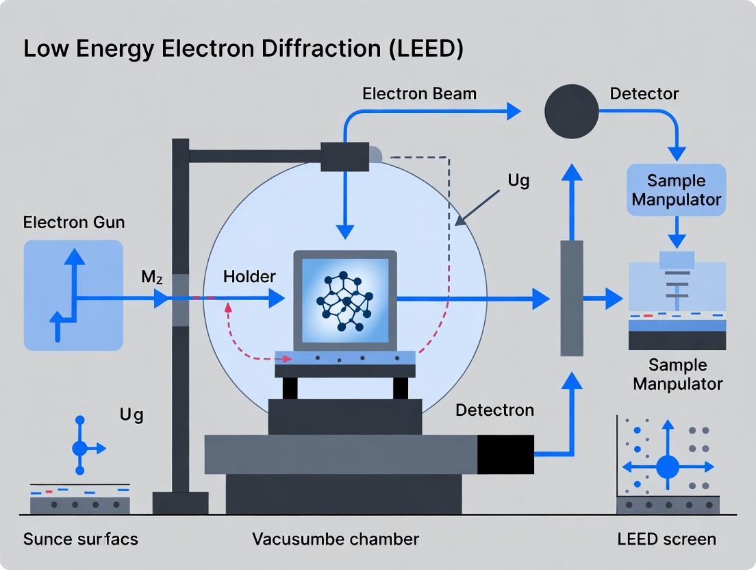
This guide explores strategies for applying Low-Energy Electron Diffraction (LEED) to beam-sensitive materials, a critical challenge in pharmaceutical surface science.
LEED on Delicate Samples: A Guide to Non-Destructive Surface Analysis for Drug Research
Abstract
This guide explores strategies for applying Low-Energy Electron Diffraction (LEED) to beam-sensitive materials, a critical challenge in pharmaceutical surface science. It provides a foundational understanding of electron-induced damage mechanisms in organic crystals, thin films, and molecular adsorbates relevant to drug development. Methodological sections detail protocols for minimizing dose through instrument modifications and operational adjustments. Troubleshooting advice helps diagnose and rectify common artifacts like blurring and fading patterns. Finally, the guide validates these approaches by comparing LEED data quality with complementary techniques like XPS and AFM, offering researchers a comprehensive framework for preserving sample integrity while acquiring reliable structural data.
Understanding Beam Damage: Why Sensitive Samples Challenge Conventional LEED
Technical Support Center: Beam-Sensitive Sample Analysis in LEED
Welcome to the Technical Support Center. This guide provides targeted troubleshooting and FAQs for researchers working with beam-sensitive materials in Low-Energy Electron Diffraction (LEED) experiments, framed within the thesis: "Advancing Methodological Frameworks for Reliable Surface Crystallography of Beam-Sensitive Systems."
Frequently Asked Questions (FAQs)
Q1: How do I know if my organic crystal sample is degrading under the electron beam during a LEED experiment? A: Signs include the gradual fading, broadening, or complete loss of diffraction spots over time, accompanied by an increase in diffuse background intensity. For quantitative assessment, monitor the integrated intensity of a primary Bragg spot as a function of cumulative electron dose (see Table 1).
Q2: What are the primary strategies for minimizing beam damage to molecular adsorbate layers? A: The core strategy is to reduce the incident electron dose. This can be achieved by: (1) using the lowest possible beam current (pA range), (2) defocusing the beam to spread the dose, (3) cooling the sample to cryogenic temperatures (often below 100 K), and (4) employing fast data acquisition (e.g., using a CCD/CMOS camera instead of a fluorescent screen).
Q3: Why do I get no diffraction pattern from my thin organic film, even at very low currents? A: This could indicate either complete beam-induced disorder or that the film is inherently amorphous. First, verify film crystallinity using an alternative, gentler technique like grazing-incidence X-ray diffraction (GIXD). If crystalline, implement a "find-and-probe" protocol: use extreme low dose to locate an area, then acquire data from a fresh, unexposed adjacent area.
Q4: How can I distinguish between electron-stimulated desorption and beam-induced chemical transformation in my adsorbate system? A: Use complementary techniques. Post-LEED analysis with X-ray Photoelectron Spectroscopy (XPS) can track changes in chemical states. Temperature-Programmed Desorption (TPD) after low-dose LEED exposure can check for remaining adsorbate mass. A decrease in adsorbate-specific XPS signal without new species suggests desorption; the appearance of new chemical states indicates transformation.
Q5: What is the "safe" electron dose for a typical metal-organic framework (MOF) thin film? A: There is no universal "safe" dose, as it depends on composition, structure, and beam energy. However, reported tolerance limits for prototypical MOFs (e.g., HKUST-1) are often below 10-100 electrons per Ų. You must establish a dose curve for your specific material (see Experimental Protocol 1).
Troubleshooting Guides
Issue: Rapid Spot Fading During Data Acquisition.
- Check 1: Measure your beam current directly at the sample position with a Faraday cup. Currents should be in the picoamp range.
- Check 2: Ensure your sample is effectively cooled. Check the thermocouple reading and the stability of the cryostat.
- Action: If the problem persists, switch to a "beam-shuttering" mode. Expose the sample only during the actual camera acquisition frame, not during positioning.
Issue: High Diffuse Background Obscuring Weak Spots.
- Check 1: This often indicates hydrocarbon contamination or beam-induced disorder. Perform a thorough sample preparation and UHV bake-out cycle.
- Check 2: Verify that your electron gun is properly aligned and filaments are not aging, causing energy spread.
- Action: Use background subtraction in image processing. Acquire a background image from a nearby non-diffracting area and subtract it from the diffraction pattern.
Issue: Inconsistent Results Between Repeated Experiments on the Same Organic Film.
- Check 1: Inconsistent film preparation (thickness, morphology) is a common root cause.
- Check 2: Variable beam history. The same sample spot should never be measured twice.
- Action: Standardize film growth protocols. Implement a sample stage mapping procedure to always probe a pristine area.
Data Presentation
Table 1: Representative Critical Electron Doses for Beam-Sensitive Sample Classes Data compiled from recent literature on LEED and related TEM studies of sensitive materials.
| Sample Class | Specific Example | Typical Critical Dose (e⁻/Ų) * | Primary Damage Manifestation | Recommended LEED Beam Energy |
|---|---|---|---|---|
| Molecular Adsorbates | NTCDA on Ag(111) | 1 - 10 | Desorption & Disordering | 30 - 50 eV |
| Organic Thin Films | Pentacene on SiO₂ | 10 - 100 | Spot Fading, Amorphization | 40 - 80 eV |
| Metal-Organic Frameworks | HKUST-1 Thin Film | 50 - 200 | Structure Collapse, Loss of Long-Range Order | 50 - 100 eV |
| Halide Perovskites | MAPbI₃ Surface | 5 - 20 | Decomposition, Loss of Iodine | 30 - 60 eV |
| Light Element 2D Materials | Phosphorene | 100 - 500 | Vacancy Formation, Lattice Distortion | 60 - 120 eV |
*Dose at which diffraction spot intensity decays to 1/e of its initial value.
Experimental Protocols
Protocol 1: Determining the Critical Electron Dose for a New Material.
- Preparation: Prepare a fresh, high-quality sample and introduce it into the UHV-LEED system.
- Initial Conditions: Cool the sample to <120 K. Align the electron gun to a pristine sample area. Set beam energy to a standard value (e.g., 70 eV) and current to the lowest measurable setting (e.g., 1 pA).
- Data Acquisition Series: Acquire a LEED image with a very short exposure time (e.g., 0.1 sec). This is the "zero-dose" reference.
- Dose Accumulation: Expose the same area continuously with the beam. At fixed time intervals (corresponding to known incremental doses, e.g., 1 e⁻/Ų), acquire a new LEED image with the same short exposure.
- Analysis: Measure the integrated intensity (I) of a key diffraction spot in each image. Plot I/I₀ vs. Cumulative Electron Dose. Fit with an exponential decay. The critical dose (D_c) is the dose at which I/I₀ = 1/e (~37%).
Protocol 2: Low-Dose, High-Resolution LEED for Organic Crystals.
- Beam Conditioning: Defocus the electron beam slightly to illuminate a larger area, reducing the current density.
- Cooling: Activate liquid nitrogen or helium cooling to stabilize the sample at the lowest possible temperature.
- Find Mode: Use an extremely defocused, low-current (<0.1 pA) beam to navigate and locate a region of interest on the sample surface.
- Probe Mode: Without exposing the located area, shift the sample stage to a neighboring, unexposed region that is expected to have identical structure.
- Acquisition: On this virgin area, focus the beam to desired resolution and acquire data using a single, short exposure on a sensitive camera. Do not take multiple exposures.
Mandatory Visualization
Diagram 1: LEED Beam Damage Assessment Workflow
Diagram 2: Key Damage Pathways for Beam-Sensitive Samples
The Scientist's Toolkit: Research Reagent Solutions
| Item | Function in Beam-Sensitive LEED Research |
|---|---|
| Cryogenic Sample Stage | Cools sample to cryogenic temperatures (often 20-150 K) to suppress diffusion and reduce kinetic energy available for damage pathways. |
| Faraday Cup | A precise instrument for direct, accurate measurement of very low electron beam currents (pA-nA) at the sample position for dose calculation. |
| Fast, Sensitive CCD/CMOS Camera | Enables acquisition of diffraction patterns with very short exposure times, minimizing the required electron dose. |
| Beam Blanker/Shutter | An electrostatic or mechanical device to block the electron beam except during actual data acquisition, preventing unnecessary exposure. |
| In-Situ Thin Film Deposition Sources (Knudsen Cells, E-beam Evaporators) | Allows for preparation of clean, controlled organic/molecular films directly in UHV, avoiding air exposure and contamination. |
| Sample Stage with Precise Translation & Mapping | Permits "find-and-probe" methodologies by enabling movement to fresh, unexposed sample areas after initial location scanning. |
| Low-Current Electron Gun with FEG Source | Provides a stable, bright beam at very low emission currents, offering better signal at lower doses compared to traditional thermionic guns. |
Technical Support Center
Troubleshooting Guide & FAQs
Q1: During our LEED experiment, the diffraction pattern from our organic thin-film sample rapidly fades and disappears. What is happening and how can we mitigate it?
A: This is classic electron-stimulated desorption (ESD) and decomposition. Primary incident electrons break bonds, causing volatile fragments (e.g., H₂, CO, CH₄) to desorb and the remaining carbonaceous material to cross-link into an amorphous layer. This destroys the long-range order needed for diffraction.
Mitigation Protocol:
- Reduce Beam Energy: Immediately lower the primary electron beam energy to the minimum required for pattern visibility (often 20-40 eV instead of 100-200 eV). This reduces penetration depth and energy transfer.
- Reduce Beam Current: Use the smallest beam current that yields a detectable pattern. Employ a channelplate intensifier.
- Lower Sample Temperature: Cool the sample to cryogenic temperatures (e.g., 100 K) using a liquid nitrogen cryostat. This reduces the mobility of fragments and desorption rates.
- Defocus the Beam: When not acquiring data, defocus or raster the beam over a larger area to spread the dose.
- Acquire Rapidly: Use a fast CCD camera to capture patterns in seconds, not minutes.
Q2: Our insulating sample (e.g., metal-organic framework, pharmaceutical salt) shows severe pattern distortion, blurring, and uncontrollable drift during LEED observation. What is the cause and solution?
A: This is electron-induced charging. The insulating surface cannot conduct away the incident electron charge, creating a local positive or negative potential that deflects subsequent electrons, distorting the diffraction geometry.
Charging Mitigation Protocol:
- Conductive Coating: For non-surface-sensitive studies, apply an ultra-thin (1-2 nm), discontinuous coating of a low-atomic-number conductor like carbon via thermal evaporation.
- Reduce Beam Energy: Work near the charge-neutralization point (often between 50-150 eV, sample dependent). Below this, secondary electron emission yield >1, leaving a net positive charge; above it, yield <1, leaving a net negative charge.
- Use a Flood Gun: Employ a very low-energy (< 5 eV) Ar⁺ ion flood gun or a separate low-energy electron flood source to neutralize surface charge. Careful tuning is required to avoid damage.
- Sample Mounting: Use conductive pastes (e.g., carbon, silver) and minimize insulating contact layers. For powders, press into a soft indium foil.
- Increase Sample Temperature: Mild heating can sometimes increase intrinsic conductivity.
Q3: For quantitative I-V LEED analysis, we need maximum pattern stability. What is the optimal workflow to minimize cumulative damage during data acquisition?
A: Follow this sequential, dose-minimizing protocol.
Experimental Protocol for Damage-Minimized I-V LEED:
- Preparation:
- Cool sample to ≤ 100 K.
- Pre-align optics at a single, non-critical energy using a sacrificial spot on the sample.
- Set beam to largest usable spot size (defocused) to reduce current density.
- Calibration:
- Briefly focus beam on a clean, stable reference metal surface (e.g., Cu(100)) to calibrate beam energy and lens settings.
- Immediately return to defocused state and move to region of interest.
- Data Acquisition:
- Program the I-V sweep (e.g., 30-300 eV in 1 eV steps).
- At each energy step, use a fast-shuttering CCD camera with an exposure time of 50-200 ms.
- Implement a beam blanker to block the beam during camera readout and stage movement.
- If possible, use a raster mode, moving the beam slightly between each energy point to expose a fresh area.
- Verification:
- After the sweep, immediately return to the starting energy and re-measure the first pattern to check for degradation. If significant change is observed, discard the dataset and repeat with lower current or exposure time.
Key Quantitative Data on Electron-Beam Damage
Table 1: Critical Damage Thresholds for Selected Material Classes
| Material Class | Example | Typical Critical Dose (Electrons/cm²) | Primary Damage Mechanism | Recommended Max Beam Energy |
|---|---|---|---|---|
| Small Organic Molecules | Pentacene, Perylene | 10¹⁵ - 10¹⁶ | ESD, Molecular Decomposition | 30 eV |
| Polymers | PMMA, Polystyrene | 10¹⁷ - 10¹⁸ | Chain Scission, Cross-linking | 50 eV |
| Metal-Organic Frameworks | MOF-5, ZIF-8 | 10¹⁶ - 10¹⁷ | Linker Desorption, Collapse | 40 eV |
| Pharmaceutical Salts | Aspirin, Theophylline Hydrate | 10¹⁶ - 10¹⁷ | Dehydration, Crystalline Amorphization | 50 eV |
| Alkali Halides | NaCl, KCl | 10¹⁸ - 10¹⁹ | Defect Formation, Charging | 100 eV |
| Graphene/Oxides | Graphene on SiO₂, TiO₂ | > 10¹⁹ | Knock-on Damage, Charging | 80 eV |
Table 2: Mitigation Strategy Efficacy
| Mitigation Technique | Reduction in Damage Rate (Approx. Factor) | Primary Limitation/Consideration |
|---|---|---|
| Cooling from 300K to 100K | 10 - 100 | Cryostat required, condensation risk |
| Reducing Beam Energy from 150 eV to 40 eV | 10 - 1000 | Reduced diffraction contrast, pattern change |
| Reducing Beam Current from 1 µA to 1 nA | 1000 | Requires high-sensitivity detector |
| Conductive Carbon Coating (2 nm) | 100 (for charging) | Obscures true surface structure |
| Fast Acquisition (< 500 ms/pattern) | 10 - 100 (vs. slow scan) | Increased noise, requires pattern averaging |
The Scientist's Toolkit: Research Reagent Solutions
Table 3: Essential Materials for Beam-Sensitive LEED Experiments
| Item | Function | Key Consideration |
|---|---|---|
| Liquid Nitrogen Cryostat | Cools sample to 80-100 K to suppress desorption and diffusion. | Ensure sample has good thermal contact with cold finger. |
| Conductive Carbon Paint | Provides electrical and thermal contact for insulating samples. | Use sparingly to avoid outgassing into UHV. |
| High-Sensitivity CCD/Channelplate Detector | Enables pattern capture at ultra-low beam currents. | Cool CCD to reduce dark current noise. |
| Indium Foil | Ductile, conductive substrate for pressing powder samples. | Pure foil minimizes contamination. |
| Electron Beam Flood Gun | Low-energy (<5 eV) source for charge neutralization. | Must be carefully balanced to avoid adding damage. |
| Fast Beam Blanker/Chopper | Electrically blocks beam between measurements to eliminate unnecessary exposure. | Switching times < 1 ms are ideal. |
| Sacrificial Calibration Crystal | A stable metal crystal (e.g., Cu, Ni, W) for pre-alignment without damaging the target sample. | Keep clean via standard sputter/anneal cycles. |
Workflow and Pathway Diagrams
Title: Damage-Minimized LEED Workflow
Title: Electron Damage Pathways to LEED Degradation
Troubleshooting Guides & FAQs
Q1: My LEED pattern disappears or degrades rapidly during observation. What are the primary parameters to check? A: This is a classic sign of beam damage. Immediately check and record:
- Beam Energy (eV): High energy increases knock-on damage. For organic/organometallic samples, start ≤ 50 eV.
- Beam Current Density (A/cm²): Measure your beam current and approximate spot size. Excess current density causes rapid heating and desorption.
- Total Exposure Dose (C/cm²): This is the integral of current density over time. Use the minimum dose needed for a usable signal. If you must observe for longer, reduce current density.
Q2: How do I systematically determine "safe" parameters for a new, unknown sample? A: Follow a standardized damage threshold protocol:
- Begin at the lowest usable beam energy (e.g., 30-40 eV) and a very low current density (e.g., 1 x 10⁻⁹ A/cm²).
- Acquire a reference LEED pattern.
- Expose a fresh sample region to a fixed, low dose (e.g., 1 x 10⁻⁶ C/cm²). Re-acquire the pattern.
- Incrementally increase the total dose on adjacent fresh spots (e.g., 5 x 10⁻⁶, 1 x 10⁻⁵ C/cm²), acquiring a pattern after each dose.
- The "safe dose" is the level before the first detectable change in pattern sharpness, spot intensity, or background appears.
Q3: My sample charging is severe at low beam energies. What can I do? A: Charging prevents pattern formation. Mitigation strategies include:
- Gradual Energy Tuning: Slowly increase beam energy in 5 eV steps to find a "conducting window."
- Reduce Current Density: Lowering the flux of electrons can reduce charge buildup.
- Sample Preparation: Use ultrathin samples (<10 nm) on conductive substrates.
- Charge Neutralization: If your instrument is equipped with a flood gun, use very low-energy electrons (≤ 5 eV) for neutralization.
Q4: How can I quantitatively compare the beam sensitivity of different materials? A: Define a Critical Dose (Dₜ) for a specific parameter change (e.g., 50% decay of a primary diffraction spot intensity). Measure Dₜ under identical beam conditions. A lower Dₜ indicates higher sensitivity.
Table 1: Typical Parameter Ranges for Beam-Sensitive Samples in LEED
| Parameter | Typical Range for Robust Samples (e.g., Metals) | Recommended Starting Range for Sensitive Samples (e.g., Molecular Films) | Measurement Technique |
|---|---|---|---|
| Beam Energy | 60 - 200 eV | 30 - 50 eV | Read from instrument display (calibrated). |
| Beam Current | 0.1 - 10 µA | 0.01 - 0.1 µA | Faraday cup measurement. |
| Beam Diameter | 0.1 - 1 mm | 0.5 - 1 mm | Controlled via aperture and lens settings. |
| Current Density | ~10⁻⁵ - 10⁻³ A/cm² | < 1 x 10⁻⁸ A/cm² | Calculated from Current / Spot Area. |
| Maximum Safe Dose | > 0.1 C/cm² | < 1 x 10⁻⁵ C/cm² | Calculated from Current Density × Exposure Time. |
Table 2: Experimental Protocol for Damage Threshold Determination
| Step | Action | Goal | Record |
|---|---|---|---|
| 1 | Prepare fresh sample spot. | Ensure pristine initial state. | Sample ID, spot location. |
| 2 | Set initial low E & low J (e.g., 40 eV, 1e-9 A/cm²). | Minimize initial damage. | E₀, J₀, t₀. |
| 3 | Acquire reference LEED image (fast shutter). | Reference data. | Image 0, exposure time. |
| 4 | Expose spot to incremental dose D₁. | Apply known stress. | D₁ = J₀ × t₁. |
| 5 | Acquire post-exposure LEED image. | Assess damage. | Image 1. |
| 6 | Repeat Steps 4-5 on fresh spots with D₂, D₃...Dₙ. | Build damage curve. | All doses and images. |
| 7 | Analyze spot intensity vs. total dose. | Determine Critical Dose Dₜ. | Dₜ value (e.g., for 10% decay). |
Experimental Protocols
Protocol: Measuring the Critical Exposure Dose (Dₜ) for a Molecular Film
Objective: Determine the total electron dose at which a chosen diffraction spot intensity decays to 50% of its initial value.
Materials: See "The Scientist's Toolkit" below.
Methodology:
- Instrument Preparation: Bake and outgas the LEED system to achieve ultra-high vacuum (<5×10⁻¹⁰ mbar). Calibrate the beam energy using a known metal surface (e.g., Au(111)).
- Sample Preparation: Grow or transfer the target molecular film onto a single-crystal substrate in situ. Verify order with a single, very brief (<1 sec) LEED exposure.
- Parameter Initialization: Set the beam energy to a predetermined low value (e.g., 40 eV). Use a large aperture to minimize current density. Measure the beam current (I) using a Faraday cup. Calculate the beam area (A) by measuring the pattern projection on a known scale. The initial current density is J = I/A.
- Reference Acquisition: On a fresh sample region, use a fast electromagnetically actuated shutter to expose the sample for exactly 0.5 seconds. Capture the LEED pattern (Reference Image R₀).
- Incremental Exposure & Data Acquisition: Move the sample stage to a pristine, adjacent region.
- Expose this region continuously for a time t₁ (e.g., 10 sec). The delivered dose is D₁ = J × t₁.
- Close the shutter and immediately capture the LEED pattern (Image I₁).
- Repeat this process on new regions for increasing exposure times t₂, t₃,...tₙ (e.g., 30 sec, 60 sec, 180 sec, 300 sec).
- Data Analysis: In each image (R₀, I₁...Iₙ), integrate the intensity of a primary diffraction spot after subtracting the local background. Normalize all intensities to the value from R₀. Plot Normalized Intensity vs. Total Exposure Dose (C/cm²). Fit the curve with an exponential decay function. The dose at which the normalized intensity equals 0.5 is reported as Dₜ.
Mandatory Visualizations
LEED Damage Threshold Experiment Workflow
Parameter-Damage-Observation Relationship
The Scientist's Toolkit
Table 3: Key Research Reagent Solutions for Beam-Sensitive LEED Studies
| Item / Reagent | Function / Rationale |
|---|---|
| Conductive Single-Crystal Substrates (e.g., HOPG, Graphene on SiC, Au(111) on mica) | Provides an atomically flat, conducting surface to minimize sample charging and facilitate ordered film growth. |
| Molecular Beam Epitaxy (MBE) or Knudsen Cell Sources | Enables precise, in-situ thermal evaporation of organic molecules under UHV conditions for clean film preparation. |
| Faraday Cup with Picoammeter | Essential for direct, accurate measurement of the incident electron beam current (I) to calculate current density (J). |
| Fast, Electromagnetically Actuated Beam Shutter | Allows precise control of exposure time (t) to deliver a defined dose (D = J×t) and acquire reference images with minimal pre-exposure. |
| Liquid Nitrogen Cryostat for Sample Stage | Cooling the sample (to 80-100 K) significantly reduces thermal desorption and diffusion-mediated damage. |
| Low-Energy Electron Flood Gun | Provides low-energy (<5 eV) electrons to neutralize positive charge buildup on insulating samples, enabling pattern acquisition. |
Technical Support Center: Troubleshooting LEED on Beam-Sensitive Samples
This support center provides guidance for researchers encountering issues with Low-Energy Electron Diffraction (LEED) analysis of beam-sensitive materials, such as organic molecular films or biological specimens. The content supports a thesis on mitigating electron-beam damage to obtain reliable surface structure data.
Troubleshooting Guides & FAQs
FAQ 1: What are the definitive visual cues in the LEED pattern that indicate the onset of beam damage to my organic thin film sample?
- Answer: The primary visual cue is a progressive loss of diffraction pattern sharpness and intensity, rather than an instantaneous disappearance. Key indicators include:
- Spot Broadening: Diffraction spots become diffuse and radially elongated. This indicates a loss of long-range order as molecules are displaced or decomposed.
- Increased Background Intensity: The diffuse background scattering between spots becomes brighter. This signifies the rise of amorphous material and disorder.
- Anisotropic Degradation: Spot fading may be direction-dependent, revealing weaker molecular bonds along specific crystal axes.
- Non-Reversible Changes: Patterns continue to degrade after beam is moved or intensity is reduced, confirming permanent damage.
FAQ 2: What quantitative metrics can I use to monitor pattern degradation in real-time to proactively halt the experiment before critical data loss?
- Answer: Implement these metrics by analyzing successive LEED images captured during exposure:
- Spot Intensity Decay Constant (I/I₀): Normalize the integrated intensity of a primary diffraction spot (I) to its initial value (I₀). A drop below 0.8 often signals significant damage.
- Full Width at Half Maximum (FWHM) Trend: Track the radial width of a spot. A sustained increase of >15-20% indicates lattice disorder.
- Signal-to-Background Ratio (S/B): Measure the peak intensity of a spot versus the nearby background. A declining ratio confirms damage.
- Total Diffracted Intensity: Sum the intensity of all diffraction spots. A steady decline suggests global degradation.
Table 1: Quantitative Metrics for LEED Pattern Degradation
| Metric | Measurement Method | Damage Threshold Indicator | Typical Onset Timeframe (for organics)* |
|---|---|---|---|
| Spot Intensity (I/I₀) | Integrated pixel intensity of a primary spot. | I/I₀ < 0.8 | Seconds to minutes |
| Spot FWHM | Radial profile fit of a primary spot. | Increase > 20% from baseline | Seconds |
| S/B Ratio | (Peak Spot Intensity) / (Adjacent Background Intensity). | Decline > 30% | Seconds |
| Pattern Ring Integral | Integrated intensity over a ring covering all spots. | Steady monotonic decrease | Minutes |
*Timeframe depends on beam energy, current density, and specific sample.
FAQ 3: My protocol requires prolonged data collection. What experimental parameters must I optimize to delay the onset of damage?
Answer: Follow this detailed methodology to establish a safe operating window:
Experimental Protocol: Determining Safe LEED Imaging Parameters
Initial Low-Dose Survey:
- Set the electron gun to the lowest usable beam current (e.g., 0.1 µA).
- Use a high phosphor screen voltage and sensitive camera.
- Acquire a reference image with a single, short exposure (< 0.5 sec).
Time-Series Experiment:
- Focus the beam on a fresh sample area.
- Program the system to acquire images at fixed intervals (e.g., every 2 seconds) for a total period (e.g., 60 seconds).
- Maintain all parameters (beam energy, current, position) constant.
Post-Processing & Analysis:
- Use image analysis software to extract the metrics in Table 1 from the time-series.
- Plot each metric versus cumulative electron dose (beam current × time / area).
Determine Safe Dose:
- Identify the electron dose at which key metrics deviate from their plateau.
- Safe Operating Dose = 50% of the dose at which I/I₀ first falls to 0.9. This provides a conservative safety margin.
Final Imaging:
- For extended scans or I-V curve measurements, ensure the total calculated dose per area remains below the Safe Operating Dose. Use multiple fresh areas as needed.
The Scientist's Toolkit: Research Reagent Solutions
Table 2: Essential Materials for LEED Studies of Beam-Sensitive Samples
| Item | Function & Relevance to Damage Mitigation |
|---|---|
| Cryogenic Sample Stage | Cools sample to ~100 K or lower. Reduces radical mobility and desorption, slowing damage progression. |
| Dosing/Evaporation Source for Noble Metals | Allows in-situ sublimation of few-atom-thick conductive layers (e.g., Au, Pt) to improve conductivity without masking LEED pattern. |
| Low-Current, High-Brightness Electron Gun | Provides sufficient beam coherence for a clear pattern at the lowest possible current density, minimizing dose. |
| Direct Electron Detector (e.g., CCD/CMOS) | Offers high quantum efficiency for detecting weak patterns, enabling use of lower beam currents. |
| Fast Beam Blanker & Automated Stage | Allows rapid shuttering of the beam and movement to fresh sample areas between measurements to distribute dose. |
Visualization of Protocols and Relationships
Diagram Title: Workflow for Determining Safe LEED Imaging Parameters
Diagram Title: Pathways from Electron Beam Impact to LEED Pattern Degradation
Practical Protocols for Low-Dose and Fast LEED Experiments
Troubleshooting Guides & FAQs
Q1: During my LEED experiment on an organic thin film, the diffraction pattern fades rapidly. What is the primary cause and how can I mitigate it? A: This is a classic symptom of beam-induced damage. The primary cause is excessive electron beam current leading to bond breaking, desorption, or structural rearrangement. Mitigation involves a systematic reduction of the beam current. Start by lowering the emission current or filament temperature at the source. Then, use the smallest aperture compatible with achieving a usable signal. Always perform a beam current measurement at the sample stage (using a Faraday cup) to establish a baseline. For highly sensitive organics, aim for a beam current in the low picoampere (pA) range.
Q2: After minimizing the beam current, my LEED pattern is too faint to analyze. How do I optimize detector sensitivity? A: When beam current is minimized, detector sensitivity must be maximized. Follow this protocol:
- Increase Screen Voltage: Gradually increase the post-acceleration voltage (typically up to 5-7 kV) to boost electron impact luminance. Do not exceed the manufacturer's limit.
- Optimize Microchannel Plate (MCP) Gain: If your system uses an MCP, incrementally increase the MCP voltage. Monitor for increased background noise.
- Camera Settings: For CCD/CMOS camera systems, increase the exposure time or gain. Use cooling to reduce dark current for long exposures. Ensure you are not saturating the sensor.
- Clean Optics: Verify that viewports, the phosphor screen, and any lenses are clean. Contamination drastically reduces signal throughput.
Q3: What is the step-by-step protocol for establishing a safe beam current threshold for a new, unknown sample? A: Use this iterative damage test protocol:
- Select a representative, non-critical area of the sample.
- Set the detector to standard sensitivity (e.g., screen voltage = 3 kV, exposure time = 0.5 s).
- Start with an ultralow beam current (e.g., 0.1 pA). Capture a reference LEED image.
- Expose the same spot continuously for a set period (e.g., 60 seconds), capturing an image every 5 seconds.
- Analyze the decay in diffraction spot intensity or increase in background halo using image analysis software.
- Repeat the process at incrementally higher beam currents (0.5 pA, 1 pA, 5 pA) on fresh spots.
- The "safe" current is one where no measurable decay occurs over a period at least 3x longer than your typical measurement time.
Q4: My lab's LEED system has a channeltron electron multiplier. How do I balance its gain with signal quality? A: Channeltron gain is controlled by its applied voltage (typically 1-3 kV). Increasing voltage increases gain but also amplifies noise and can shorten the detector's lifespan. Procedure:
- With sample in place and beam on, monitor the output signal on an oscilloscope or the system's readout.
- Start at the manufacturer's recommended voltage (e.g., 1.8 kV).
- Gradually increase voltage until the signal-to-noise ratio (SNR) is satisfactory for your measurement speed. Do not operate at the maximum voltage for prolonged periods.
- If the signal is still weak, return to beam current minimization troubleshooting first. The channeltron is the last amplification stage.
Data Presentation
Table 1: Typical Beam Current Ranges and Applications for LEED
| Sample Type | Recommended Beam Current Range | Typical Detector Settings (Screen Voltage) | Rationale |
|---|---|---|---|
| Stable Metals & Alloys | 100 pA - 10 nA | 1 - 3 kV | High current provides strong signal; damage negligible. |
| Semiconductors (Si, Ge) | 10 - 500 pA | 2 - 4 kV | Moderate sensitivity; requires balance. |
| Oxide Surfaces (TiO2) | 1 - 100 pA | 3 - 5 kV | Risk of electron-stimulated desorption. |
| Molecular Organic Films | 0.1 - 5 pA | 5 - 7 kV | Extreme beam sensitivity necessitates minimal current and maximal detection. |
| 2D Materials (Graphene on substrate) | 5 - 50 pA | 4 - 6 kV | Sublimation and defect formation possible at high currents. |
Table 2: Troubleshooting Matrix: Symptom, Likely Cause, and Immediate Action
| Symptom | Likely Cause | Immediate Troubleshooting Action |
|---|---|---|
| Pattern fading over time | Excessive beam current | 1. Measure beam current with Faraday cup. 2. Reduce filament emission or aperture size. |
| Faint pattern, high noise | Suboptimal detector sensitivity | 1. Incrementally increase screen voltage/MCP gain. 2. Increase camera exposure time. |
| Uneven illumination/vignetting | Misaligned beam or dirty optics | 1. Check beam alignment through apertures. 2. Inspect viewport and screen cleanliness. |
| High background, diffuse halo | Sample charging or contamination | 1. Ensure sample is properly grounded. 2. Check sample preparation for hydrocarbons. |
| No pattern at high sensitivity | Beam off, shutter closed, or severe misalignment | 1. Verify gun and high voltage status. 2. Check mechanical shutters. 3. Realign electron gun. |
Experimental Protocols
Protocol: Quantitative Beam Damage Threshold Measurement Objective: To determine the maximum electron dose (current x time) a sample can tolerate before significant structural degradation. Materials: Sample, LEED system with measurable beam current, Faraday cup, imaging system. Procedure:
- Insert the Faraday cup and measure the beam current (
I) at the intended working distance. - Insert the sample and locate a fresh area. Obtain a sharp LEED pattern at the lowest usable beam current.
- Define a Region of Interest (ROI) encompassing a primary diffraction spot and a background region.
- Start continuous exposure. Record an image sequence (
t=0, 10, 20, 30...seconds). - For each image
t, measure the integrated intensityS(t)of the spot ROI and the backgroundB(t). Calculate net intensityI_net(t) = S(t) - B(t). - Plot
I_net(t) / I_net(0)vs. Electron Dose (D = I * t / A, where A is the irradiated area). - Fit the curve. The dose where intensity falls to 1/e (~37%) of its initial value is often defined as the damage threshold dose.
Protocol: Detector Sensitivity Calibration Objective: To establish standardized detector settings for reproducible low-current measurements. Materials: Standard sample (e.g., clean, well-ordered Ni(111) or Au(111) surface), LEED system. Procedure:
- Prepare the standard sample to give a bright, sharp pattern at a known, moderate beam current (e.g., 1 nA).
- Set detector to a baseline configuration (e.g., Screen Voltage = 2.0 kV, MCP = Off, Camera Gain = 1, Exposure = 1s). Capture reference image.
- Parameter Sweep: Systematically vary one parameter at a time while recording the resulting peak spot intensity and background noise.
- Screen Voltage: Increase in 500 V steps from 1.0 to 7.0 kV.
- Camera Exposure: Increase in logarithmic steps (0.1s, 0.5s, 1s, 5s).
- For each setting, calculate the Signal-to-Noise Ratio (SNR).
- Create a lookup table of settings that yield a target SNR (e.g., SNR > 10) for a given beam current. This table guides setup for unknown sensitive samples.
Diagrams
Workflow for Beam-Sensitive LEED
Damage Threshold Determination Logic
The Scientist's Toolkit: Research Reagent Solutions
| Item | Function in Beam-Sensitive LEED Research |
|---|---|
| Faraday Cup | A metal cup for precisely measuring the incident electron beam current at the sample position. Critical for quantitative dose control. |
| Standard Calibration Sample (e.g., Au(111)) | A stable, well-characterized surface used to calibrate detector sensitivity and align the LEED optics without risk of damage. |
| Low-Current Electron Gun | An electron source capable of stable emission in the picoampere range, essential for studying organics and 2D materials. |
| High-Gain Detector (MCP/CCD) | A microchannel plate (MCP) and/or a cooled CCD/CMOS camera to amplify and capture faint diffraction patterns from low-current beams. |
| Sample Grounding Kit | Conductive paste, clips, or wires to ensure the sample is electrically grounded, preventing charge buildup that distorts the LEED pattern. |
| In-Situ Sample Cleaning Tools | Electron beam flashers, ion sputter guns, or gas dosers for cleaning standard samples without breaking vacuum, ensuring measurement consistency. |
| Beam Blanker | An electrostatic or mechanical device to deflect or block the beam between measurements, minimizing total exposure to the sample. |
Troubleshooting Guides & FAQs
Q1: During Fast-LEED data acquisition, my diffraction pattern appears faint and noisy, even with reasonable beam current. What are the primary causes and solutions?
A: This is often due to excessive readout speed or incorrect gain settings on your detector. For beam-sensitive samples, the goal is to capture enough signal before damage occurs.
- Solution A: Reduce the readout speed (e.g., from 40 MHz to 10 MHz) to improve signal-to-noise ratio (SNR) per frame.
- Solution B: Systematically increase the detector gain while monitoring for the onset of amplification noise. Use the Frame Averaging protocol (see below) to integrate multiple short-exposure frames.
- Solution C: Verify sample preparation and cleanliness. A faint pattern can also indicate surface contamination absorbing/scattering the electron beam.
Q2: I observe a progressive blurring and loss of diffraction spot intensity during a single measurement sequence. Is this sample damage?
A: Yes, this is a classic signature of beam-induced damage. Fast-LEED is designed to mitigate this.
- Solution: Immediately halt the measurement. Re-evaluate your acquisition parameters using the "Dose-Limited Exposure" protocol:
- Drastically reduce the electron beam current (e.g., to 0.1 nA from 1 nA).
- Decrease the exposure time per frame to the minimum detectable level (e.g., 0.05-0.1 seconds).
- Increase the number of frames for averaging proportionally. The total integrated dose for a usable averaged pattern should be below the damage threshold.
Q3: After frame averaging, my spots are sharp, but the background appears patchy and non-uniform. What causes this artifact?
A: This is typically caused by sample drift or charging during the acquisition of the frame sequence.
- Solution A (Drift): Ensure sample stability by allowing sufficient time for thermal equilibrium after sample transfer. Implement a frame alignment (registration) algorithm during post-processing. Most Fast-LEED software includes a cross-correlation function to align successive frames before averaging.
- Solution B (Charging): For insulating samples, reduce beam energy further if possible (e.g., to 20-30 eV). Consider using an even lower incident current. The Fast-LEED approach of ultra-short exposures is beneficial here, as it prevents charge buildup during a single frame.
Q4: How do I determine the optimal number of frames to average for my specific beam-sensitive sample?
A: There is a trade-off between SNR improvement and total accumulated electron dose. A systematic protocol is required.
- Pilot Test: On a non-critical region of your sample, acquire a sequence of, for example, 100 frames with exposure time t.
- Progressive Analysis: Generate averaged images from the first N=10, 20, 50, and 100 frames.
- Quantify: Measure the SNR (peak spot intensity / local background standard deviation) and spot sharpness (full width at half maximum, FWHM) for each averaged image.
- Plot & Decide: Create a table (see below). The optimal N is where SNR gain plateaus significantly before visual degradation (increased FWHM) occurs in the final averaged image, indicating the onset of damage over the full sequence.
Table 1: Frame Averaging Optimization Pilot Test (Example Data)
| Number of Frames (N) | Total Dose (Arb. Units) | Spot SNR (Ratio) | Spot FWHM (pixels) | Visual Assessment |
|---|---|---|---|---|
| 10 | 10 | 5.2 | 3.1 | Very noisy, spots discernible |
| 20 | 20 | 8.1 | 2.9 | Improved, usable for strong spots |
| 50 | 50 | 12.5 | 2.8 | Good, clear pattern |
| 100 | 100 | 14.0 | 3.5 | SNR slightly better, but spots broader (damage) |
Conclusion for this example: N=50 is optimal, providing good SNR with minimal broadening.
Experimental Protocol: Fast-LEED with Frame Averaging for Dose-Limited Samples
Objective: To obtain a quantitative LEED pattern from a beam-sensitive surface while minimizing total electron dose.
Materials & Setup:
- UHV system with LEED optics.
- Fast, low-noise CCD or CMOS detector (directly viewing phosphor screen or via fiber optic).
- Beam-sensitive sample (e.g., organic thin film, metal-organic framework).
- Computer with Fast-LEED control and acquisition software.
Procedure:
- Sample Preparation & Alignment:
- Prepare and characterize the sample using standard surface science techniques.
- Align the sample at the manipulator's eucentric point.
- Set the LEED gun to a low energy (e.g., 30-80 eV) and the lowest achievable beam current (e.g., 0.05-0.2 nA). Record the exact value.
Parameter Calibration (Find Minimum Exposure):
- In the software, set a very short single-frame exposure time (e.g., 0.02 seconds).
- Acquire a single frame. Visually inspect if any diffraction spots are detectable above noise.
- Gradually increase exposure time in small increments until the faintest spots are just perceptible in the live, single-frame image. This is your minimum usable exposure time, t_min.
Damage Threshold Test (Optional but Recommended):
- On a sacrificial area, acquire a continuous video stream at t_min.
- Monitor the intensity of a primary diffraction spot in real-time.
- Note the time (or frame count) at which the spot intensity drops to 50% of its initial value. This defines the damage frame limit, N_max.
Fast-LEED Data Acquisition:
- Move to a fresh, untouched region of interest.
- Program the acquisition sequence:
- Exposure time per frame: tmin (e.g., 0.05 s).
- Number of frames: N << Nmax (if known), otherwise start with N=50-100.
- Delay between frames: Minimal (software-limited, e.g., 0.01 s).
- Initiate the rapid, automated acquisition sequence. The total experiment time is ~N * (t_min + delay).
Post-Processing & Frame Averaging:
- Alignment: Run a cross-correlation algorithm to align all frames to a reference (e.g., the first frame) based on the positions of bright features or the direct beam. This corrects for minor sample drift.
- Averaging: Generate the final output image by calculating the per-pixel arithmetic mean of all aligned frames.
- Analysis: Perform spot finding, intensity integration, and background subtraction on the high-SNR averaged image.
Visualization: Fast-LEED Workflow for Beam-Sensitive Samples
The Scientist's Toolkit: Key Reagent & Material Solutions
Table 2: Essential Research Reagents & Materials for Fast-LEED on Sensitive Samples
| Item | Function in Fast-LEED Experiments |
|---|---|
| Low-Emission, High-Coherence Electron Gun | Provides a stable, focused beam at very low currents (pA to nA range) to initiate measurement before damage. |
| Low-Noise, High-Quantum Efficiency Detector (e.g., CCD/CMOS) | Enables detection of faint diffraction patterns from single ultra-short exposures. High QE is critical for low-dose imaging. |
| UHV-Compatible Sample Holders with Precise Cooling/Heating | Allows temperature control. Cryogenic cooling (to 100 K or lower) can significantly reduce beam damage rates for many organic materials. |
| High Dynamic Range Phosphor Screen | Converts electron diffraction pattern to photons for detection. Must have linear response and high efficiency to preserve weak spot information. |
| In-Situ Sample Preparation Sources (e.g., MBE, Evaporators) | For depositing thin films of sensitive materials (organic semiconductors, biomolecules) directly in UHV to avoid contamination. |
| Fast Beam Blanker/Electrostatic Deflector | Cuts off the electron beam between exposures with millisecond precision, preventing unnecessary irradiation during readout delays. |
| Software with Real-Time Processing & GPU Acceleration | Controls rapid acquisition, performs live frame alignment and running average calculations, allowing for immediate feedback on data quality. |
Sample Preparation and Handling to Intrinsic Robustness
Technical Support Center: Troubleshooting LEED Analysis of Beam-Sensitive Samples
Troubleshooting Guides & FAQs
Q1: My organic thin-film sample shows rapid degradation and loss of LEED pattern within seconds. What are the primary preparation factors to check? A: Immediate degradation indicates excessive beam exposure or inadequate sample stabilization. First, verify your preparation protocol against the following critical parameters:
- Sample Thickness: Ensure ultrathin films (<10 nm) are used to minimize charging and bulk damage.
- Substrate Temperature During Deposition/Measurement: Maintain the substrate at a temperature below the sample's desorption or decomposition point. For many organics, this is often near or below 150K.
- Deposition Rate: Use a slow, controlled deposition rate (typically 0.1-0.5 nm/min) to promote ordered growth.
- Initial Beam Exposure: Use the smallest possible beam current (e.g., 0.1 nA) and defocus the beam during alignment. Use a beam blanker and expose only for image acquisition.
Q2: I observe persistent charging on my insulating sample, distorting the LEED pattern. How can I mitigate this? A: Charging is common with organic films and insulators. Implement a tiered approach:
- Reduce Beam Current: Decrease to the lowest usable current (often 0.05-0.2 nA).
- Adjust Beam Energy: Slightly increase the electron beam energy (e.g., to 90-120 eV) as higher energy electrons may penetrate better and reduce surface charge buildup.
- Sample Preparation: Use conductive substrates (e.g., graphene-coated metals, highly ordered pyrolytic graphite (HOPG)) or grow films on a pre-covered metal surface.
- Post-Preparation: For suitable samples, deposit an ultra-thin, transparent capping layer (e.g., 1-2 nm of graphene or amorphous carbon) in-situ.
Q3: What in-situ preparation checks should I perform before introducing the sample to the LEED beam? A: Follow this pre-exposure checklist:
- Confirm base pressure in preparation chamber is < 5 x 10⁻¹⁰ mbar.
- Verify sample cleaving, heating, or deposition occurred under ultra-high vacuum (UHV) conditions.
- For deposited films, use Low-Energy Electron Microscopy (LEEM) or Auger Electron Spectroscopy (AES) in a connected chamber (if available) to check order and composition before LEED.
- Plan and rehearse all beam alignment steps to minimize active beam time on the sample.
Q4: My metal-organic framework (MOF) sample shows varying robustness. How can I quantify its intrinsic beam tolerance? A: You can establish a "Critical Dose" for your material using a systematic decay experiment.
Experimental Protocol: Determining Critical Electron Dose
- Prepare a fresh, well-characterized sample spot.
- Set LEED parameters to a fixed, low beam current (I) and energy (E). Measure the beam spot size (A) to calculate current density (j = I/A).
- Acquire a reference LEED image with a very short exposure time (t), e.g., 0.1 seconds.
- Expose the same spot continuously to the beam. At fixed time intervals (Δt), acquire a new LEED image using the identical short exposure.
- Analyze the decay of diffraction spot intensity (I) versus total exposure dose (D = j * t_total). Fit the curve to an exponential decay model.
- Define the critical dose (D_c) as the dose at which the diffraction spot intensity falls to 1/e (~37%) of its initial value.
Quantitative Data: Typical Critical Doses for Sample Classes Table 1: Comparative Beam Sensitivity of Materials in LEED (at ~100 eV)
| Material Class | Example | Typical Critical Dose (e⁻/nm²) | Recommended Max Beam Current | Optimal Substrate Temp. |
|---|---|---|---|---|
| 2D Covalent Organic Frameworks | Boroxine-linked COF | 10 - 100 | < 0.1 nA | Room Temperature |
| Self-Assembled Monolayers | Alkanethiols on Au(111) | 100 - 1,000 | 0.1 - 0.5 nA | < 150 K |
| Metal-Organic Frameworks | Cu-BTC thin film | 10² - 10³ | 0.2 - 0.8 nA | < 200 K |
| Small Organic Molecules | PTCDA on Ag(111) | 10³ - 10⁴ | 0.5 - 1 nA | < 130 K |
| Graphene / 2D Inorganics | Graphene on SiC | > 10⁸ | 1 - 10 nA | Room Temperature |
Q5: Are there computational methods to predict a material's beam sensitivity during the drug co-crystal design phase? A: Yes, early-stage computational screening can guide synthesis. Use Density Functional Theory (DFT) to calculate:
- Binding Energy per Atom/Molecule: Lower average binding energies often correlate with lower displacement thresholds.
- Molecular Orbital Eigenvalues: The energy of the Highest Occupied Molecular Orbital (HOMO) can indicate susceptibility to electronic excitation.
- Radiolysis Susceptibility: Simulate the cleavage energy of the weakest bond in the structure (e.g., a ligand-metal bond in a MOF).
The Scientist's Toolkit: Research Reagent Solutions
Table 2: Essential Materials for Robust Sample Preparation
| Item | Function & Relevance to Robustness |
|---|---|
| HOPG (Highly Ordered Pyrolytic Graphite) | An atomically flat, conductive substrate that minimizes charging and provides a template for ordered growth of 2D films. |
| Graphene-coated TEM grids | Provides a suspended, ultra-thin, conductive support for fragile nanocrystals, allowing LEED-like transmission studies. |
| UHV-compatible Molecular Evaporator (Knudsen Cell) | Enables controlled, thermal deposition of organic molecules with precise rate control for uniform, ordered films. |
| Cryogenic Sample Holder (LHe or LN₂ cooled) | Allows sample temperature maintenance below 150K, drastically reducing diffusion, desorption, and beam-induced damage. |
| In-situ Sputter & Anneal Kit | For substrate preparation. Atomically clean and ordered substrate surfaces (e.g., metal single crystals) are critical for epitaxial, robust film growth. |
| Beam Blanker / Fast Shutter | An electronic shutter that blocks the beam except during data acquisition, limiting total exposure. Essential for dose-controlled experiments. |
| UHV-compatible Transfer Case | Allows safe, contamination-free transfer of air-sensitive crystals from a glovebox to the UHV system load lock. |
Experimental Workflow & Pathway Diagrams
Pre-LEED Workflow for Robust Data
Electron-Sample Interaction Pathways
Step-by-Step Workflow for a First Measurement on a New Sensitive Material
This technical support center is framed within a thesis dedicated to overcoming the challenges of beam-sensitive samples in Low-Energy Electron Diffraction (LEED) research. The following guides and FAQs provide a structured protocol and troubleshooting advice for researchers, scientists, and drug development professionals performing initial measurements on novel, sensitive materials.
Experimental Protocol: First Measurement Workflow
Objective: To obtain a valid LEED pattern from a new, beam-sensitive material while minimizing electron beam-induced damage.
Detailed Methodology:
Pre-experimental Planning & Sample Preparation:
- Material Characterization: Prior to LEED, use complementary techniques (e.g., XPS, AFM) to understand baseline composition and morphology.
- Substrate Preparation: Ensure the substrate (e.g., single crystal metal) is clean and well-ordered, verified by its own sharp LEED pattern.
- Material Deposition: Deposit the sensitive material onto the substrate under controlled conditions (UHV, correct temperature). Aim for sub-monolayer to few-monolayer coverage initially.
- In-situ Transfer: Transfer the sample to the LEED stage without breaking vacuum.
Initial LEED Parameter Setup for Sensitivity Testing:
- Start with Ultra-Low Beam Conditions: Set the electron gun to the lowest possible beam current (e.g., ≤ 0.1 nA) and a relatively high electron energy (e.g., 80-120 eV). High energy can sometimes reduce total dose for a given current.
- Detector Settings: Use a high gain/sensitivity setting on the phosphor screen or CCD camera.
- Cool the Sample: If possible, cool the sample stage to cryogenic temperatures (e.g., 100 K or lower) to inhibit diffusion and damage processes.
The "Look-and-Check" Iterative Procedure:
- Brief Exposure: Expose the sample to the electron beam for a very short duration (≤ 1 second).
- Acquire Image: Capture the LEED pattern immediately.
- Beam Blank: Blank the beam immediately after acquisition.
- Assess: Examine the captured image for signs of diffraction. If no pattern is visible, very slightly increase exposure time or beam current (increment by ≤ 20%).
- Repeat the "Look-and-Check" cycle until a faint pattern is observed.
- Critical: If the pattern degrades (spots broaden or fade) between consecutive exposures, the previous exposure parameters represent the damage threshold. All subsequent data must be acquired at parameters below this threshold.
Systematic Data Acquisition:
- Once safe parameters are identified, acquire a series of patterns across a range of energies (e.g., 40-150 eV in 5 eV steps) to confirm periodicity and for subsequent I-V analysis.
- For each energy, use the minimum exposure time required to obtain a usable signal-to-noise ratio.
Post-measurement Validation:
- Re-measure a reference area of the substrate or a previously stable material to confirm the instrument alignment and performance has not drifted.
- Perform a post-mortem analysis of the measured spot (e.g., with XPS or AES) to quantify any beam-induced chemical changes.
Troubleshooting Guides & FAQs
Q1: I see no diffraction pattern from my sensitive organic layer, even after several "Look-and-Check" cycles. What should I do? A1: Follow this checklist:
- Verify Substrate: Ensure your substrate alone produces a sharp, bright LEED pattern.
- Check Coverage: Your initial deposit may be too thin or too patchy. Consider slightly increasing coverage.
- Order: The film may be amorphous or polycrystalline. Confirm growth parameters aim for crystalline order.
- Increase Sensitivity: Maximize detector gain. Use the most sensitive channelplate/CCD settings.
- Last Resort Increment: Only after exhausting the above, make a small increase in beam current (e.g., from 0.1 nA to 0.12 nA) and restart the "Look-and-Check" protocol.
Q2: The diffraction spots fade or broaden during my measurement. How can I salvage the experiment? A2: This is clear evidence of beam damage. Immediately:
- Blank the beam.
- Move the sample to a fresh, unexposed region (if possible).
- Restart the experiment using beam parameters at least 50% lower than those that caused the observed damage.
- Consider further cooling the sample stage.
- Adopt a "single-shot" approach: for each energy, expose a virgin sample area only once and then move on.
Q3: My pattern has high background noise, obscuring the diffraction spots. How can I improve contrast? A3:
- Reduce Beam Current: A lower current decreases the diffuse scattering contribution.
- Clean the Sample/Chamber: High background often stems from adsorbed hydrocarbons. Perform a chamber bake-out and sample degassing/cleaning cycle.
- Check for Static Discharge: Ensure proper grounding of the sample and holder.
- Use a Smaller Beam Spot: If your system allows it, focus the beam to a smaller area to reduce scattering from disordered regions.
Q4: How do I quantitatively determine the "safe" electron dose for my material?
A4: Calculate the electron dose (D) per measurement:
D (C/cm²) = Beam Current (A) × Exposure Time (s) / Beam Area (cm²)
Establish a damage threshold by monitoring the decay of diffraction spot intensity (I) versus cumulative dose. The safe dose is typically an order of magnitude below the dose where I drops to 90% of its initial value (I₀).
Table 1: Quantitative Beam Parameter Guidance for Sensitive Materials
| Material Class | Suggested Starting Beam Current | Suggested Starting Energy | Max Recommended Dose (Est.) | Critical Sample Temperature |
|---|---|---|---|---|
| Soft Organic Crystals | 0.05 - 0.1 nA | 80 - 100 eV | 10⁻³ - 10⁻² C/cm² | ≤ 100 K |
| Metal-Organic Frameworks (MOFs) | 0.1 - 0.3 nA | 90 - 120 eV | 10⁻² - 10⁻¹ C/cm² | ≤ 150 K |
| Halide Perovskites | 0.2 - 0.5 nA | 70 - 90 eV | 10⁻¹ - 1 C/cm² | ≤ 100 K |
| 2D Polymers & Graphene-like | 0.5 - 1 nA | 120 - 150 eV | 1 - 10 C/cm² | Room Temp or ≤ 150 K |
| Reference: Clean Metal Surface | 1 - 10 nA | 60 - 150 eV | > 100 C/cm² | Room Temp |
Workflow Visualization
Title: First LEED Measurement Workflow for Sensitive Materials
Title: Troubleshooting: No Diffraction Pattern
The Scientist's Toolkit: Key Research Reagent Solutions
Table 2: Essential Materials & Reagents for Sensitive Sample LEED
| Item | Function in Experiment |
|---|---|
| Single Crystal Substrates (Au(111), Cu(100), Graphene on SiC, etc.) | Provide a clean, atomically flat, and well-ordered base for growing sensitive materials. Their known diffraction pattern serves as a crucial reference. |
| High-Purity Evaporation Sources (Organic powders, metal rods in crucibles) | Enable the deposition of ultra-pure, contaminant-free thin films of the sensitive material under UHV conditions. |
| Cryogenic Coolant (Liquid Nitrogen or Helium) | Used to cool the sample manipulator. Reduces thermal energy, suppressing beam-induced diffusion, desorption, and decomposition. |
| In-situ Sample Cleaver/Scraper | Allows for the creation of fresh, clean surfaces of bulk sensitive materials inside the UHV chamber, avoiding air exposure. |
| Low-Current, High-Stability Electron Gun | The core component for generating the low-intensity, focused electron beam required to probe sensitive materials without immediate damage. |
| Channelplate/CCD Detector System | Amplifies and captures weak diffraction signals with high efficiency, allowing for the use of minimal electron doses. |
| UHV-Compatible Transfer Pod | Enables safe, vacuum-sealed transfer of air-sensitive samples from preparation or synthesis chambers to the LEED analysis chamber. |
Diagnosing and Solving Common LEED Artifacts on Sensitive Surfaces
Troubleshooting Guide & FAQs
FAQ 1: Why are my LEED spots blurring or fading during data acquisition on my organic thin film?
Answer: This is a classic sign of beam-induced sample degradation. The primary cause is electron-stimulated desorption (ESD) and radiolysis, where the incident electron beam breaks bonds, desorbs surface species, or causes structural rearrangement. For molecular films, this often manifests as a loss of long-range order, leading to spot broadening and intensity decay.
Correction:
- Reduce Beam Current: Lower the emission current or use a smaller beam spot size. Start at the lowest current that provides a discernible pattern.
- Minimize Exposure Time: Use a fast-scan camera and avoid pausing the beam on one area. Employ beam blanking when not acquiring data.
- Cool the Sample: Use a liquid nitrogen or helium cryostat to cool the sample stage (often to below 100 K). This reduces molecular mobility and desorption rates.
- Increase Sample Distance (R): While this reduces resolution, it spreads the current density, lowering the dose per unit area.
FAQ 2: My diffraction spots disappear completely after a few seconds. Is my sample destroyed?
Answer: Complete and rapid spot loss indicates catastrophic beam damage. The sample is likely altered in the irradiated area. For highly sensitive materials like pharmaceuticals or metal-organic frameworks, the crystalline order can be completely amorphized.
Correction:
- Defocused Beam Mode: Defocus the incident beam to spread the energy over a larger area before focusing for measurement.
- Raster the Beam: If possible, continuously move the sample or the beam during measurement to expose fresh areas.
- Pre-Check with Low Dose: First, use an ultra-low current (< 0.1 nA) for a very short pulse to confirm a pattern exists before standard imaging.
- Validate with Complementary Techniques: Cross-verify sample integrity with non-destructive techniques like optical microscopy or AFM pre- and post-LEED.
FAQ 3: How can I quantify the beam damage to adjust my parameters?
Answer: Perform a beam damage threshold measurement. Monitor the integrated intensity of a primary diffraction spot as a function of cumulative electron dose.
Experimental Protocol:
- Set the sample at a standard temperature (e.g., 100 K).
- Choose a specific beam current (I) and beam area (A). Calculate current density (J = I/A).
- Acquire sequential LEED images with fixed time intervals (Δt).
- For each image, measure the intensity (I) of a key diffraction spot.
- Calculate the cumulative dose for each point: Dose = J * t (where t = n * Δt).
- Plot Normalized Intensity (I/I₀) vs. Cumulative Dose.
Typical Damage Thresholds for Sensitive Materials:
| Material Class | Approximate Critical Dose (electrons/cm²) at 100 K | Primary Degradation Mode |
|---|---|---|
| Organic Molecular Crystals (e.g., Pentacene) | 10¹⁵ – 10¹⁶ | Radiolysis, ESD |
| Pharmaceutical Cocrystals | 10¹⁴ – 10¹⁵ | Bond cleavage, loss of hydration |
| Metal-Organic Frameworks (Thin Films) | 10¹³ – 10¹⁴ | Linker decomposition, collapse |
| Alkali Halide Surfaces (Reference) | > 10¹⁸ | Thermal disorder, defect creation |
FAQ 4: What are the instrumental causes of blurry spots, and how do I distinguish them from sample damage?
Answer: Instrumental issues like misalignment, space charge, and thermal drift can also cause blurring. Key differentiator: Damage is time- and dose-dependent; instrumental issues are typically constant or intermittent regardless of exposure time on a fresh sample spot.
Correction Checklist:
- Poor Vacuum (>10⁻⁹ mbar): Causes scattering and adsorption. Improve UHV conditions.
- Filament/Grid Instability: Check filament age and alignment. Ensure stable power supplies.
- Sample Charging: For insulating samples, use a lower beam energy (e.g., 20-40 eV) or a flood gun for charge neutralization.
- Mechanical Drift: Ensure sample stage is stable and cool-down/heating cycles are settled.
Diagnostic Protocol:
- Move the sample to a fresh, unexposed area. If the pattern is immediately sharp, previous area was damaged.
- Switch to a standard, robust sample (e.g., clean Si(111)-7x7). If spots remain blurry, the issue is instrumental.
- Observe the pattern in real-time. A gradual fade indicates damage; a sudden shimmer or shift indicates electrical instability or drift.
The Scientist's Toolkit: Key Research Reagent Solutions
| Item | Function & Application in Beam-Sensitive LEED |
|---|---|
| Cryogenic Sample Holder | Cools samples to cryogenic temperatures (≤100 K) to suppress thermal desorption and reduce damage rates. Essential for organic films. |
| Beam Blanker / Chopper | Electrically deflects the beam off the sample between measurements. Crucial for minimizing total exposure. |
| Low-Current Filament (e.g., LaB₆) | Provides a stable, high-brightness source allowing usable signal at lower total emission currents compared to standard tungsten. |
| Indium Foil / GaIn Eutectic | Used for mounting fragile, non-conductive samples to a sample plate to ensure optimal thermal contact and heat dissipation. |
| Degradable Reference Sample (e.g., NaCl) | A well-characterized, moderately sensitive crystal used to calibrate and benchmark the damage threshold of the instrument under specific settings. |
| Fast Acquisition CCD/CMOS Camera | Reduces the required acquisition time per image, lowering the dose needed for a usable pattern. |
| Charge Neutralization Flood Gun | A low-energy electron or ion source that neutralizes positive charge buildup on insulating samples, preventing field-induced distortion and blurring. |
Diagrams
Title: LEED Spot Degradation Troubleshooting Decision Tree
Title: Pathways of Electron Beam Damage in Sensitive Samples
Managing Sample Charging on Insulating Pharmaceutical Compounds
Technical Support & Troubleshooting Center
Frequently Asked Questions (FAQs)
Q1: Why does my insulating pharmaceutical sample appear blurry, unstable, or drift excessively during LEED observation? A: This is a classic symptom of sample charging. The incident electron beam causes a buildup of negative charge on the insulating surface, deflecting subsequent electrons and distorting the LEED pattern. For beam-sensitive organics, this is often accompanied by degradation.
Q2: What are the quickest methods to mitigate charging during initial setup? A: 1) Reduce Beam Energy: Start with a lower incident beam energy (e.g., 20-50 eV). 2) Minimize Dose: Use the lowest beam current and shortest exposure time possible. 3) Apply a Conductive Coating: Sputter a sub-nanometer layer of a light, conductive metal (e.g., Au, Pt) only if surface chemistry is not the primary study. 4) Ensure Good Grounding: Check that your sample holder and stage are properly grounded.
Q3: How can I distinguish between sample charging and sample decomposition in LEED patterns? A: Charging typically causes pattern distortion, blurring, and rapid, continuous drift. Decomposition leads to a gradual loss of diffraction spot intensity and increase in background haze, often irreversibly. If reducing beam parameters temporarily restores the pattern, charging is the primary issue.
Q4: Is there a way to measure the degree of charging quantitatively? A: Yes. Monitor the shift in the position of the (0,0) specular beam on the LEED screen as a function of beam energy or time. A systematic, energy-dependent shift indicates charging. The following table summarizes metrics:
Table 1: Quantitative Indicators of Sample Charging in LEED
| Metric | Stable, Conductive Sample | Charging Insulating Sample |
|---|---|---|
| Spot Position Stability | < 0.5% drift over 60s | > 2% drift, often continuous |
| Spot Sharpness (FWHM) | Constant, defined spots | Increased, blurred, fluctuating |
| Required Beam Energy for Pattern | 30-200 eV (standard) | Often >100 eV to penetrate surface potential |
| Pattern Reproducibility | High across multiple scans | Low, pattern varies with exposure history |
Q5: What advanced techniques are suitable for highly insulating, sensitive pharmaceutical crystals? A: 1) Near-Ambient Pressure (NAP) LEED: Introducing a low-pressure (e.g., 0.1 mbar) inert gas can help dissipate charge. 2) Electron Flood Gun: Use a low-energy (< 5 eV) flood of electrons or ions to neutralize surface charge. 3) Sample Cooling: Cooling the sample to cryogenic temperatures (e.g., 100 K) can reduce conductivity but must be tested for condensation risks.
Detailed Experimental Protocols
Protocol 1: Optimized Sample Preparation for Insulating Pharmaceuticals Objective: To prepare a thin, uniform film of an insulating pharmaceutical compound while maximizing conductivity pathways.
- Substrate Selection: Use a substrate with high intrinsic conductivity (e.g., highly ordered pyrolytic graphite - HOPG, or a conductive semiconductor wafer). Clean via standard UHV protocols (sputter/anneal).
- Film Deposition: Employ physical vapor deposition (PVD) in a UHV preparation chamber. Sublime the pharmaceutical powder from a Knudsen cell held at the minimum temperature required for sublimation.
- Thickness Control: Use a quartz crystal microbalance (QCM) to monitor deposition rate. Aim for ultrathin films (< 10 nm) to minimize bulk charging.
- In-situ Annealing: Gently anneal the film at a temperature below its decomposition point (determined by separate TGA/DSC) to improve crystallinity and contact with the substrate.
- Transfer: Transfer the sample under UHV to the LEED analysis chamber to avoid contamination.
Protocol 2: Charge-Neutralized LEED Imaging Procedure Objective: To acquire a stable LEED pattern from a beam-sensitive insulating compound.
- Initial Conditions: Set the LEED optics to a low incident beam energy (30 eV) and the minimum emission current that allows spot detection.
- Neutralizer Setup: If available, activate the electron flood gun. Set its energy to 1-3 eV, and adjust its current until pattern stability is achieved (monitor spot drift).
- Gradual Parameter Increase: Slowly increase the primary LEED beam energy in 5 eV increments, pausing for 30 seconds at each step to observe stability. Do not exceed the energy threshold where spots begin to distort.
- Data Acquisition: Use a pulsed beam mode or a fast, low-dose CCD camera to capture the pattern. Take multiple short exposures instead of one long one.
- Post-Exposure Check: Return to the initial low-energy condition and re-image the sample to check for beam-induced damage.
The Scientist's Toolkit: Research Reagent Solutions
Table 2: Essential Materials for Managing Charging on Insulating Pharmaceuticals
| Item | Function | Key Consideration |
|---|---|---|
| HOPG Substrate | Provides an atomically flat, conductive surface for thin film growth. | Cleave immediately before UHV insertion to ensure freshness. |
| Conductive Silver Epoxy | Electrically bonds the sample/substrate to the sample holder. | Use sparingly; ensure it does not outgas in UHV. Cure fully. |
| Ultra-Thin Gold/Pt Sputter Target | For depositing a nanoscale conductive cap layer. | Use magnetron sputtering at low power for controlled, thin (<1 nm) films. |
| Cryogenic Sample Holder | Cools sample to reduce beam damage and potentially increase conductivity. | Must have a dedicated, well-shielded electrical feedthrough for grounding. |
| Electron Flood Gun (Integral) | Provides low-energy electrons to neutralize positive surface charge. | Calibrate its energy to be lower than the work function of your sample to avoid secondary effects. |
| Quartz Crystal Microbalance (QCM) | Precisely monitors the thickness of vapor-deposited films. | Calibrate for the specific material being deposited. |
Experimental Workflow & Logical Diagrams
Diagram 1: Decision Pathway for Charge Mitigation Strategy
Diagram 2: Protocol for Charge-Managed LEED Experiment
Technical Support Center: LEED on Beam-Sensitive Samples
This support center provides guidance for researchers facing challenges in obtaining high-quality Low-Energy Electron Diffraction (LEED) data from beam-sensitive samples, such as organic molecular films, pharmaceuticals, or biological crystals.
Troubleshooting Guides & FAQs
Q1: My LEED pattern fades rapidly during acquisition. What immediate steps should I take? A: This is a classic sign of electron beam damage. Immediately:
- Reduce Beam Current: Lower the emission current or filament temperature to the minimum required to see a pattern.
- Increase Acquisition Speed: Switch your detector to the fastest acquisition mode (e.g., highest gain, shortest integration time).
- Cool the Sample: If available, lower the sample temperature (e.g., to 100 K or lower) to reduce diffusion and degradation rates.
- Defocus the Beam: Slightly defocus the primary beam to spread the current over a larger area, reducing the dose per unit area.
Q2: How do I quantitatively determine the maximum safe electron dose for my thin organic film? A: Perform a systematic dose-dependent degradation experiment.
- Protocol: Acquire a series of LEED patterns (I-V curves or single energy) from a fresh sample spot with increasing exposure time. For each pattern, measure the intensity of a primary Bragg spot and the background noise. Plot normalized spot intensity vs. total electron dose (current × time / area). The "safe dose" is often defined as the dose causing a 10-20% decrease in diffraction intensity.
Q3: What is the optimal strategy to balance SNR and total dose for a 2D protein crystal? A: Use a dose-fractionation approach combined with frame averaging.
- Protocol:
- Divide your maximum allowable total dose (from Q2's experiment) into N small fractions (e.g., 10-100 frames).
- Acquire each frame on a fresh, unexposed area of the sample by translating the sample stage between exposures.
- Align and sum all frames digitally during post-processing. This averages the signal while the noise averages out as √N, improving SNR without exceeding the critical dose per location.
Q4: My background intensity is high, obscuring weak diffraction spots. How can I improve contrast? A: High background often comes from inelastic scattering or sample charging.
- Checklist:
- Sample Grounding: Ensure your sample is in optimal electrical contact with the holder.
- Beam Energy: Adjust the primary beam energy. Sometimes a slightly higher energy (e.g., 150-200 eV) can improve contrast for some insulating layers.
- Post-Filtering: Apply a high-pass or band-pass filter during image processing to subtract slow-varying background intensity.
- Surface Cleanliness: Verify that hydrocarbon contamination is minimized, as it contributes to a diffuse background.
Table 1: Critical Electron Doses for Various Beam-Sensitive Materials
| Material Class | Example | Typical Critical Dose (e⁻/Ų) [for >10% intensity loss] | Recommended LEED Beam Current (nA) | Optimal Sample Temp. |
|---|---|---|---|---|
| Organic Semiconductors | Pentacene thin film | 0.5 - 2 | 0.5 - 1 | 100 K |
| Pharmaceutical Cocrystals | Aspirin-Caffeine | 1 - 5 | 1 - 2 | 150 K |
| 2D Protein Crystals | S-Layer proteins | 0.1 - 1 | 0.1 - 0.5 | 80 K (Cryo) |
| Self-Assembled Monolayers | Alkanethiols on Au | 10 - 50 | 2 - 5 | 300 K |
| Perovskite Thin Films | MAPbI₃ | 5 - 20 | 1 - 3 | 200 K |
Table 2: SNR Optimization Strategies Comparison
| Strategy | Key Parameter Adjustments | Expected SNR Change | Relative Total Dose Per Area | Best For |
|---|---|---|---|---|
| Current Reduction | Lower beam current by 80% | Decrease by ~50% | Dramatic Decrease (80%) | Initial scout imaging, dose limit finding |
| Frame Averaging (Same Spot) | Average N=25 frames | Increase by √N = 5x | Linear Increase (25x) | Robust, non-sensitive surfaces |
| Dose Fractionation (Fresh Areas) | Average N=25 frames from fresh spots | Increase by √N = 5x | No Change (per spot) | Very sensitive, homogeneous samples |
| Cooling (300K to 100K) | Lower sample temperature | Increase by 2-10x (varies) | Decrease (allows longer exposure) | Samples where damage is thermally activated |
Experimental Protocols
Protocol: Determining the Critical Electron Dose
- Preparation: Prepare a fresh, uniform sample. Introduce it into the LEED system and achieve UHV conditions.
- Calibration: Calibrate the LEED screen/flux and measure the beam current at the sample using a Faraday cup. Calculate the current density (I/A) at the sample plane.
- Baseline Acquisition: On a fresh spot, acquire a reference LEED pattern or I-V curve for a key diffraction spot using the lowest viable beam current and fastest detector setting. Record the exposure time (t₀).
- Dose Ramp: On a new, adjacent spot, acquire a sequence of patterns. For each subsequent pattern, increase the exposure time (e.g., t₀, 2t₀, 5t₀, 10t₀...). Ensure the sample is not moved during this sequence.
- Analysis: For each pattern, measure the peak intensity (I) of a chosen spot and the nearby background (B). Calculate SNR = (I-B)/σB, where σB is the standard deviation of the background. Plot Normalized Spot Intensity (I/I₀) vs. Total Electron Dose (current density × exposure time).
- Determination: The critical dose (D_c) is identified as the dose where the normalized intensity drops to a predefined threshold (e.g., 0.8 or 1/e of its initial value).
Protocol: Dose-Fractionated LEED Acquisition for Homogeneous Samples
- Pre-Set Parameters: Based on Protocol 1, define your single-frame dose (Dframe) to be well below Dc (e.g., D_c / 5). Define the total number of frames N needed for your target SNR.
- Map the Sample: Use the sample manipulator to define a regular grid of positions over a large, homogeneous area.
- Automated Acquisition: Program an automated script to: a. Move the sample to a new, unexposed grid position. b. Acquire a single LEED frame with exposure set to deliver D_frame. c. Repeat steps a-b N times.
- Post-Processing: Use image alignment algorithms (e.g., cross-correlation) to correct for minor stage drift or misalignment between frames. Sum all aligned frames to generate the final, high-SNR composite image.
Visualization
Diagram Title: Workflow for LEED Dose vs. SNR Optimization
Diagram Title: Key Factors Affecting LEED SNR and Total Dose
The Scientist's Toolkit: Research Reagent Solutions
Table 3: Essential Materials for LEED Studies of Sensitive Samples
| Item | Function & Relevance |
|---|---|
| Cryogenic Sample Holder (LHe/LN₂) | Cools samples to 80-150 K, drastically reducing the rate of electron-beam-induced damage and diffusion, enabling longer exposures. |
| Low-Current Electron Gun | Provides stable, finely adjustable beam currents in the low nA or even pA range, essential for dose-controlled experiments. |
| Faraday Cup | A calibrated device for directly measuring the absolute electron beam current incident on the sample, required for dose calculation. |
| Fast, Sensitive CCD/CMOS Detector | Enables acquisition of single-electron counting or very low-light images, allowing shorter exposures per frame. |
| UHV-Compatible Sample Translation Stage | Allows precise movement to fresh sample areas for dose-fractionation protocols without breaking vacuum. |
| Conductive Sample Adhesives (e.g., Graphite Tape, Ag Paste) | Ensures good electrical and thermal contact between the sample and holder, mitigating charging and improving cooling. |
| Sputter/Ion Source & Annealing Stage | For in-situ substrate cleaning and preparation, ensuring a clean, ordered starting surface before depositing sensitive overlayers. |
| In-Situ Organic Molecular Evaporator | Allows thermal deposition of pure, clean organic/pharmaceutical thin films directly in the UHV system, avoiding air exposure. |
Troubleshooting Guides & FAQs
Q1: At what point in a LEED exposure series should I abort the experiment due to sample damage? A: Abort the experiment when the diffraction spot intensity (I) of a key substrate peak drops by more than 30% from its initial value (I₀) or when the spot full-width at half-maximum (FWHM) increases by over 15%. These thresholds typically indicate the onset of irreversible, order-destroying damage. Proceeding further yields unreliable structural data.
Q2: What are the definitive visual signs in the LEED pattern that damage is irreversible? A: Irreversible damage is indicated by: 1) The complete disappearance of higher-order fractional or superlattice spots, leaving only the (1x1) substrate pattern. 2) The transformation of sharp, distinct spots into diffuse, donut-shaped halos. 3) A rapid, exponential increase in background intensity between spots, signaling amorphous carbon buildup or complete disordering.
Q3: How do I differentiate between electron-induced damage and simple surface contamination? A: Use the following protocol: First, perform a low-dose, wide-area survey scan with Auger Electron Spectroscopy (AES). High carbon and oxygen signals suggest contamination. If contamination is low, initiate a controlled damage test: expose a fresh sample area to a fixed, low electron dose (e.g., 1x10⁻⁵ C/cm² at 80 eV) and acquire a subsequent LEED pattern. Contamination often develops gradually and may be partially reversible with mild annealing (e.g., 450-550 K), while beam damage is progressive and irreversible. Key metrics are compared below:
| Observation | Likely Contamination | Likely Beam Damage |
|---|---|---|
| Spot Broadening | Gradual, uniform | Rapid, spot-specific |
| Background Intensity | General increase, uniform | Strong increase near spots |
| Reversibility | Sometimes with annealing | Never |
| AES Trend | C/O signal rises steadily | C signal may rise, substrate signals decay |
Q4: My molecular monolayer sample shows rapid fading. Is there a protocol to salvage data before total degradation? A: Yes, implement a "Fast-Scan & Abort" protocol immediately:
- Pre-cool: Cool the sample to 100 K or below to retard diffusion-driven damage.
- Reduce Dose: Lower beam energy to 30-50 eV and defocus slightly.
- Rapid Acquisition: Take a single, short-exposure (≤ 0.5 sec) LEED image. Do not attempt averaging.
- Abort & Move: Immediately deflect the beam and translate the sample to a pristine spot.
- Iterate: Repeat steps 3-4 to collect data from multiple fresh areas. Combine data post-experiment.
Q5: What are the key experimental parameters to log for determining damage thresholds? A: You must log the following for every LEED image or series:
- Cumulative Electron Dose: Beam Current (A) × Exposure Time (s) / Irradiated Area (cm²). This is the primary damage driver.
- Beam Energy (eV): Higher energy can increase penetration but also damage.
- Sample Temperature (K): Critical for assessing diffusion effects.
- Spot Intensity & FWHM: For at least one substrate and one adsorbate peak.
- Background Intensity: Measured in a representative region between spots.
| Parameter | Typical Safe Range for Organics | Critical Damage Threshold | Measurement Tool |
|---|---|---|---|
| Cumulative Dose | < 1 x 10⁻⁵ C/cm² | > 5 x 10⁻⁵ C/cm² | Faraday cup, beam current meter |
| Beam Energy | 30 - 80 eV | > 100 eV (for soft materials) | LEED gun setting |
| Sample Temperature | 80 - 120 K (cryo-cooled) | > 300 K (for diffusion-sensitive) | Cryostat thermocouple |
| Intensity Decay (I/I₀) | > 0.8 | < 0.7 | LEED spot profile analysis |
The Scientist's Toolkit: Research Reagent Solutions
| Item | Function / Explanation |
|---|---|
| Highly Ordered Pyrolytic Graphite (HOPG) | A standard, atomically flat, and conductive calibration substrate. Used to benchmark beam damage effects on a well-known, relatively robust surface. |
| Molecular Beam Epitaxy (MBE)-Grown Ultrathin Oxide Films (e.g., 2 ML MgO/Ag(001)) | Provides a reproducible, clean model substrate for adsorbing beam-sensitive molecules. The uniform surface minimizes confounding defects. |
| Cryogenic Cooling System (Liquid N₂ or He) | A critical tool to reduce thermal diffusion and desorption, effectively "freezing" the sample to prolong its integrity under electron beam exposure. |
| In-Situ Thermal Annealing Stage | Allows for controlled heating to remove volatile contaminants (if done pre-exposure) or to study temperature-dependent damage mechanisms. |
| Faraday Cup or Picoammeter | Essential for directly measuring the incident electron beam current to accurately calculate the cumulative electron dose, the key damage metric. |
Experimental Protocol: Determining a Sample-Specific Damage Threshold
Objective: To quantitatively determine the maximum tolerable electron dose for a new beam-sensitive sample before irreversible damage occurs.
Materials: Sample, LEED/AES system with dose measurement, cryocooler, data acquisition software.
Methodology:
- Preparation: Clean the sample via standard sputter-anneal cycles. Confirm cleanliness with AES. Cool to 120 K.
- Initial Characterization: On a fresh area, acquire a reference LEED pattern at standard conditions (e.g., 70 eV, 0.1 µA, 0.5 sec). Measure reference intensities (I₀) and FWHM for 3-5 distinct spots.
- Dose-Controlled Exposure: On a new, adjacent fresh area, expose the surface to a fixed, low electron flux. Acquire a LEED pattern after cumulative doses of: 1x10⁻⁷, 5x10⁻⁷, 1x10⁻⁶, 5x10⁻⁶, 1x10⁻⁵ C/cm². Use the same imaging parameters each time.
- Data Analysis: For each dose step, plot normalized intensity (I/I₀) and normalized FWHM (W/W₀) versus cumulative dose.
- Threshold Identification: The damage threshold is defined as the dose at which I/I₀ drops below 0.7 or W/W₀ exceeds 1.15 for the most sensitive superlattice spot. All future experiments must stay below this dose.
Visualizations
Decision Flow for Aborting a LEED Experiment
Electron Beam Damage Pathways in Organic Samples
Ensuring Data Fidelity: Cross-Validation with Complementary Surface Techniques
Technical Support Center: Troubleshooting Guides and FAQs
FAQ Topic: General Measurement & Correlation
Q1: During a correlated LEED-XPS experiment on an organic thin film, my LEED pattern disappears after minimal XPS acquisition time, but the XPS survey shows little change. What is happening? A1: This is a classic sign of electron-beam-induced damage specific to the surface order. LEED is sensitive to the long-range periodic order of the surface layer, which can be disrupted by secondary electrons generated during XPS analysis, even when the bulk composition (as probed by XPS) appears stable. The primary mechanism is the breaking of weak bonds (e.g., van der Waals, hydrogen bonds) that stabilize the molecular lattice.
Protocol for Diagnosis:
- Pre-characterization: Acquire a reference LEED pattern at the lowest possible electron beam energy (e.g., 30-40 eV) and beam current.
- Establish Damage Threshold: Expose the sample spot to the XPS source (typically Al Kα) for progressively longer durations (e.g., 0s, 30s, 60s, 120s).
- Intermittent LEED Check: After each XPS exposure interval, briefly switch back to LEED (using the same, pre-aligned sample position) to capture the pattern intensity.
- Quantify Degradation: Measure the spot intensity of a primary diffraction spot versus XPS exposure time to create a damage profile.
Q2: I observe a continuous shift in the C 1s peak position to higher binding energy during XPS measurement of my perovskite sample, making state assignment difficult. How can I manage this? A2: This is likely due to positive surface charging that is not fully compensated by the flood gun, exacerbated by the insulating nature of the sample. The shift indicates a dynamic change in the sample's electrical state during measurement.
Troubleshooting Protocol:
- Optimize Charge Neutralization: Systematically adjust the flood gun electron current and energy while monitoring the C 1s or adventitious carbon peak position (set to 284.8 eV) in real-time. Find the setting that stabilizes the peak position.
- Reduce Dose: Switch to a faster acquisition mode (lower resolution, fewer sweeps) for the core-level spectra.
- Reference Protocol: Acquire spectra in a "burst" mode: collect a very short scan (e.g., 5-10 seconds) of the peak of interest, then move the spectrometer to the reference peak (e.g., Au 4f from a sputtered foil in electrical contact), and alternate. This provides a real-time charge reference track.
Q3: When attempting to correlate specific surface reconstructions (via LEED) with oxidation states (via XPS), the act of measuring one technique seems to alter the information for the other. What is the optimal sequence of operations? A3: The guiding principle is to perform the most beam-sensitive technique first, with minimal dose, and proceed to more penetrating/less-sensitive techniques. For beam-sensitive samples, the sequence is critical.
Detailed Experimental Protocol:
- Initial LEED (Low Dose): Locate area of interest. Use lowest possible beam current (< 0.5 µA), highest screen voltage, and fastest exposure time (camera) to just capture the pattern. Immediately deflect the beam away.
- XPS Survey & Fast Core-Levels: Move sample to XPS position. Take a rapid survey scan (1-2 min) and immediately acquire critical core-level peaks (e.g., O 1s, C 1s, metal peaks) with minimal scans/step.
- Optional: Further Analysis: If the sample proves robust, proceed with longer acquisitions or other techniques (e.g., AES, sputtering).
- Final LEED Verification: Return to LEED position to confirm surface order integrity post-XPS. This validates the correlation or quantifies the damage.
FAQ Topic: Data Interpretation & Artifacts
Q4: My data shows a clear change in the O 1s XPS spectrum after repeated LEED observation. How can I determine if this is a real reduction or an artifact like differential charging? A4: You must distinguish between chemical reduction and an artifact where different chemical components charge differently.
Diagnostic Protocol:
| Observation | Suggests Real Reduction | Suggests Differential Charging Artifact |
|---|---|---|
| Peak Shift | All O 1s components shift coherently to lower BE. | Components shift incoherently; lattice O may shift differently than hydroxide. |
| Valence Band | Fermi edge emerges; band gap changes. | Valence band shape distorts but no clear Fermi edge emergence. |
| Metallic Peak | Appearance of a low-BE metal state (e.g., Mo^0, Sn^0). | No new metal state, only shifting of existing states. |
| Flood Gun Response | Peak shape/position changes are irreversible and persist after flood gun adjustment. | Peak shape/position can be restored by optimizing flood gun parameters. |
Q5: What are the key material parameters to monitor for beam damage in a correlated experiment? A5: Track these quantitative indicators to establish a damage threshold for your sample.
Beam Damage Monitoring Parameters Table:
| Parameter | Technique | How to Measure | Damage Indicator |
|---|---|---|---|
| Diffraction Spot Intensity | LEED | Integrated pixel intensity of a primary spot. | Decrease > 20% from initial value. |
| Spot Profile (FWHM) | LEED | Full width at half maximum of a spot. | Increase > 15% indicating disorder. |
| Peak Position Shift | XPS | Binding Energy of a known reference peak. | Drift > 0.1 eV during acquisition. |
| Peak FWHM Increase | XPS | Width of a core-level peak. | Increase > 5% indicating multiple states/damage. |
| Stoichiometry Ratio | XPS | Ratio of atomic concentrations (e.g., O/Metal). | Change > 10% from initial survey. |
The Scientist's Toolkit: Research Reagent Solutions & Essential Materials
| Item | Function in Correlated LEED-XPS on Beam-Sensitive Samples |
|---|---|
| Low-Current Electron Gun (for LEED) | Minimizes primary electron dose to preserve surface order during diffraction pattern acquisition. |
| Monochromatic Al Kα X-ray Source | Provides higher spectral resolution and reduces sample damage compared to non-monochromatic sources, by eliminating Bremsstrahlung radiation. |
| Low-Energy Electron Flood Gun | Neutralizes positive charge build-up on insulating samples during XPS, critical for accurate binding energy measurement. |
| In-Situ Sample Preparation (Cleaver, Heater, Sputter Gun) | Allows for creation and regeneration of clean, ordered surfaces within UHV, essential for reliable correlation studies. |
| Cryogenic Sample Stage (≤ 100 K) | Cools samples to reduce diffusion and desorption rates, significantly mitigating electron-beam and X-ray induced damage. |
| Fast Entry Load Lock | Minimizes air exposure when transferring air-sensitive samples (e.g., organics, batteries) into the UHV system. |
| Digital CCD Camera for LEED | Enables rapid pattern capture with high sensitivity, reducing required electron beam exposure time. |
| Internal Sputterable Conducting Foils (Au, Ag) | Provides a reliable in-situ reference for binding energy calibration and charge referencing. |
Experimental Workflow and Signaling Pathways
Title: Correlated LEED-XPS Workflow for Beam-Sensitive Samples
Title: Beam Damage Pathways and Observable Effects in LEED and XPS
Technical Support Center: Troubleshooting LEED Analysis of Beam-Sensitive Samples
This support center provides guidance for researchers using Low-Energy Electron Diffraction (LEED) on beam-sensitive materials, with a focus on benchmarking against Atomic Force Microscopy (AFM) and Optical Spectroscopy to validate results and diagnose issues.
Frequently Asked Questions (FAQs)
Q1: During LEED analysis, my organic thin-film sample appears to degrade rapidly, showing a fading diffraction pattern. How can I confirm this is electron-beam damage and not poor sample preparation? A: This is a classic sign of beam sensitivity. First, immediately reduce the electron beam current to the lowest possible setting (e.g., ≤ 1 nA). To confirm damage without further electron exposure, employ a correlative workflow:
- Use in-situ Optical Spectroscopy (if available): Acquire a UV-Vis or photoluminescence spectrum from the same sample spot before and after a short, low-dose LEED measurement. A spectral shift or intensity drop confirms photophysical degradation.
- Use ex-situ Atomic Force Microscopy (AFM): On a separately prepared but identical sample, define a grid of measurement points with a micro-indenter or using fiduciary markers. Perform LEED at one point under your standard conditions. Then, use AFM to scan the grid. Compare the topography and phase signal at the exposed point versus unexposed points. Swelling, material loss, or altered mechanical properties indicate beam damage.
Q2: When benchmarking LEED surface periodicity against AFM, the lattice constants show a discrepancy (~5%). How should I resolve this? A: A consistent discrepancy requires systematic troubleshooting:
- Calibration: Verify the calibration of both instruments using a known standard (e.g., highly oriented pyrolytic graphite (HOPG) or a cleaved mica surface for AFM; a clean Si(100) or Au(111) surface for LEED).
- AFM Tip Artifact: Worn or contaminated AFM tips can distort measurements. Perform the AFM scan with a new, sharp tip and ensure you are using a non-contact or tapping mode to avoid sample deformation.
- LEED Voltage Accuracy: Ensure the accelerating voltage reading on the LEED system is precise. Small errors can affect the calculated lattice constant from diffraction spot positions.
- Sample Area Difference: LEED probes a larger area (mm) than AFM (µm). Your sample may have long-range disorder or multiple domains. Use AFM to scan several large (e.g., 10x10 µm) areas to check for domain uniformity before correlating with the LEED data.
Q3: My optical spectroscopy data (Raman/Photoluminescence) from a sample shows no change after LEED, but the LEED pattern itself degrades. What does this mean? A: This indicates that the electron beam is causing damage primarily to the long-range order and surface structure probed by LEED, while leaving the bulk molecular fingerprint probed by optical techniques relatively intact. This is common in weakly bound molecular assemblies. The protocol is:
- Use AFM to confirm the loss of surface periodicity (as in Q1).
- Consider using Tip-Enhanced Raman Spectroscopy (TERS), which combines AFM with nanoscale Raman. This can potentially detect very localized chemical changes at the electron-impact sites that conventional optical spectroscopy misses due to its large probe area.
- Conclude that your sample's long-range crystallinity is more beam-sensitive than its short-range chemical bonds. Your benchmarking validates the need for ultra-low-dose LEED or alternative surface-sensitive optical methods.
Q4: What is a definitive experimental protocol to benchmark LEED results for a novel beam-sensitive material? A: Follow this correlated, multi-technique protocol:
Phase 1: Pre-LEED Characterization (Baseline)
- Optical Spectroscopy: Record UV-Vis absorption and/or photoluminescence spectra. Note peak positions and intensities.
- AFM: In tapping mode, image multiple large-area (≥ 5x5 µm) regions. Document topography, phase contrast, and domain structure. Measure lattice parameters on well-ordered terraces.
Phase 2: Controlled LEED Exposure
- Prepare multiple identical sample batches.
- On the LEED sample, define a "test region." Use the lowest possible electron flux and shortest exposure time (e.g., use a beam blanker, pulse the beam). Capture the LEED pattern.
- On a separate, identical sample, expose it to your standard LEED conditions for comparison.
Phase 3: Post-LEED Correlative Analysis
- Optical Spectroscopy: Re-measure the exact sample area (if possible) or an identically prepared and exposed sample. Look for spectral changes.
- AFM: Image the exposed region(s) and surrounding unexposed areas. Look for topographic changes (material loss, aggregation) and changes in nanomechanical properties (phase imaging).
Phase 4: Data Reconciliation
- Compare lattice constants from AFM (pre-LEED) and LEED. Resolve discrepancies using calibration standards.
- Correlate the threshold of optical/AFM changes with the electron dose. Establish a "safe" dose for your material.
Quantitative Data Comparison Table
Table 1: Benchmarking Parameters for AFM, Optical Spectroscopy, and LEED
| Parameter | Atomic Force Microscopy (AFM) | Optical Spectroscopy (e.g., Raman/PL) | Low-Energy Electron Diffraction (LEED) |
|---|---|---|---|
| Primary Information | Topography, Nanomechanical Properties | Molecular Fingerprint, Electronic Structure | Surface Crystallinity, Lattice Constants |
| Spatial Resolution | ~1 nm (lateral), ~0.1 nm (vertical) | ~0.5-1 µm (conventional); ~10 nm (TERS) | ~0.1-1 mm (probe area); Ångstroms (resolution) |
| Probing Depth | 1-10 nm (surface) | 50 nm - 1 µm (depends on technique & material) | 0.5-5 nm (high surface sensitivity) |
| Beam/Dose Type | Mechanical force (low, typically non-destructive) | Photons (varies; can cause photodegradation) | Electrons (highly damaging to sensitive samples) |
| Key Benchmarking Metric | Lattice constant from atomic resolution, Surface roughness | Spectral peak position/intensity pre- vs. post-LEED | Diffraction spot sharpness & position vs. electron dose |
| Typical Calibration Standard | HOPG, Mica, calibrated gratings | Silicon peak (520.7 cm⁻¹ for Raman), Neon/Argon lamps | Si(100)-(7x7), Au(110)-(1x2) |
| Damage Threshold (Typical) | Very High (potential tip-induced deformation) | Medium-High (laser power/ wavelength dependent) | Very Low (≈ 1-10 electrons/Ų for organics) |
Experimental Protocols
Protocol 1: Correlative AFM-LEED Analysis for Damage Assessment
- Sample Preparation: Fabricate your beam-sensitive sample (e.g., organic semiconductor thin film) on a conductive substrate suitable for both AFM and LEED (e.g., ITO or Au-coated Si).
- Pre-Characterization (AFM): Using tapping mode AFM with a sharp silicon probe (k ~ 40 N/m, f₀ ~ 300 kHz), image a 10x10 µm area. Identify a region of interest (ROI) with uniform morphology. Perform a high-resolution 1x1 µm scan within the ROI. Measure lattice parameters if possible.
- Fiducial Marker Creation: Gently make a micro-indentation or locate a unique topographic feature near the ROI using the AFM tip (at low force) to enable re-location.
- LEED Exposure: Transfer the sample to the LEED chamber. Using the transfer arm, position the sample so the fiducial marker is in view of the viewport. Using a low-current (≤ 1 nA), well-focused beam, expose the ROI for a precisely timed interval (e.g., 5 seconds). Capture the LEED pattern. Record the exact beam energy, current, and exposure time to calculate electron dose.
- Post-Exposure AFM: Transfer the sample back (ensuring atmospheric exposure does not contaminate it). Re-locate the fiducial marker and the exposed ROI using AFM. Re-image the identical 1x1 µm area under identical AFM settings.
- Analysis: Compare pre- and post-exposure images for changes in height, roughness, and domain structure. Use cross-sectional analysis to quantify material loss or swelling.
Protocol 2: In-situ Photoluminescence Monitoring During LEED This protocol requires a viewport on the LEED chamber aligned to the sample and fiber-coupled spectrometer.
- Setup: Align an optical fiber connected to a spectrophotometer with the LEED sample position. Use a focusing lens to ensure the excitation light (from a compatible LED or laser diode) and collection path are coincident with the electron beam impact point.
- Baseline Measurement: With the electron beam off, acquire a photoluminescence (PL) spectrum from the pristine sample spot using low excitation power to avoid photobleaching.
- Dosed Measurement: Turn on the LEED electron beam at the chosen low-dose parameters. During or immediately after exposure, acquire a new PL spectrum from the same spot without moving the sample.
- Iteration: Repeat step 3 for increasing cumulative electron doses.
- Analysis: Plot PL intensity (integrated or at a key peak) versus cumulative electron dose. The dose at which the intensity drops by 10% (D10) is a critical damage threshold metric for your material.
Visualizations
Correlative Workflow for LEED Benchmarking
Troubleshooting Logic for Degrading LEED Patterns
The Scientist's Toolkit: Research Reagent Solutions
Table 2: Essential Materials for Benchmarking Experiments
| Item | Function & Relevance to Benchmarking |
|---|---|
| Highly Oriented Pyrolytic Graphite (HOPG) | AFM Calibration Standard. Provides an atomically flat, inert surface with a known lattice constant (0.246 nm) to calibrate AFM scanner dimensions and verify tip condition before measuring unknown samples. |
| Gold-coated Silicon Wafer (Au(111)) | Multi-technique Substrate. Provides a clean, conductive, and flat surface that can yield a known LEED pattern for instrument calibration. Also excellent for AFM and as a substrate for sensitive thin films. |
| Calibrated Diffraction Grating (e.g., 1 µm pitch) | AFM Lateral Calibration. A traceable standard for verifying the absolute accuracy of AFM lateral measurements, critical for resolving discrepancies with LEED lattice constants. |
| Neon or Argon Emission Lamp | Optical Spectrometer Calibration. Provides sharp, known spectral lines (e.g., Ne at 540.06 nm, 585.25 nm) to calibrate wavelength accuracy in spectrophotometers used for damage assessment. |
| Low-Current Faraday Cup | LEED Dose Quantification. Essential for directly measuring the incident electron beam current (in nA or pA) at the sample position. This allows accurate calculation of electron dose (e.g., electrons/cm²), the key parameter for damage studies. |
| Conductive, Epoxy-Based Silver Paint | Sample Grounding. Ensures effective electrical grounding of insulating or semi-insulating samples in the LEED system to prevent charging, which distorts the diffraction pattern and can be mistaken for damage. |
| Non-Contact AFM Probes (Si, f₀~300 kHz) | Non-destructive Topography. High-frequency, sharp tips for tapping mode AFM minimize lateral forces, allowing accurate pre- and post-LEED imaging of delicate, beam-sensitive surfaces without inducing tip-based damage. |
| Cryogenic Sample Holder (Optional) | Damage Mitigation. Cooling the sample to cryogenic temperatures (e.g., 100 K) during LEED can significantly reduce the rate of radiation damage for many organic and biological samples, allowing for longer data acquisition. |
Technical Support Center: LEED on Beam-Sensitive Organic Films
Troubleshooting Guides & FAQs
Q1: Our Low-Energy Electron Diffraction (LEED) pattern from a pentacene thin film degrades and disappears within seconds of beam exposure. What are the primary mitigation strategies? A1: Immediate degradation indicates high beam sensitivity. Implement these actions in order:
- Reduce Beam Current: Lower the emission current to the minimum required to see a pattern (e.g., 0.1-0.5 µA).
- Increase Beam Energy: Slightly increase the electron energy (e.g., to 90-120 eV). Higher energy electrons may transmit through the sample with less energy deposition.
- Use Defocused Beam: Defocus the incident beam to spread the energy over a larger area.
- Implement Fast Imaging: Use a CCD camera to capture patterns with single, short exposures (<1 sec) rather than continuous viewing.
- Cool the Sample: Use a liquid nitrogen cryostat to cool the sample stage to ~100 K. This reduces molecular mobility and radical diffusion.
Q2: How do we quantitatively distinguish between a true thermal order-disorder transition and beam-induced disordering? A2: This requires a controlled experiment. Follow this protocol:
- Variable Temperature (VT-LEED) Protocol: Acquire a series of short-exposure LEED patterns while ramping temperature without beam exposure between measurements.
- Beam Dose Test: At a fixed temperature (below suspected transition), expose a fresh spot to the beam for a controlled time (t) and capture a final pattern. Repeat for increasing t.
- Data Analysis: Plot the diffraction spot intensity (I) vs. temperature (from step 1) and I vs. beam dose (from step 2).
Table 1: Distinguishing Thermal vs. Beam-Induced Effects
| Observation | Suggests Thermal Transition | Suggests Beam-Induced Damage |
|---|---|---|
| Transition Temperature | Sharp, reproducible at specific T | Broad, depends on beam current/dose |
| Spot Broadening | Increases symmetrically near T_c | Increases linearly with exposure time |
| Reversibility | Reversible upon cooling (hysteresis possible) | Irreversible; new spot pattern may appear |
| Dose Dependence | Independent of measurement speed | Faster measurement shows higher apparent T_c |
Q3: What specific sample preparation steps minimize initial disorder for thermally evaporated organic films? A3:
- Substrate Preparation: Clean Au(111) or graphene on metal substrates via repeated Ar+ sputter (1 keV, 15 min) and anneal (720 K for Au) cycles until a sharp LEED pattern is obtained.
- In-Situ Deposition: Evaporate organic material (e.g., pentacene, perylene) from a thoroughly outgassed Knudsen cell. Use a low deposition rate (0.1-0.5 Å/min).
- Substrate Temperature: Hold the substrate at a defined temperature during deposition (often 300-350 K for room-temperature studies).
- Post-Deposition Annealing: Gently anneal the as-deposited film by heating 10-20 K above the deposition temperature for 5-10 minutes to facilitate ordering.
Q4: Our quantitative spot intensity analysis shows unexplained fluctuations. What are potential instrumental causes? A4:
- Cause 1: Unstable filament or high-voltage supply. Check for drift in the beam current.
- Cause 2: Charging on the insulating organic film. Ensure beam energy is in a range that balances secondary electron emission (often 90-150 eV for organics).
- Cause 3: Sample drift during exposure. Verify sample mounting and thermal stability.
- Solution Protocol: Perform a reference measurement on a stable, conductive substrate (e.g., clean MoS2) using identical settings to isolate sample effects from instrumental noise.
The Scientist's Toolkit: Research Reagent Solutions
Table 2: Essential Materials for Organic Thin Film LEED Studies
| Material / Reagent | Function & Critical Specification |
|---|---|
| Single Crystal Substrates (Au(111), Graphene/Ir(111)) | Provides a well-defined, atomically flat, and conductive template for organic film growth. Must be UHV-compatible. |
| High-Purity Organic Source Material (e.g., Pentacene, C60) | The model compound for thin film formation. Purity >99.9% is critical to prevent impurity-induced disorder. |
| UHV-Compatible Knudsen Cell Evaporator | Allows for thermal evaporation of organics with precise, controlled rates. Requires a dedicated temperature controller. |
| Liquid Nitrogen Cryostat & Sample Holder | Enables sample cooling to reduce beam damage and study low-temperature phases. Cooling must be vibrationally isolated. |
| Fast, Low-Noise CCD Camera for LEED | Captures diffraction patterns with millisecond exposures, minimizing total electron dose on the sample. |
| Sputter Ion Gun (Argon Source) | For in-situ substrate cleaning prior to film deposition. Must operate at low energies (0.5-1.5 keV) for delicate surfaces. |
Experimental Protocols & Methodologies
Protocol: VT-LEED for Order-Disorder Transition Determination
- Prepare a clean, ordered monolayer of the organic film on a suitable substrate as per Q3.
- Locate a fresh sample area. Set the LEED beam to ultra-low dose conditions (e.g., 80-120 eV, defocused, 0.2 µA).
- Using a resistive heater or cryostat, set the sample to a starting temperature well below the expected transition (e.g., 100 K).
- At each temperature step (recommended step: 2-5 K), allow 2 minutes for thermal equilibration.
- Rapidly acquire a LEED image (single shot, <0.5 sec exposure). Immediately deflect or turn off the beam after capture.
- Increase temperature and repeat steps 4-5.
- Analyze the integrated intensity (I) and full-width-at-half-maximum (FWHM) of a primary diffraction spot versus temperature (T). The transition is indicated by a sharp drop in I and increase in FWHM.
Protocol: Beam Damage Threshold Measurement
- At a fixed temperature, prepare a fresh sample area.
- Define a specific beam condition (e.g., 95 eV, focused, 0.5 µA).
- Expose the sample spot continuously.
- Capture a LEED image every 2 seconds for a total of 60 seconds.
- Plot the normalized spot intensity versus total electron dose (calculated from beam current and exposure time).
- Define the damage threshold as the dose at which the intensity drops to 1/e of its initial value. Use this to set safe imaging parameters.
Visualization: Experimental Workflow
Diagram Title: Workflow for LEED on Beam-Sensitive Films
Diagram Title: Electron Beam Interaction Pathways in Organic Films
Technical Support Center
Troubleshooting Guides & FAQs
Q1: During my LEED experiment on an organic molecular crystal, the diffraction pattern fades within seconds. What is happening and how can I mitigate this? A: This is classic beam-induced damage. The primary electron beam is decomposing or disordering your sample. Mitigation strategies include:
- Reduce Beam Current: Lower the incident electron flux. Start at the lowest possible current (e.g., 0.1 nA) and increase only as needed for signal detection.
- Lower Beam Energy: Operate at the threshold energy for diffraction (often between 20-40 eV for organics) to minimize penetration and energy deposition.
- Cryo-Cooling: Cool the sample to cryogenic temperatures (below 100 K). This drastically reduces molecular mobility and diffusion of radicals.
- Fast Acquisition: Use a direct electron detector to capture patterns with very short exposure times (e.g., 0.1-0.5 seconds) before damage manifests.
Q2: My acquired LEED patterns from a metal-organic framework (MOF) sample show high background noise, obscuring the diffraction spots. How do I improve signal-to-noise? A: High background often stems from inelastic scattering from a contaminated or degrading surface.
- In-situ Cleaning: If possible, employ gentle in-situ annealing or plasma cleaning after initial characterization to remove surface adsorbates without damaging the bulk structure.
- Post-Processing: Apply background subtraction algorithms in your analysis software. Model the diffuse background (polynomial or rolling-ball) and subtract it to highlight elastic diffraction spots.
- Deflector Grid Tuning: Ensure the LEED optics' suppressor grids are correctly biased to filter out inelastically scattered electrons. Re-optimize these voltages for your specific sample and beam conditions.
- Verify Sample Conductivity: Ensure your sample is adequately grounded. Charge accumulation can deflect low-energy electrons, increasing diffuse scattering.
Q3: When I attempt low-current/low-dose strategies, my diffraction patterns are too faint for analysis. What tools can help? A: This is the core trade-off: dose versus data quality. Modern detectors and computational methods are key.
- Direct Electron Detector (DED): Upgrade from a phosphor screen/CCD to a DED. They offer higher detective quantum efficiency (DQE) at low doses, capturing more signal per electron.
- Event-Driven Acquisition: Some DEDs can count individual electron events, providing noise-free data at very low fluxes.
- Data Averaging & Integration: Acquire multiple sequential frames of the same pattern and average them. Use spherical integration of diffraction rings (for polycrystalline samples) to boost signal from weak patterns.
Q4: How can I pre-assess if my novel pharmaceutical compound is too beam-sensitive for informative LEED analysis? A: Implement a pre-screening protocol.
- Visual Monitoring: Record a video of the sample surface in the LEED display at standard beam conditions. Note the time to visible changes (blurring, fading).
- Critical Dose Measurement: Systematically acquire patterns over time. Plot the intensity of a key diffraction spot versus cumulative electron dose. The dose at which the intensity decays to 1/e (~37%) of its initial value is the critical dose (Dc). This quantifies sensitivity.
| Sample Type | Typical Critical Dose (Dc) Range (electrons/Ų) | Recommended Max Dose for Data Acquisition |
|---|---|---|
| Organic Molecular Crystals | 1 - 10 | < 0.3 * Dc |
| Metal-Organic Frameworks | 10 - 100 | < 0.5 * Dc |
| Self-Assembled Monolayers | 0.1 - 5 | < 0.2 * Dc |
| Pure Metal Surfaces | > 10,000 | Standard protocols apply |
Experimental Protocol: Determining Critical Dose (Dc)
- Setup: Align LEED optics on a fresh sample region. Select a representative primary diffraction spot.
- Measurement: Use a fixed, known beam current (I). Acquire sequential LEED images with a short, fixed exposure time (t). Record the total elapsed time.
- Analysis: For each image, measure the integrated intensity (I) of the selected spot. Calculate the cumulative electron dose (D) for each frame:
D = (I * t * frame_number) / irradiated_area. - Fitting: Plot Spot Intensity vs. Cumulative Dose (D). Fit the curve with an exponential decay function:
I(D) = I0 * exp(-D / Dc). The fitted parameter Dc is the critical dose. - Protocol Setting: For future experiments on this material, set your total acquisition dose to a fraction of Dc (e.g., 20-30%) to ensure data integrity.
Visualizing the Damage Mitigation Strategy Workflow
Title: Workflow for LEED on Beam-Sensitive Samples
Title: The Fundamental Trade-off in LEED of Sensitive Materials
The Scientist's Toolkit: Research Reagent Solutions
| Item | Function in Beam-Sensitive LEED Experiments |
|---|---|
| Cryogenic Sample Holder | Cools samples to temperatures as low as 20 K. Reduces thermal motion and radical diffusion, dramatically increasing critical dose (Dc). |
| Direct Electron Detector (DED) | Counts individual electrons with high efficiency. Enables clear pattern acquisition at electron doses far below the critical damage threshold. |
| Low-Current Electron Gun | Provides stable, finely controllable beam currents in the picoampere to nanoampere range, essential for dose-controlled experiments. |
| Conductive Substrate (HOPG, Graphene) | An ultra-flat, highly conductive support. Mitigates charging for insulating samples and can be used for sample sublimation/preparation. |
| In-situ Plasma Cleaner | Allows gentle, controlled cleaning of the sample surface in the vacuum chamber to remove contaminants that contribute to background noise. |
| E-beam Evaporator | Used to deposit a thin, ultra-fine metal (e.g., Pt, Au) mesh or coating onto insulating samples to provide a conduction path, preventing charging. |
| Anti-contamination Cold Trap | A liquid nitrogen-cooled surface inside the chamber that traps hydrocarbons and water vapor, preventing them from condensing on the sample and causing contamination-driven damage. |
Conclusion
Successfully applying LEED to beam-sensitive samples demands a paradigm shift from standard high-current practice to a meticulous, low-dose methodology. By understanding the damage mechanisms (Intent 1), implementing optimized instrument protocols (Intent 2), and adeptly troubleshooting in real-time (Intent 3), researchers can extract valuable surface structural information without compromising sample integrity. Crucially, validating LEED findings with complementary techniques (Intent 4) is essential for building confidence in data from fragile systems like organic semiconductors or drug candidate layers. These strategies directly enable the study of pristine surface structures in pharmaceutical development, paving the way for more reliable characterization of drug morphology, polymorph stability, and surface-mediated reactions—ultimately contributing to more predictable drug formulation and performance.