Mastering XPS Analysis with Avantage Software: A Comprehensive Guide for Biomedical Researchers
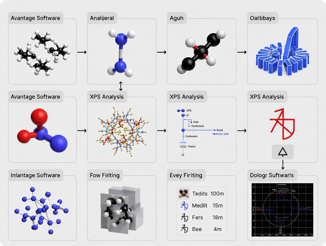
This article provides researchers, scientists, and drug development professionals with a detailed guide to Thermo Scientific Avantage software for X-ray Photoelectron Spectroscopy (XPS) analysis.
Mastering XPS Analysis with Avantage Software: A Comprehensive Guide for Biomedical Researchers
Abstract
This article provides researchers, scientists, and drug development professionals with a detailed guide to Thermo Scientific Avantage software for X-ray Photoelectron Spectroscopy (XPS) analysis. We explore the core principles of XPS data interpretation, demonstrate practical workflows for surface characterization of biomaterials and drug formulations, address common data processing challenges with proven solutions, and validate Avantage's performance against key analytical metrics. Learn how to leverage this powerful software to derive reliable, publication-ready chemical state information for your research.
What is Avantage Software? Core Principles of XPS Data Interpretation for Surface Science
Application Notes
Note 1: High-Throughput Screening of Pharmaceutical Tablet Coatings In drug development, consistent tablet coating is critical for controlled release and stability. Avantage software enables rapid, automated XPS analysis of coating uniformity and chemical composition across batch samples. A study of 50 enteric-coated tablets showed Avantage's automated mapping reduced analysis time by 70% compared to manual point-and-shoot methods, while quantifying critical coating elements (C, O, N, and specific polymer functional groups).
Table 1: XPS Analysis of Enteric Coating Uniformity (n=50 tablets)
| Metric | Manual Analysis | Avantage Automated Mapping | Improvement |
|---|---|---|---|
| Avg. Time per Tablet | 45 minutes | 13.5 minutes | 70% reduction |
| Spatial Resolution Achieved | 100 µm spot | 10 µm pixel size | 10x finer detail |
| Detection of Coating Thickness Variation | >10% thickness difference | >2% thickness difference | 5x more sensitive |
| Key Elements Quantified | C, O | C, O, N, specific functional group ratios (C-O/C=O) | Added chemical state specificity |
Note 2: Investigating Biomaterial Surface Aging for Implantable Devices For implantable drug-eluting devices, surface oxide layer stability on alloys (e.g., Nitinol, Titanium) is paramount. Avantage’s depth profiling and peak-fitting protocols were used to model oxide layer growth and contamination adsorption over accelerated aging. Data from a 30-day study showed a non-linear growth of carbonaceous contamination layer, critical for predicting device shelf-life and biocompatibility.
Table 2: Depth Profile of Ti6Al4V Alloy Surface After 30-Day Aging
| Depth (nm) | Atomic % Ti (Metallic) | Atomic % Ti (Oxide) | Atomic % C (Adventitious) | O/Ti Ratio |
|---|---|---|---|---|
| 0 (Surface) | 2.1 | 15.3 | 45.6 | 2.1 |
| 5 | 8.7 | 32.4 | 18.9 | 1.9 |
| 10 | 25.6 | 21.1 | 5.3 | 1.5 |
| 20 | 68.9 | 4.2 | 1.8 | 0.8 |
Experimental Protocols
Protocol 1: Automated Multi-Sample Coating Uniformity Workflow in Avantage Objective: To standardize high-throughput XPS screening of coating thickness and chemistry on pharmaceutical tablets.
- Sample Mounting: Secure up to 24 tablets in the Thermo Scientific AutoTray sample holder. Ensure surfaces are level.
- Avantage Method Setup: a. In the Experiment Designer, create a new Multi-Sample Map method. b. Define the sample holder geometry and select all tablet positions. c. For each position, apply a Large Area Survey (800 µm spot) followed by a High-Resolution Region Scan (100 µm spot) on the tablet center and four predefined edge points. d. Set acquisition parameters: Pass Energy 50 eV for surveys, 20 eV for high-resolution scans of C 1s, O 1s, N 1s. e. Enable Charge Compensation (Flood Gun) for all measurements.
- Automated Acquisition: Queue and run the sequence. Avantage automatically handles stage movement, focus, and data collection.
- Batch Processing: a. Use the Batch Processor to apply a standard Avantage Data System template to all spectra. b. Template includes: Shirley background subtraction, sensitivity factors, and peak-fitting models for C-C/C-H (284.8 eV), C-O (286.5 eV), O=C-O (289.0 eV).
- Data Export: Export atomic percentages and peak component ratios for all points to a CSV file for statistical analysis.
Protocol 2: Depth Profiling of Oxide Layers on Metallic Biomaterials Objective: To characterize the composition and thickness of native oxide layers and adsorbed contamination.
- Sample Preparation: Cut alloy sample (e.g., 10x10 mm). Clean ultrasonically in isopropanol for 5 minutes and dry under nitrogen stream.
- Avantage Method Setup: a. Create a Depth Profile experiment. b. Set initial surface analysis: High-resolution scans of relevant core levels (e.g., Ti 2p, O 1s, C 1s, Al 2p). c. Configure the ion gun: Use Ar⁺ ions at 1 keV, 1 µA current, raster over a 2x2 mm area. Set etch cycle time to 30 seconds. d. Set the sequence to alternate between etch cycles and analysis of selected high-resolution peaks (Snapshot mode).
- Acquisition: Run the profile for a predetermined total time (e.g., 400 seconds for ~20 nm depth).
- Data Processing: a. Use the Depth Profile tool to align spectra based on the C 1s adventitious carbon peak (284.8 eV) for initial etch cycles. b. Fit high-resolution Ti 2p spectra for each depth using a doublet separation of 5.7 eV, separating metallic Ti (453.8 eV) and TiO₂ (458.8 eV) components. c. Use the Sputter Rate Calculator with a Ta₂O₅ standard to convert etch time to approximate depth (nm).
- Reporting: Generate plots of atomic concentration vs. depth and oxide/metal ratio vs. depth using the built-in graphing tools.
Diagrams
Diagram 1: Avantage High-Throughput Screening Workflow
Diagram 2: Surface Aging Study Pathway for Implants
The Scientist's Toolkit: Key Research Reagent Solutions for XPS Analysis
Table 3: Essential Materials for Reliable XPS Surface Analysis
| Item | Function & Importance |
|---|---|
| Thermo Scientific Avantage Software | Central hub for instrument control, experiment design, automated data acquisition, advanced processing (peak fitting, depth profiling, mapping), and reporting. |
| Reference Standards (Au, Ag, Cu Foils) | For binding energy scale calibration and spectrometer performance verification. Au 4f7/2 peak at 84.0 eV is a common reference. |
| Argon Gas (High Purity, 99.999%) | Source for ion gun used for sample cleaning and depth profiling (sputtering). Essential for removing adventitious carbon or profiling interfaces. |
| Conductive Adhesive Tabs (e.g., Cu, C) | For mounting insulating samples (e.g., polymers, coated tablets) to minimize sample charging during analysis. |
| Charge Neutralization Flood Gun (Integrated) | Electron source to compensate for positive charge buildup on insulating samples, enabling analysis of non-conductive materials. |
| Ultrasonic Cleaner & Solvents (IPA, Acetone) | For reproducible sample cleaning to remove loose contamination prior to introduction into the UHV analysis chamber. |
| Certified Sputter Rate Standards (e.g., Ta₂O₅, SiO₂) | Thin films of known thickness used to calibrate the ion gun sputter rate, converting etch time to approximate depth (nm). |
| Multi-Element Check Sample | A sample with known, stable surface composition used for daily or weekly checks of instrument sensitivity factors and quantitative accuracy. |
This application note details the core analytical workflow within Thermo Scientific Avantage software for X-ray Photoelectron Spectroscopy (XPS). The broader thesis posits that rigorous control over spectral processing—from acquisition to quantification—is foundational for reliable material characterization in research and drug development. Avantage provides an integrated environment to execute this workflow with precision, ensuring that derived atomic concentrations are both accurate and reproducible.
Core Workflow: From Acquisition to Quantification
The foundational process in XPS analysis follows a defined sequence. The logical relationship between these steps is outlined below.
Diagram 1: Core XPS data processing workflow.
Detailed Protocols & Methodologies
Protocol: Optimized Spectra Acquisition in Avantage
Objective: To acquire high signal-to-noise (S/N) spectra suitable for quantitative analysis.
- Sample Preparation: Mount sample securely on holder using conductive tape or clips. Insert into spectrometer load lock.
- Preliminary Survey Scan: In Avantage, set a wide energy range (e.g., 0-1200 eV), pass energy of 150 eV, step size of 1.0 eV, and 2 scans for rapid overview.
- High-Resolution Regional Scan:
- Navigate to the 'Acquisition' panel.
- Select the element/region of interest from the survey.
- Set pass energy to 20-50 eV for optimal resolution.
- Adjust step size to 0.05-0.1 eV.
- Set dwell time and number of scans to achieve desired S/N (e.g., 10-20 scans). Use the software's preview function.
- Charge Neutralization: For insulating samples, ensure the flood gun is activated and optimized using Avantage's automatic charge compensation routine.
- Data Saving: Save spectra in Avantage's native
.vmsformat for full processing history.
Protocol: Background Subtraction
Objective: To remove the inelastic background signal, isolating the primary photoelectron peaks.
- Load Spectrum: Open the high-resolution region spectrum in the 'Processing' tab.
- Select Background Type: The choice is critical and depends on sample morphology.
- Linear: Rarely used for quantitative work.
- Shirley (Integral): Default for homogeneous materials. Accounts for inelastic scattering.
- Tougaard: More accurate for in-depth composition analysis or polymers. Available in advanced Avantage modules.
- Smart Background: Avantage's automated method, useful for standard cases.
- Application: Select the background type. Manually adjust the start and end points (BE) of the background region to bracket the peak(s) of interest. The software automatically calculates and subtracts the background.
Protocol: Peak Fitting & Deconvolution
Objective: To mathematically resolve overlapping chemical states into individual component peaks.
- Define Peak Shape: In the 'Peak Fit' tab, select a line shape. A mix of Gaussian-Lorentzian (e.g., 70% G, 30% L) is standard for most materials.
- Add Components: Add a component for each suspected chemical state. Initial positions can be guided by literature or database values (accessible within Avantage).
- Apply Constraints: Use software constraints judiciously:
- Fix full width at half maximum (FWHM) to be equal for peaks from the same element in similar bonding environments.
- Fix spin-orbit doublet separations and area ratios (e.g., 2:1 for p orbitals, 3:2 for d orbitals).
- Optimize Fit: Execute the iterative fitting algorithm. Manually adjust component position, height, and FWHM if necessary to minimize the residual (difference between raw data and fit).
- Validate Fit: The residual should be flat and featureless. The fit should be chemically plausible.
Protocol: Quantifying Atomic Concentration
Objective: To calculate the relative atomic concentration of detected elements.
- Extract Peak Areas: After fitting, the software reports the area (counts*eV) for each component or whole region.
- Apply Sensitivity Factors: Avantage uses built-in relative sensitivity factors (RSFs). The formula applied is:
C_x = (I_x / S_x) / Σ(I_i / S_i)whereC_xis atomic concentration of element X,I_xis the measured peak area, andS_xis the RSF. - Report Results: Concentrations are typically reported as atomic percent (at.%). The software generates a quantitative report table.
Data Presentation: Comparative Analysis of Background Methods
The choice of background significantly impacts derived peak areas and final atomic concentrations, as demonstrated in the table below.
Table 1: Impact of Background Subtraction Method on C 1s Peak Area and Calculated Atomic % for a Polymer Sample.
| Background Method | C 1s Peak Area (a.u.) | O 1s Peak Area (a.u.) | Calculated C % (at.%) | Calculated O % (at.%) | Best For |
|---|---|---|---|---|---|
| Shirley | 45,321 | 12,588 | 75.8 | 24.2 | Homogeneous solids, metals, oxides |
| Linear | 48,955 | 12,588 | 77.5 | 22.5 | Quick overview (not quantitative) |
| Tougaard (λ=1) | 41,220 | 12,588 | 74.1 | 25.9 | Polymers, inelastic loss analysis |
| Smart (Avantage) | 44,850 | 12,588 | 75.6 | 24.4 | Standard automated quantification |
The Scientist's Toolkit: Research Reagent Solutions & Essential Materials
Table 2: Essential Materials for XPS Sample Preparation & Analysis.
| Item | Function in XPS Analysis |
|---|---|
| Conductive Carbon Tape | Mounts powdered or non-conductive samples; provides a path to ground to mitigate charging. |
| Indium Foil | Ductile metal foil for mounting irregularly shaped samples; provides good electrical and thermal contact. |
| Argon Gas (99.999%) | Used in ion source for sample cleaning (sputtering) and depth profiling to remove surface contamination. |
| Charge Neutralization Flood Gun | Integrated electron source to compensate for positive surface charge on insulating samples during analysis. |
| Certified Reference Materials | Standards (e.g., clean Au, Ag, Cu foils) for instrument performance validation and energy scale calibration. |
| Ultra-High Vacuum (UHV) Compatible Solvents | (e.g., HPLC-grade isopropanol) For ultrasonic cleaning of samples and sample holders without introducing contaminants. |
| XPS Database Software | (e.g., NIST XPS Database, commercial libraries) Provides reference binding energies for peak identification and fitting. |
Visualization of the Peak Fitting Decision Pathway
The process of deciding on a peak model involves logical checks to ensure a chemically and physically valid result.
Diagram 2: Logical pathway for XPS peak fitting and validation.
Within the framework of a thesis dedicated to advancing X-ray Photoelectron Spectroscopy (XPS) analysis methodologies, mastering the Thermo Scientific Avantage software interface is paramount. This software serves as the central hub for data acquisition, processing, and interpretation. For researchers in drug development, this enables precise surface characterization of novel pharmaceutical compounds, polymer excipients, and biomaterial coatings, providing critical data on elemental composition, chemical state, and layer thicknesses that correlate with performance and stability.
Core Interface Components: Workspace, Viewer, and Toolbars
The Workspace
The Workspace is the primary project management area. It organizes data in a hierarchical tree structure, encompassing samples, spectra, processed data, and reporting elements.
Key Functions:
- Project Tree: Manages all data files within an analysis session.
- Sample Navigation: Allows grouping and batch processing of related spectra.
- Data State Tracking: Differentiates between raw, processed, and fitted data sets.
The Spectra Viewer
This is the main visualization and interaction pane for spectral data.
Key Functions:
- Multi-spectra Overlaying: Directly compare control and treated samples.
- Interactive Inspection: Zoom, region selection, and real-time coordinate reading.
- Layer Display: For depth profile data visualization.
The Processing Toolbars
Context-sensitive toolbars provide access to data manipulation routines essential for quantitative analysis.
Primary Toolbars:
- Spectrum Toolbar: Core operations like background subtraction, smoothing, and peak labeling.
- Quantification Toolbar: Access to sensitivity factors, atomic concentration calculations, and line-shape definitions.
- Peak Fitting Toolbar: Sophisticated routines for deconvoluting complex chemical states using Gaussian-Lorentzian curves.
Table 1: Quantitative Data Output from Avantage Standard Analysis
| Data Type | Typical Output | Precision (Relative %) | Primary Toolbar Source |
|---|---|---|---|
| Atomic Concentration | Elemental % | 1-5% | Quantification Toolbar |
| Peak Position (BE) | Binding Energy (eV) | ±0.1 eV | Spectrum Toolbar |
| Peak Area (Intensity) | Counts-eV/s | 2-8% | Peak Fitting Toolbar |
| FWHM | Line width (eV) | ±0.15 eV | Peak Fitting Toolbar |
| Depth Profile Sputter Time | Seconds per layer | <5% | Spectra Viewer (Layer Mode) |
Experimental Protocols for Key Analyses
Protocol 3.1: Chemical State Analysis of a Drug-Excipient Interface
Objective: To identify the chemical states of carbon and nitrogen at the interface between an Active Pharmaceutical Ingredient (API) and a polymeric coating.
Materials & Methods:
- Sample Preparation: Spin-coat a 100 nm poly(lactide-co-glycolide) (PLGA) film onto a silicon wafer. Deposit the API via controlled sublimation.
- Data Acquisition: Acquire high-resolution spectra for C 1s and N 1s regions using a monochromatic Al Kα source (1486.6 eV), pass energy of 50 eV, and step size of 0.1 eV.
- Avantage Processing Workflow: a. Workspace: Import spectra and create a "Drug-Excipient Interface" sample group. b. Spectra Viewer: Overlay C 1s spectra from pure API, pure PLGA, and the interface sample. c. Processing Toolbars: i. Apply a Smart background subtraction (Spectrum Toolbar). ii. Calibrate spectra to the adventitious C 1s peak at 284.8 eV. iii. Using the Peak Fitting Toolbar, create a synthetic component model for C 1s: C-C/C-H (284.8 eV), C-O (286.5 eV), O-C=O (288.9 eV), and π-π* satellite (~291 eV). iv. Constrain FWHM within chemically reasonable limits (0.8-1.2 eV difference between components). v. Iterate fit until χ² is minimized.
- Data Interpretation: Quantify the relative area of the O-C=O component from the PLGA and the API-specific N 1s state to determine interfacial mixing.
Protocol 3.2: Thin-Film Oxide Thickness Measurement
Objective: To determine the thickness of a silicon oxide (SiO₂) layer on a drug delivery microdevice component.
Materials & Methods:
- Sample: Silicon wafer with a thermally grown oxide layer.
- Data Acquisition: Acquire survey scan and high-resolution Si 2p spectra.
- Avantage Processing Workflow:
a. Quantification Toolbar: Calculate atomic concentrations from survey scan using standard RSF values.
b. Peak Fitting Toolbar: Deconvolute the Si 2p high-resolution spectrum into substrate Si⁰ (99.3 eV) and oxide Si⁴⁺ (103.8 eV) doublets.
c. Integrated Calculation: Use the
Layer Thicknesscalculator (accessed via Tools menu). Input the measured intensities (peak areas) of the Si⁴⁺ (oxide) and Si⁰ (substrate) peaks, their inelastic mean free paths (IMFP), and the take-off angle. - Calculation: The software applies the standard overlayer thickness equation: d = λ * sin(θ) * ln( (I_ox / I_sub) * (R_sub / R_ox) + 1 ), where λ is IMFP, θ is take-off angle, I is intensity, and R is the relative sensitivity factor.
Logical Workflow Diagram
Diagram 1: Avantage XPS Data Analysis Workflow
The Scientist's Toolkit: Essential Research Reagents & Materials
Table 2: Key Materials for XPS Surface Analysis in Drug Development
| Item | Function & Relevance in XPS Analysis |
|---|---|
| Monocrystalline Silicon Wafer | An atomically flat, conductive substrate for spin-coating polymer or API films. Provides a consistent background for quantification. |
| Adventitious Carbon Reference | Ubiquitous hydrocarbon contamination used as an internal energy scale reference (C 1s at 284.8 eV) for charge correction. |
| ISO-Calibration Standards | Certified materials (e.g., Au, Ag, Cu) for verifying instrument energy scale and resolution performance, ensuring data validity. |
| Argon Gas (99.999% purity) | Used for charge neutralization (flood gun) and for ion beam sputtering to perform depth profiling through coatings. |
| Ultra-High Purity Solvents (e.g., HPLC-grade Toluene, Isopropanol) | For substrate cleaning to minimize unwanted organic contamination that complicates C 1s spectral interpretation. |
| Reference Compounds (e.g., Polytetrafluoroethylene - PTFE, Polyethylene terephthalate - PET) | Well-characterized polymers with known chemical states used to validate peak-fitting models and relative sensitivity factors (RSFs). |
Application Notes: Data Formats in Avantage for XPS Analysis
In the context of research utilizing Thermo Scientific Avantage software for X-ray Photoelectron Spectroscopy (XPS) analysis, effective data management and collaboration are paramount. Avantage generates and utilizes proprietary data formats to store complex spectral information, which must be understood for proper data curation, sharing, and publication. The transition from raw experimental data to collaborative insight hinges on mastering these formats and their export pathways.
The primary native data formats are:
- VGD (VG Datafile): The fundamental raw data container. It stores all the original, unprocessed spectral data collected from the instrument, including counts, binding energies, and acquisition parameters. It is the definitive source for any reprocessing.
- VMS (Avantage Datafile): The primary working project file. It contains processed spectra (with calibrations, background subtractions, peak fits), quantification results, reports, and references to the original VGD files. It is the central file for daily analysis within Avantage.
For collaboration outside the Avantage ecosystem, data must be exported into universal, open formats. Failure to properly archive VGD/VMS files and their exported counterparts can compromise research reproducibility, a cornerstone of scientific integrity in fields like materials science and drug development, where surface chemistry analyzed by XPS informs on catalyst efficiency or biomaterial compatibility.
Quantitative Comparison of Avantage Data Formats & Exports
Table 1: Core Characteristics of Essential Avantage Data Formats
| Format | Primary Function | Data Content | Readability | Mutability |
|---|---|---|---|---|
| VGD | Raw Data Archival | Raw counts, instrument parameters, spectral metadata. | Avantage Software only. | Immutable (read-only). |
| VMS | Analysis & Processing | Processed spectra, peak models, quantification, reports, links to VGDs. | Avantage Software only. | Fully editable. |
| VGS (ASCII) | Data Export | XY data (Binding Energy vs. Counts) for a single spectrum. | Any text editor or plotting software. | Editable as text. |
| VAMAS (ISO 14976) | Standardized Export | XY data plus comprehensive experimental parameters. | Specialist software (CasaXPS, SpecsLab, etc.). | Editable with difficulty. |
Table 2: Export Format Suitability for Collaboration
| Export Format | Recommended For | Key Advantage | Key Limitation |
|---|---|---|---|
| ASCII (.txt, .csv) | Plotting in graphing tools (Origin, Excel), simple data exchange. | Universally readable, simple structure. | Loss of rich metadata, no processing history. |
| VAMAS (.vms) | Collaboration with other XPS experts, journal submission. | Preserves critical experimental metadata, ISO standard. | Not human-readable, requires compatible software. |
| Image (.tif, .emf) | Inclusion in presentations, reports, and draft manuscripts. | Visual representation, accessible to all. | No underlying numerical data for further analysis. |
| PDF Report | Sharing final results with non-specialists or for archival. | Self-contained, formatted summary of findings. | Data is not extractable for re-analysis. |
Detailed Protocols for Data Handling and Export
Protocol 3.1: Creating a Collaborative Data Package from an Avantage VMS Project
Objective: To generate a complete, reproducible data package from an Avantage session for sharing with external collaborators or for archival, ensuring all necessary components are included.
Materials:
- Computer with Thermo Scientific Avantage Software (v5.9923 or later).
- Processed VMS project file.
- Associated VGD raw data files.
Procedure:
- Project Consolidation: Open the VMS file in Avantage. Navigate to
File > Save As. Check the option "Include VG Data Files" (or equivalent). Save under a new name (e.g.,ProjectX_Complete.vms). This ensures all raw data is embedded within the single VMS bundle. - Export Processed Spectra for Analysis:
- Select the processed spectrum or region in the Spectrum Window.
- Navigate to
File > Export > Spectrum. - In the dialog box, set "Save as type:" to "VAMAS Format (*.vms)".
- Choose a descriptive filename and location. This file is now suitable for import into other XPS data processing software.
- Export Numerical Data for Plotting:
- Repeat the export step, but set "Save as type:" to "Text (Tab Delimited) (*.txt)".
- This creates a simple two-column ASCII file for universal plotting.
- Export the Quantitative Report:
- Ensure the quantification table is visible.
- Navigate to
File > Export > Reportor copy the table directly to clipboard for pasting into a spreadsheet.
- Package Contents: The final collaborative package should contain: (a) The consolidated
.vmsfile, (b) Exported.vms(VAMAS) files for key spectra, (c) Exported.txtfiles for key spectra, (d) AREADME.txtfile explaining the contents and processing steps.
Protocol 3.2: Batch Export of Multiple Spectra to ASCII Format
Objective: To efficiently export numerical data from multiple spectra within a VMS file for bulk external analysis or plotting, saving time and ensuring consistency.
Procedure:
- In the Avantage Workspace Explorer, select multiple spectra by holding
Ctrland clicking on the desired spectrum items. - Right-click on the selected group and choose "Export" from the context menu.
- In the export wizard, select "Text (Tab Delimited)" as the format.
- Specify an output directory. Avantage will create individual
.txtfiles for each exported spectrum, using the spectrum names as filenames. - Verify the output files by opening one in a text editor to confirm it contains two columns of data (e.g., Binding Energy and Intensity).
Visualization of Data Flow and Decision Pathway
XPS Data Flow from Acquisition to Collaboration
The Scientist's Toolkit: Essential Research Reagent Solutions for XPS Sample Preparation
While XPS is a surface analysis technique, sample preparation is often critical, especially in drug development research (e.g., analyzing polymer coatings, implant surfaces, or catalyst materials).
Table 3: Key Materials for XPS Sample Preparation
| Material/Reagent | Function in XPS Research | Example & Notes |
|---|---|---|
| Solvent Series | Ultrasonic cleaning of substrates to remove organic contaminants prior to film deposition or analysis. | Sequential baths of toluene, acetone, and isopropanol (IPA) of electronic grade. Removes machining oils, fingerprints. |
| Plasma Cleaner (O₂, Ar) | Generates a reactive plasma for ultra-cleaning surfaces or for subtle surface modification (etching, functionalization). | Argon plasma for gentle sputter-cleaning of sensitive organics. Oxygen plasma for removing hydrocarbon layers. |
| Spin Coater | Creates uniform thin films of polymer or biomaterial solutions on flat substrates for surface property analysis. | Used to prepare model surfaces of drug-eluting coatings or biocompatible polymers for XPS characterization. |
| Reference Samples | Essential for energy scale calibration and quantification verification. | Clean, sputtered Au foil (for Au 4f7/2 at 84.0 eV), Cu foil (for Cu 2p3/2 at 932.67 eV), and highly oriented pyrolytic graphite (for C 1s at 284.8 eV). |
| Conductive Adhesive Tape (Carbon) | Mounting electrically insulating samples to minimize surface charging during XPS analysis. | Double-sided carbon tape provides both adhesion and electrical contact to the sample holder. |
| Argon Gas (High Purity) | Used in the integrated ion gun for depth profiling (sputter etching) to reveal in-depth chemical composition. | 99.999% purity argon minimizes introduction of contaminants during the depth profile experiment. |
Within the rigorous demands of modern biomedical research, surface analysis is a cornerstone for understanding material-biology interactions. The central thesis underpinning this application note is that Avantage software for X-ray Photoelectron Spectroscopy (XPS) is an indispensable tool for generating, interpreting, and reporting high-fidelity chemical state information, which is the critical differentiator between descriptive surface analysis and actionable biomedical insight. While XPS provides elemental composition, it is the precise chemical state data—revealing bonding environments, oxidation states, and functional groups—that deciphers the true nature of biomaterial surfaces, protein coronas, implant interfaces, and drug delivery systems. This document details the protocols and applications that demonstrate this thesis in practice.
Core Applications & Quantitative Data
The following table summarizes key biomedical surface analysis scenarios where Avantage’s chemical state resolution is paramount.
Table 1: Critical Biomedical Surface Analysis Scenarios and Avantage Output
| Biomedical Application | Key Element(s) Analyzed | Descriptive Analysis (Atomic % Only) | Chemical State Analysis via Avantage (Critical Insight) |
|---|---|---|---|
| Implant Biocompatibility (e.g., Ti-6Al-4V) | Titanium (Ti), Oxygen (O) | Ti: 15%, O: 50%, C: 35% | Quantifies % of TiO₂ (biocompatible oxide) vs. TiO/sub>x or Ti⁰ (potentially corrosive). Predicts in-vivo stability. |
| Protein Corona on Nanoparticle | Nitrogen (N), Carbon (C), Sulfur (S) | N: 5% (confirms protein presence) | Deconvolutes N 1s into amine (-NH₂), amide (-CONH-), and protonated ammonium (-NH₃⁺), mapping protein orientation/conformation. |
| Drug-Polymer Coating Stability | Fluorine (F), Silicon (Si) | F: 2% (confirms drug presence) | Distinguishes covalent C-F bonds (intact drug) from ionic/migratory F (degradation/leaching). |
| Antimicrobial Surface Efficacy | Silver (Ag) | Ag: 3% (total silver) | Differentiates Ag⁰ (nanoparticle reservoir) from Ag⁺ (biocidal ion), enabling dose-response correlation. |
| Plasma Polymer Functionalization | Carbon (C) | C: 85% | Quantifies relative concentrations of C-C/C-H, C-O, C=O, and COOR functionalities, confirming intended surface chemistry. |
Detailed Experimental Protocols
Protocol 1: Analyzing Protein Corona Formation on a Drug Delivery Nanoparticle
Objective: To quantify the chemical composition and bonding states of proteins adsorbed onto PLGA nanoparticles using Avantage.
Materials & Reagents:
- Synthesized PLGA nanoparticles with encapsulated agent.
- Bovine Serum Albumin (BSA) solution (1 mg/mL in PBS).
- Phosphate Buffered Saline (PBS), pH 7.4.
- Ultra-pure water (18.2 MΩ·cm).
- Centrifuge and microcentrifuge tubes.
- Clean silicon wafer substrates.
Procedure:
- Incubation: Dilute nanoparticle suspension to 1 mg/mL in PBS. Mix 1:1 with BSA solution. Incubate at 37°C for 1 hour with gentle agitation.
- Isolation: Centrifuge at 20,000 RCF for 30 minutes to pellet corona-coated nanoparticles. Carefully discard supernatant.
- Washing: Resuspend pellet in 1 mL PBS. Repeat centrifugation and washing step twice to remove loosely associated proteins.
- Sample Preparation: After final wash, resuspend pellet in a minimal volume of ultra-pure water (~50 µL). Deposit suspension onto a clean silicon wafer and allow to dry in a laminar flow hood.
- XPS Data Acquisition:
- Instrument: Use a monochromatic Al Kα X-ray source.
- Settings: Pass Energy 50 eV for survey scans, 20 eV for high-resolution regions.
- Regions: Acquire survey spectrum (0-1200 eV). Acquire high-resolution spectra for C 1s, N 1s, O 1s, and S 2p.
- Charge Neutralization: Use a combined low-energy electron/ion flood gun.
- Avantage Data Processing:
- Load spectra into Avantage. Apply a linear background subtraction.
- For C 1s, set the adventitious hydrocarbon (C-C/C-H) peak to 284.8 eV for charge correction.
- Critical Chemical State Fitting: Deconvolute the high-resolution spectra using Avantage’s peak fitting module.
- C 1s: Fit components for C-C/C-H (284.8 eV), C-O (286.5 eV), C=O (288.0 eV), and O-C=O (289.0 eV).
- N 1s: Fit components for amine (-NH₂, ~399.2 eV) and amide (-CONH-, ~400.0 eV). A component at ~401.5 eV may indicate protonated amines.
- Use the N 1s amide peak area and the known stoichiometry of BSA to estimate relative surface coverage.
Protocol 2: Assessing Oxidation State of an Antimicrobial Silver Coating
Objective: To determine the ratio of metallic silver (Ag⁰) to ionic silver (Ag⁺) on a plasma-deposited antimicrobial coating.
Materials & Reagents:
- Plasma-coated test substrate (e.g., medical catheter segment).
- Reference materials: High-purity silver foil (for Ag⁰), Ag₂O powder (for Ag⁺).
- Conductive carbon tape.
Procedure:
- Sample Mounting: Mount the coated substrate and reference materials on a standard sample bar using conductive carbon tape.
- XPS Data Acquisition:
- Instrument Settings: As in Protocol 1.
- Critical Region: Acquire high-resolution Ag 3d spectrum with excellent statistics (>100,000 counts in main peak).
- Avantage Data Processing & Quantification:
- Charge correct spectrum to the C 1s hydrocarbon peak at 284.8 eV.
- Analyze the Ag 3d₅/₂ peak.
- Chemical State Fitting:
- Load reference spectra from the pure Ag⁰ foil and Ag₂O powder into Avantage’s Spectral Data Bank.
- Use Avantage’s "Touch-to-Peak" or "Component Fitting" tool. Apply a Lorentzian-Gaussian mix (e.g., 30% L).
- Fit the sample's Ag 3d₅/₂ peak with two doublet-constrained components (spin-orbit splitting ~6.0 eV, ratio ~3:2).
- The component with a binding energy near 368.2 eV is assigned to Ag⁰. The component shifted to higher BE (~367.8-368.0 eV for Ag₂O, can be higher for AgO) is assigned to Ag⁺.
- Quantification: Avantage directly reports the percentage of the total Ag 3d peak area attributable to each chemical state. Report the Ag⁺/(Ag⁰+Ag⁺) ratio.
Visualization of Workflows and Relationships
Title: From Sample to Insight: The Avantage Chemical State Workflow
Title: Chemical State Analysis of Protein Corona
The Scientist's Toolkit: Essential Research Reagents & Materials
Table 2: Key Reagents and Materials for Biomedical XPS Sample Preparation
| Item | Function in Biomedical Surface Analysis |
|---|---|
| Clean Silicon Wafers | An atomically smooth, low-background substrate for depositing nanoparticle or protein samples for XPS analysis. |
| Phosphate Buffered Saline (PBS), pH 7.4 | Standard physiological buffer for simulating biological fluids during protein corona formation or dissolution studies. |
| Ultra-Pure Water (Type 1, 18.2 MΩ·cm) | For final rinsing of samples to remove residual salts and buffers that create interfering XPS signals. |
| Conductive Carbon Tape | For mounting non-conductive biomedical samples (polymers, tissues on substrate) to prevent charging during XPS. |
| Low-Energy Electron/Ion Flood Gun | Integrated charge neutralization system essential for analyzing insulating biomaterials (polymers, ceramics). |
| Certified XPS Reference Materials | Pure elements (Au, Ag, Cu) and compounds (TiO₂, Ag₂O) for energy scale calibration and chemical state verification within Avantage. |
| Plasma Cleaner (Ar/O₂) | For in-situ cleaning of substrates and, crucially, for gentle surface treatment of sensitive samples to remove adventitious carbon without altering bulk chemistry. |
Step-by-Step Avantage Workflows: From Raw Data to Publication-Ready Results
Application Notes
Surface characterization is a critical step in the development and quality control of polymer biomaterials used in drug delivery, medical implants, and biosensors. X-ray Photoelectron Spectroscopy (XPS) is the premier technique for quantifying elemental surface composition and identifying chemical bonding states at the top 5-10 nm of a material. Within the context of a broader thesis on Avantage software for XPS analysis research, this workflow demonstrates how the software's advanced data processing and mapping capabilities transform raw spectral data into actionable insights for material scientists. For polymers like Poly(lactic-co-glycolic acid) (PLGA) and Poly(ethylene glycol) (PEG) coatings, key parameters include the verification of coating purity, detection of surface contaminants, quantification of copolymer ratios, and assessment of coating uniformity. Avantage's sophisticated peak fitting, quantification algorithms, and chemical state mapping are indispensable for these tasks, enabling researchers to correlate surface chemistry with biological performance and manufacturing variables.
Protocols
Protocol 1: Sample Preparation and Mounting for XPS Analysis
Objective: To prepare polymer biomaterial samples for contamination-free, reliable XPS analysis.
- Clean Handling: Use powder-free nitrile gloves and clean, blunt tweezers for all sample manipulations.
- Substrate Preparation: For coating analysis, apply the PLGA or PEG solution onto a clean, conductive substrate (e.g., silicon wafer, gold-coated slide). Spin-coating is recommended for uniform thin films.
- Drying/Curing: Dry samples under vacuum desiccation for a minimum of 24 hours to remove residual solvent and atmospheric contaminants.
- Mounting: Affix the sample to the XPS sample holder using double-sided conductive carbon tape. Ensure the analysis area is flat and securely attached to prevent charging.
- Transfer: If available, use an inert atmosphere transfer vessel to move samples from the preparation glovebox to the XPS introduction chamber to minimize airborne hydrocarbon contamination.
Protocol 2: XPS Data Acquisition for Polymer Surfaces
Objective: To collect high-quality survey and high-resolution spectra for quantitative surface analysis.
- Instrument Setup: Use a monochromatic Al Kα X-ray source (1486.6 eV). Set the pass energy to 160 eV for survey scans (0-1350 eV) and 20-50 eV for high-resolution regional scans.
- Charge Neutralization: Engage the low-energy electron flood gun and argon ion source to compensate for surface charging on insulating polymer samples. Adjust parameters to achieve a known adventitious carbon C 1s peak position at 284.8 eV.
- Data Collection:
- Perform a minimum of three survey scans from different spots (~1 mm²) to assess homogeneity.
- Acquire high-resolution spectra for all elements of interest: C 1s, O 1s, N 1s (if applicable), and any expected contaminants (Si 2p, Na 1s).
- Dwell Time & Scans: Use a dwell time of 50-100 ms and accumulate 5-10 scans for high-resolution regions to ensure a good signal-to-noise ratio.
Protocol 3: Data Processing in Avantage Software
Objective: To quantify elemental composition and identify chemical states using Avantage.
- Import & Calibration: Import spectral data. Calibrate the energy scale using the adventitious carbon C 1s peak (C-C/C-H) at 284.8 eV.
- Quantification: From the survey spectrum, apply the "Quantify" function. Use the "Scofield" relative sensitivity factors (RSFs) provided in the Avantage library. Generate an atomic percentage (At.%) table.
- Peak Fitting (High-Resolution C 1s for PLGA):
- Background Subtraction: Apply a Smart (Shirley) background to the C 1s region.
- Component Definition: Add Voigt (70% Gaussian, 30% Lorentzian) line shapes for expected bonds:
- C1: C-C/C-H (hydrocarbon) at 284.8 eV.
- C2: C-O (ether/alcohol) at ~286.5 eV.
- C3: O-C=O (ester) at ~288.9 eV.
- Constraint Setting: Constrain the Full Width at Half Maximum (FWHM) to be equal for all components. Fit the spectrum iteratively until the residual is minimized.
- Mapping Analysis: For coating homogeneity, load an XPS map dataset. In Avantage, select a specific chemical state peak (e.g., ester carbon at 288.9 eV) and generate a "Chemical State Map" to visualize its distribution across the sample surface.
Protocol 4: Calculating PLGA Copolymer Ratio from XPS Data
Objective: To determine the lactic to glycolic acid (LA:GA) ratio in PLGA from the O 1s spectrum.
- Acquire High-Resolution O 1s Spectrum: Follow Protocol 2.
- Peak Fit O 1s: Fit the O 1s peak with two components:
- O1: Ester oxygen (O=C) at ~532.0 eV.
- O2: Ether oxygen (O-C) at ~533.3 eV.
- Calculate Ratio: The area of the O1 component corresponds to both carbonyl oxygens in the ester. The LA:GA ratio can be inferred from the relative intensities of C-O and C=O in the C 1s spectrum or by comparing the O1s fit to known standards calibrated via NMR.
Data Tables
Table 1: Theoretical vs. Experimental Atomic Composition of Common Polymer Biomaterials
| Polymer | Theoretical Composition (At.%) | Typical Experimental XPS (At.%) | Key Contaminants Often Detected |
|---|---|---|---|
| PEG (Pure) | C: 66.7%, O: 33.3% | C: 65-68%, O: 32-35% | Si (<1%), Na (<0.5%) |
| PLGA (50:50 LA:GA) | C: 60.0%, O: 40.0% | C: 58-62%, O: 38-42% | N (<0.5%, from synthesis), Si |
| PLGA (75:25 LA:GA) | C: 62.5%, O: 37.5% | C: 61-64%, O: 36-39% | N (<0.5%), Si |
| Polylactic Acid (PLA) | C: 60.0%, O: 40.0% | C: 58-62%, O: 38-42% | Hydrocarbon (C-C) from degradation |
Table 2: Characteristic XPS Binding Energies for Polymer Functional Groups
| Functional Group | Chemical State | Approx. C 1s B.E. (eV) | Approx. O 1s B.E. (eV) |
|---|---|---|---|
| Hydrocarbon | C-C, C-H | 284.8 | - |
| Ether/Alcohol | C-O | 286.3-286.5 | 533.0-533.5 |
| Carbonyl (Ester) | O-C=O | 288.7-289.0 | 531.8-532.2 |
| Acetal (PEG) | O-C-O | 286.5-286.7 | 533.0-533.3 |
Diagrams
The Scientist's Toolkit
Table 3: Essential Research Reagent Solutions for Polymer Biomaterial XPS Analysis
| Item | Function in Workflow |
|---|---|
| Silicon Wafer Substrates | Provides an atomically smooth, conductive, and clean surface for spin-coating polymer films, minimizing sample charging during XPS analysis. |
| High-Purity Solvents (Chloroform, TFE, Acetone) | Used to dissolve polymers (e.g., PLGA, PEG) for solution casting and to clean substrates and sample holders to prevent contamination. |
| Conductive Carbon Tape | Used to mount insulating polymer samples to the XPS sample stub, providing a path for charge neutralization. |
| Certified XPS Reference Foils (Au, Cu, Ag) | Used for periodic calibration and performance verification of the XPS instrument's energy scale and resolution. |
| Avantage Software Database (RSF Library) | Contains the relative sensitivity factors and reference spectra essential for accurate quantification and peak identification. |
| Low-Energy Electron Flood Gun Source | A critical component of the XPS instrument that provides electrons to neutralize positive charge buildup on insulating polymer surfaces. |
| Inert Atmosphere Transfer Module | A vacuum or nitrogen-filled vessel that allows samples to be moved from a glovebox to the XPS without exposure to air, preserving clean surfaces. |
Within the broader thesis on Avantage software for X-ray Photoelectron Spectroscopy (XPS) analysis, this application note details its critical role in nanomedicine R&D. Avantage’s sophisticated data processing, quantitative elemental/chemical state analysis, and depth profiling capabilities enable researchers to accurately characterize the surface composition of drug-loaded nanoparticles (NPs) and liposomes. This is essential for correlating material properties with drug loading efficiency, stability, and targeted release mechanisms.
Key Analytical Questions & XPS Capabilities
XPS, powered by Avantage software, addresses core questions:
- Surface Purity: What is the elemental composition at the nanoparticle surface?
- Drug Confirmation: Is the active pharmaceutical ingredient (API) present on the surface?
- Coating Integrity: Are stabilizing polymers (e.g., PEG) or targeting ligands successfully conjugated?
- Degradation/Stability: Are there signs of surface oxidation or chemical degradation after storage?
Experimental Protocols
Protocol 3.1: Sample Preparation for XPS Analysis
- Objective: To prepare a homogeneous, dry film of nanoparticles/liposomes for reproducible XPS analysis.
- Materials: Aqueous suspension of drug-loaded NPs/liposomes, silicon wafer, filter membrane, or indium foil; micro-pipette; vacuum desiccator.
- Procedure:
- Clean a substrate (e.g., 1x1 cm silicon wafer) with solvents and plasma clean for 2 minutes.
- Pipette 20-50 µL of the nanocarrier suspension onto the substrate.
- Allow to air-dry in a clean, dust-free environment for 60 minutes.
- Transfer the sample to a vacuum desiccator for a minimum of 12 hours to remove residual water.
- Mount the dried sample on the XPS holder using conductive double-sided tape or clips.
Protocol 3.2: XPS Data Acquisition & Avantage Processing Workflow
- Objective: To acquire and process high-quality spectra to determine atomic percentages and chemical states.
- Instrument Setup: Use a monochromatic Al Kα X-ray source (1486.6 eV). Charge neutralization is mandatory for insulating samples.
- Acquisition Parameters: Survey spectrum: Pass Energy 160 eV, step size 1.0 eV. High-resolution regions: Pass Energy 20-50 eV, step size 0.1 eV. Analysis area: 300-700 µm spot.
- Avantage Software Workflow:
- Load Spectra & Calibrate: Reference adventitious carbon C 1s peak to 284.8 eV.
- Elemental Identification: Use survey spectrum to identify all elements present (Table 1).
- Peak Fitting: For high-resolution spectra, apply Smart Background and use Avantage’s peak fitting toolbox. Constrain peaks based on known chemical states (e.g., C-C/C-H, C-O, C=O for carbon; P-O-C, phosphate for phosphorus in liposomes).
- Quantification: Use relative sensitivity factors (RSF) provided by the software to calculate atomic concentrations.
Data Presentation
Table 1: Exemplary XPS Surface Composition Data for Nanocarriers
| Sample Description | C (at%) | O (at%) | N (at%) | P (at%) | Specific Element (e.g., Drug) | Key Finding |
|---|---|---|---|---|---|---|
| PLGA Nanoparticle (Blank) | 72.5 | 27.1 | 0.4 | 0.0 | - | Surface composition matches polymer. |
| Doxorubicin-loaded PLGA NP | 70.8 | 26.5 | 1.2 | 0.0 | N (1.2 at%) | Surface N confirms presence of doxorubicin. |
| PEGylated Liposome | 68.2 | 27.8 | 0.0 | 3.2 | P (3.2 at%) | High P signal confirms lipid headgroups. |
| Ligand-Targeted Liposome | 65.4 | 28.1 | 2.5* | 3.0 | N (2.5 at%) | Presence of N confirms ligand conjugation. |
*Nitrogen from targeting peptide ligand.
Visualization of Analysis Workflow
Diagram Title: XPS Analysis Workflow for Nanocarriers Using Avantage
The Scientist's Toolkit: Essential Research Reagents & Materials
| Item | Function in Analysis |
|---|---|
| Silicon Wafer Substrate | Provides an atomically flat, clean, and conductive surface for depositing nanocarriers to minimize spectral interference. |
| Aluminum Kα X-ray Source | Standard monochromatic X-ray source (1486.6 eV) for ejecting core electrons, providing high-resolution spectra. |
| Charge Neutralizer (Flood Gun) | Compensates for positive charge buildup on non-conductive samples (e.g., polymer NPs), preventing peak shifting/broadening. |
| Avantage Software Suite | Comprehensive platform for spectral processing, quantitative analysis, peak deconvolution, and generating publication-ready data. |
| Conductive Adhesive Tape/Clips | Ensures stable electrical contact between sample and holder, reducing charging artifacts. |
| Vacuum Desiccator | Removes residual solvents and water from dried nanocarrier films, preventing vacuum degradation and water vapor interference. |
Application Notes
Within the context of a thesis utilizing Avantage software for XPS analysis, this protocol provides a method to quantitatively investigate the protein corona that forms spontaneously on biomaterial implant surfaces upon exposure to biological fluids. This corona dictates the host immune response, cellular adhesion, and ultimately, implant success or failure. Avantage software is critical for deconvoluting the complex chemical state information from the heterogeneous organic layer, enabling precise quantification of adsorbed protein composition and conformation.
The workflow involves the controlled incubation of implant material coupons (e.g., Ti-6Al-4V, 316L stainless steel, PEEK) in simulated biological fluids, followed by rigorous rinsing to remove loosely bound proteins. The resultant corona is analyzed using X-ray Photoelectron Spectroscopy (XPS), with data processed through Avantage to quantify elemental ratios (e.g., N/C, O/C) and identify chemical states (e.g., amide, hydrocarbon) indicative of specific protein footprints.
Protocols
Protocol 1: Sample Preparation and Protein Corona Formation
Objective: To form a reproducible protein corona on implant surface coupons.
- Material Preparation: Cut implant material into 10mm x 10mm coupons. Sequentially polish and clean coupons via sonication in acetone, ethanol, and ultrapure water (18.2 MΩ·cm) for 15 minutes each. Sterilize using UV-ozone treatment for 30 minutes.
- Protein Solution Preparation: Prepare simulated body fluid (SBF) supplemented with a defined protein cocktail. A standard model includes:
- Bovine Serum Albumin (BSA): 45 mg/mL
- Fibrinogen: 3 mg/mL
- Immunoglobulin G (IgG): 1 mg/mL
- Apolipoprotein A-I: 0.5 mg/mL
- Dissolve in DPBS, pH 7.4. Filter sterilize using a 0.22 µm PES syringe filter.
- Incubation: Immerse each material coupon in 5 mL of protein solution within a sterile 12-well plate. Incubate at 37°C with gentle orbital shaking (50 rpm) for 60 minutes.
- Rinsing: Carefully remove coupon with ceramic tweezers. Dip-rinse 5x in 50 mL of fresh DPBS (pH 7.4) to remove non-adherent proteins. Gently blot the edge on a lint-free wipe. Dry under a gentle stream of ultrapure nitrogen gas.
Protocol 2: XPS Analysis and Avantage Data Processing
Objective: To acquire and quantify XPS data from the protein corona layer.
- Instrument Setup: Load samples into XPS instrument. Use a monochromatic Al Kα X-ray source (1486.6 eV). Operate at 15 kV and 10 mA.
- Survey Scan: Acquire a wide survey scan (0-1350 eV) with a pass energy of 160 eV and step size of 1.0 eV to identify all elements present.
- High-Resolution Scans: Acquire high-resolution spectra for C 1s, N 1s, O 1s, and any material-specific peaks (e.g., Ti 2p, Fe 2p). Use a pass energy of 20 eV and step size of 0.1 eV. Use charge neutralization for non-conductive materials.
- Avantage Processing: a. Calibration: Calibrate spectra to the hydrocarbon (C-C/C-H) peak in the C 1s spectrum at 285.0 eV. b. Quantification: Use the Quantification wizard. Apply a Shirley background. Use relative sensitivity factors (RSFs) supplied by the instrument manufacturer. c. Peak Fitting for C 1s: Deconvolute the C 1s spectrum using the Peak Fit module. Apply constraints guided by known protein binding energies: * C-C/C-H: 285.0 eV (fixed) * C-N/C-O: 286.5 eV (±0.2 eV) * N-C=O (Amide): 288.1 eV (±0.2 eV) * O-C=O (Carboxyl): 289.0 eV (±0.2 eV) d. Thickness Estimation: Use the Overlayer Thickness calculator, utilizing the substrate signal attenuation (e.g., Ti or Fe) and the C/ N signals from the protein layer.
Data Presentation
Table 1: XPS Elemental Composition of Protein Corona on Different Implant Materials
| Material | Atomic % C | Atomic % N | Atomic % O | Atomic % Substrate | N/C Ratio | O/C Ratio | Estimated Corona Thickness (Å) |
|---|---|---|---|---|---|---|---|
| Ti-6Al-4V | 68.2 ± 2.1 | 12.5 ± 0.8 | 17.1 ± 1.5 | 2.2 ± 0.5 (Ti) | 0.183 | 0.251 | 32 ± 5 |
| 316L SS | 70.5 ± 3.0 | 11.8 ± 1.0 | 16.9 ± 1.8 | 0.8 ± 0.3 (Fe) | 0.167 | 0.240 | 38 ± 7 |
| PEEK | 75.1 ± 1.5 | 10.2 ± 0.5 | 14.7 ± 0.9 | - | 0.136 | 0.196 | 45 ± 4 |
| Control (Clean Ti) | 25.4 ± 5.0 | 0.1 ± 0.1 | 58.3 ± 3.0 | 16.2 ± 2.0 (Ti) | 0.004 | 2.295 | - |
Table 2: C 1s Peak Deconvolution for Protein Corona on Ti-6Al-4V
| Component | Binding Energy (eV) | Atomic % of C 1s | Assignment & Implication |
|---|---|---|---|
| C-C / C-H | 285.0 | 52.1 ± 3.0 | Hydrophobic backbone/regions; indicative of protein denaturation. |
| C-N / C-O | 286.5 | 28.7 ± 2.5 | Amino and hydroxyl groups; shows presence of polypeptide chains. |
| N-C=O (Amide) | 288.1 | 15.5 ± 1.5 | Amide bond from protein backbone; high ratio suggests native-like structure. |
| O-C=O | 289.0 | 3.7 ± 0.5 | Acidic residues; may indicate orientation of specific proteins. |
Diagrams
XPS Workflow for Protein Corona Analysis
Protein Corona Formation Logic & Impact
The Scientist's Toolkit
Table 3: Key Research Reagent Solutions for Corona Studies
| Item | Function in Protocol |
|---|---|
| Titanium Alloy (Ti-6Al-4V) Coupons | Standard orthopedic/dental implant material; provides a reproducible, biologically relevant substrate. |
| Simulated Body Fluid (SBF) | Ionic solution mimicking human blood plasma; provides physiologically relevant incubation conditions. |
| Defined Protein Cocktail (BSA, Fibrinogen, IgG, ApoA1) | Models key blood proteins that compete for surface adsorption; allows controlled compositional studies. |
| Dulbecco's Phosphate Buffered Saline (DPBS), pH 7.4 | Isotonic rinsing buffer; removes loosely associated proteins without disrupting the hard corona. |
| Avantage Software with ESCA Database | Essential for accurate background subtraction, peak fitting, and quantification of complex organic overlayers. |
| Monochromated Al Kα X-ray Source | Provides high-resolution, narrow XPS peaks essential for resolving subtle chemical state differences in proteins. |
This application note is framed within a broader thesis on extending the analytical capabilities of Avantage software for XPS. A core research pillar is optimizing non-destructive and destructive depth profiling methodologies to decode complex vertical stratification in functional thin films, which is critical for materials science and advanced drug delivery system characterization.
Key Depth Profiling Methodologies: Protocols & Data
Table 1: Comparison of Primary XPS Depth Profiling Techniques
| Technique | Principle | Depth Resolution | Destructive? | Typical Applications | Key Advantage in Avantage |
|---|---|---|---|---|---|
| Angle-Resolved XPS (ARXPS) | Varies electron take-off angle to change sampling depth. | 1-5 nm (topmost layers) | No | Polymer surfaces, self-assembled monolayers, gate oxides. | Built-in modeling for layer thickness and composition. |
| Gas Cluster Ion Beam (GCIB) Sputtering | Erosion using clusters (Arn+, n=1000-5000) to minimize damage. | 5-10 nm | Yes | Organic semiconductors, bioactive coatings, polymer multilayers. | Sputter rate calibration for soft materials; damage minimization algorithms. |
| Monoatomic Ion Beam Sputtering | Erosion using single ions (Ar+, Cs+). | 2-5 nm (inorganics) | Yes | Inorganic stacks, metal oxides, nitride layers. | High sputter rate databases; excellent for inorganic matrices. |
| Tungsten Needle Profiling | Mechanical crater creation via a fine needle. | ~100 nm (for thick layers) | Yes | Thick polymer films, soft laminated structures. | Profile alignment and crater depth measurement tools. |
Detailed Experimental Protocols
Protocol 1: Non-Destructive ARXPS for a Polymer Blend Surface Layer
- Objective: Determine thickness and composition of a surface-enriched component in a polymer thin film.
- Avantage Setup:
- Mount sample on a stage allowing precise rotation (typically 0° to 70° relative to analyzer).
- In
Acquisitionmode, define a multi-angle experiment. Collect high-resolution spectra of key elemental peaks (e.g., C 1s, O 1s, F 1s) at angles: 0°, 30°, 45°, 60°, 70°. - Use charge neutralization appropriate for the polymer.
- Data Processing in Avantage:
- Process all spectra: calibrate to adventitious C 1s (284.8 eV), apply smart backgrounds, peak fit relevant chemical states.
- Navigate to
Advanced Processing>ARXPSmodule. - Input the atomic concentrations for a specific element from each angle.
- Select a model (e.g., uniform layer overlaying substrate) and use the software's iterative algorithm to calculate overlayer thickness and composition.
Protocol 2: Destructive Depth Profile using Ar-GCIB for a Drug-Loaded PLGA Film
- Objective: Obtain concentration depth profiles of drug and polymer components to assess homogeneity.
- Avantage Setup:
- Insert sample into the FAB/Sputter source chamber. Select
Gas Cluster Ion Source. - Set parameters: Ar2000+, 10 keV beam energy, 1 mm2 raster size. Perform a sputter rate test on a reference spot.
- In
Experiment Designer, create a cyclic sequence: a) Sputter for a time calculated from test rate (e.g., 30s/cycle ≈ 5 nm), b) Move to analysis position, c) Acquire high-resolution C 1s, O 1s, N 1s (from drug) spectra. - Repeat for 20-40 cycles.
- Insert sample into the FAB/Sputter source chamber. Select
- Data Processing in Avantage:
- Use
Profileprocessing workspace. Align all spectra to correct for minor charging shifts. - Apply consistent peak fitting to all cycles. Quantify chemical states corresponding to PLGA (C-C/C-H, C-O, O-C=O) and drug (specific C-N, N-C=O).
- Plot concentrations vs. sputter time/depth. Use
3D Chemical State Imagingto visualize distribution.
- Use
Visualization of Workflows
ARXPS Analysis Workflow for Thin Films
GCIB Sputtering Depth Profiling Protocol
The Scientist's Toolkit: Key Research Reagent Solutions
Table 2: Essential Materials for Depth Profiling Studies
| Item | Function & Relevance |
|---|---|
| Reference Sputter Rate Standards (e.g., Ta₂O₅, SiO₂/Si, Ion-implanted Si) | Calibrate erosion rates for different beam conditions, converting sputter time to depth. Critical for quantitative profiling. |
| Conductive Adhesive Tapes (Carbon, Copper) | Provide stable, non-contaminating electrical contact for insulating samples, mitigating charging during long profiles. |
| Charge Neutralization Flood Gun (Low-energy e-/Ar+) | Integrated system in modern XPS. Essential for stabilizing potential on insulating films (e.g., polymers, oxides) during analysis. |
| Ultrathin Polymer Reference Films (PS, PMMA) | Used to validate GCIB sputter conditions and assess ion beam damage metrics for organic materials. |
| In-situ Cleaving/Scraping Stage | Allows preparation of clean, uncontaminated cross-sections or fresh subsurface areas for complementary analysis within the vacuum. |
| Avantage Software "Depth Profile" & "ARXPS" Processing Modules | Dedicated software toolkits for data alignment, model-based quantification, 3D visualization, and seamless integration of sputter parameters. |
Within the context of a comprehensive thesis on X-ray Photoelectron Spectroscopy (XPS) data analysis using Thermo Scientific Avantage software, the generation of publication-quality figures is paramount. For researchers, scientists, and professionals in drug development and material science, effective visual communication of complex XPS data—such as spectra overlays, quantitative atomic percentage tables, and chemical state maps—is essential for elucidating surface composition and chemical states. This Application Note provides detailed protocols for leveraging Avantage software’s advanced figure customization tools to create clear, informative, and visually compelling graphics for peer-reviewed publications and presentations.
The Scientist's Toolkit: Essential Research Reagent Solutions
| Item | Function in XPS Analysis |
|---|---|
| Avantage Data System Software | Primary software for XPS data acquisition, processing, quantitative analysis, and figure generation. Provides tools for peak fitting, overlays, and mapping. |
| Monochromated Al Kα X-ray Source | Standard excitation source providing high-energy resolution X-rays for core-level electron ejection. |
| Low-Energy Electron/Ion Flood Gun | Essential for charge compensation on insulating samples (e.g., polymers, pharmaceutical coatings). |
| Certified Reference Materials (e.g., Au, Cu, Ag foils) | Used for spectrometer calibration (binding energy scale, intensity response). |
| Argon Gas Cluster Ion Source (GCIS) | For depth profiling of organic and delicate samples while preserving chemical state information. |
| High-Precision Sample Mounting Stubs | Ensures reproducible and electrically stable sample positioning in the analysis chamber. |
Protocols for Creating Customized Spectra Overlays
Objective: To visually compare multiple spectra (e.g., from different samples, treatment conditions, or acquisition times) on a single, clearly labeled axis.
Detailed Methodology:
- Data Selection: In the Avantage workspace, select the processed spectra (peak-fitted, background-subtracted) to be overlaid. These can be from different sample spots, depths, or time points.
- Initiate Overlay Tool: Navigate to
Display > Overlay Spectraor use the corresponding toolbar icon. - Customization: Use the
Overlay Propertiesdialog to:- Normalize Spectra: Choose normalization to
Peak Height,Peak Area, or a specificRegion Maximumto facilitate direct comparison of line shapes. - Adjust Offset: Apply a vertical offset (
Y Shift) between spectra for clarity. A 5-10% offset is typically sufficient. - Customize Line Styles: Differentiate spectra using line color, thickness (1.5-2.0 pt for publication), and style (solid, dash, dot). Maintain consistency with other figures in your manuscript.
- Labeling: Add clear, legible legend entries via
Edit Legend. Place the legend in an unobtrusive area (e.g., top-right corner). Ensure font size is readable upon figure export.
- Normalize Spectra: Choose normalization to
- Export: Export the final overlay as a high-resolution (≥ 600 DPI) vector graphic (
.epsor.svg) for publication or as.tif/.pngfor presentations.
Data Presentation: Spectra Overlay Parameters
| Parameter | Recommended Setting | Purpose |
|---|---|---|
| Normalization | Region Maximum | Aligns spectra for shape/chemical state comparison |
| Line Width | 1.5 - 2.0 pt | Ensures visibility in printed formats |
| Legend Font Size | 10 - 12 pt | Clear, non-dominant labeling |
| Export Format | .eps (Vector) | Prevents loss of quality during scaling |
| Export Resolution | 600 DPI (Raster) | Journal publication standard |
Protocols for Generating and Formatting Atomic % Tables
Objective: To present quantitative surface composition data derived from survey spectra or region scans in a concise, professional table.
Detailed Methodology:
- Quantification: Ensure quantification parameters are consistent: Use
Instrument Relative Sensitivity Factors (RSFs)provided with Avantage. Apply aShirleyorSmartbackground subtraction to all relevant core-level peaks. - Generate Table: In the
Quantificationtab orResultspane, select the samples/analyses for comparison. Right-click and selectExport Quantification ResultsorCreate Table. - Format in Avantage: Use the table editor to:
- Round Numbers: Round atomic percentages to one decimal place (e.g., 72.3% C, 18.7% O).
- Organize Columns: Arrange by sample name/condition, then by element (often in descending atomic % order).
- Include Error Metrics: Add columns for standard deviation (from multiple spots/analyses) or fitting error.
- Finalize Externally: For maximum control, export the data (
.csv) and format in spreadsheet or word processing software. Use clean, sans-serif fonts (Arial, Helvetica), minimal gridlines, and consistent decimal alignment.
Data Presentation: Atomic % Composition of a Drug-Loaded Polymer Coating
| Sample | C 1s (at.%) | O 1s (at.%) | N 1s (at.%) | F 1s (at.%) | Cl 2p (at.%) |
|---|---|---|---|---|---|
| Polymer Blank | 74.2 ± 0.5 | 24.1 ± 0.4 | 1.7 ± 0.2 | 0.0 | 0.0 |
| 1% Drug Load | 70.8 ± 0.6 | 23.5 ± 0.5 | 2.5 ± 0.3 | 2.1 ± 0.2 | 1.1 ± 0.1 |
| 5% Drug Load | 66.3 ± 0.8 | 22.9 ± 0.5 | 3.1 ± 0.3 | 6.5 ± 0.4 | 1.2 ± 0.1 |
Protocols for Creating Informative Chemical State Maps
Objective: To visualize the spatial distribution of specific chemical states or elements across a sample surface.
Detailed Methodology:
- Data Acquisition: Acquire a
Stage Image(optical or SEM) of the region of interest. Define aMap Areaand collect aSpectrum Image(multi-channel acquisition across the surface). - Chemical State Definition: In the processed map dataset, define
Chemical State Imagesbased on:- Peak Position: Set a binding energy window around a fitted component (e.g., C-C at 284.8 eV vs. C=O at 288.5 eV).
- Peak Area/Height: Create an image representing the intensity of a specific chemical state.
- Colorization & Overlay:
- Apply a perceptually uniform color scale (
Viridis,Plasma) to single maps for intensity representation. - To overlay two chemical states, assign each to a primary color channel (e.g., C-C in red, C=O in green). Use
Display > Overlay Imagesand adjust transparency (Alpha) for clarity.
- Apply a perceptually uniform color scale (
- Annotation: Add a scale bar, a concise title, and a color intensity scale. For overlays, include a key identifying the color associated with each chemical state.
Integrated Protocol: From Data to Publication Figure
Mastering the figure customization tools within the Avantage software suite enables researchers to transform complex XPS datasets into unambiguous, high-impact visual narratives. By adhering to the detailed protocols for spectra overlays, atomic percent tables, and chemical state maps outlined herein, scientists can effectively communicate subtle changes in surface chemistry critical to fields ranging from drug delivery system characterization to biomaterial development. These practices ensure that visual data presentation meets the rigorous standards of scientific peer review.
Solving Common Avantage Challenges: Peak Fitting, Charge Correction, and Data Artifacts
Within the framework of XPS analysis using Thermo Scientific Avantage software, achieving accurate chemical state identification and quantification hinges on the quality of peak fitting. A common source of error in XPS data interpretation stems from the improper selection of line shapes (Gaussian-Lorentzian mixtures, or GL ratios) and the injudicious application of constraints. This application note provides a structured protocol for troubleshooting poor fits, grounded in the physical principles of photoemission and the practical functionalities of the Avantage environment.
Theoretical Background: The Voigt Function and GL(%) Ratio
The intrinsic line shape of an XPS peak is a Voigt function, a convolution of Gaussian and Lorentzian components. The Gaussian broadening arises from instrumental factors and phonon broadening. The Lorentzian component reflects the core-hole lifetime (natural line width).
- Lorentzian Fraction (L or %L): Often expressed as a percentage (0-100%). A higher %L indicates a shorter core-hole lifetime.
- Gaussian-Lorentzian Sum (GL): Avantage uses a sum-form product, where the GL ratio defines the proportion of each component. A GL(30) signifies 30% Gaussian and 70% Lorentzian.
Table 1: Typical GL(%) Values for Common XPS Peaks
| Element & Core Level | Typical GL(%) Range | Primary Broadening Influence | Justification |
|---|---|---|---|
| C 1s (Adventitious) | 20-35 | Mixed | Short lifetime, moderate instrumental broadening. |
| O 1s (Metal Oxides) | 25-40 | Lifetime | Relatively short core-hole lifetime in oxides. |
| Au 4f | 25-35 | Lifetime | Metallic gold has a well-defined natural width. |
| Si 2p | 10-30 | Mixed | Varies with chemical state (elemental vs. oxide). |
| Transition Metals (e.g., Fe 2p3/2) | 10-30 | Multiplet Splitting | Often requires multiplets; residual is fit with low %L. |
| Polymers (C 1s) | 70-90 (High Gaussian) | Instrumental/Disorder | High disorder and charging effects dominate. |
Protocols for Systematic Peak Fitting in Avantage
Protocol 3.1: Initial Setup and Baseline Selection
- Data Preparation: Import spectrum into Avantage. Apply a standard charge correction (e.g., adventitious C 1s set to 284.8 eV).
- Background Subtraction: Select an appropriate background (Shirley, Tougaard, or Smart background). For most routine analyses, the Shirley background is recommended.
- Region Definition: Define the fitting region to extend sufficiently on either side of the peak complex to capture all components and a flat background.
Protocol 3.2: Iterative Line Shape Optimization
- Initial Component Placement: Add components at suspected binding energy positions based on chemical knowledge.
- Apply a Standard GL(%) Constraint: Start with a common value (e.g., GL(30)) for all components in a single chemical series.
- Initial Fit: Perform a fit, allowing binding energy (BE), height, and full width at half maximum (FWHM) to vary.
- Line Shape Troubleshooting:
- Symptom: Poor fit in peak tails. If tails are underestimated, increase the Lorentzian fraction (e.g., move from GL(30) to GL(20)). If tails are overestimated, decrease it (move to GL(40)).
- Symptom: Consistent misfit across all peaks. Re-evaluate the background type.
- Symptom: Good fit for one component, poor for another in the same region. Unlink the GL(%) parameter for different chemical states. For example, a metallic component may have a different %L than its oxide.
- Refine with Constraints: Apply physically meaningful constraints.
- FWHM Constraints: Link FWHM for components representing the same chemical state in a spin-orbit doublet (e.g., Au 4f~7/2~ and Au 4f~5/2~). Allow FWHM to differ between different chemical states (e.g., metal vs. oxide).
- Area Ratios: Constrain spin-orbit doublet areas to their theoretical ratios (e.g., 4:3 for 3d, 2:1 for 4f~7/2~:4f~5/2~).
- BE Separation: Fix BE separations for spin-orbit doublets and known chemical shifts where justified by literature.
Protocol 3.3: Validation of Fit Quality
- Residual Analysis: The residual (difference spectrum) should be flat and featureless, with magnitude comparable to the noise level.
- Physical Plausibility Check: Verify that derived parameters (BE, FWHM, GL%) are within typical ranges for the material (see Table 1).
- Chi-squared (χ²) Monitoring: Use Avantage's goodness-of-fit parameter as a relative guide during iteration, not an absolute truth. A significant increase in χ² after a change indicates a worse fit.
Visualization of the Peak Fitting Decision Workflow
Title: XPS Peak Fitting Troubleshooting Workflow in Avantage
The Scientist's Toolkit: Key Reagents & Materials for XPS Sample Preparation
Table 2: Essential Research Reagent Solutions for XPS Analysis
| Item | Function/Description | Critical Application Note |
|---|---|---|
| Solvent Series (Iso-propanol, Acetone, Toluene) | Sequential ultrasonic cleaning to remove organic contaminants from sample surfaces. | Use HPLC or spectroscopy grade to avoid residue. Follow a less-to-more aggressive solvent sequence. |
| Argon Gas (Research Grade, 99.9999%) | For inert atmosphere transfer and sample storage. Essential for sputter cleaning in the preparation chamber. | Prevents adventitious hydrocarbon re-deposition and surface oxidation of air-sensitive samples. |
| In-situ Sputter Source (Ar⁺ ions) | Gentle surface cleaning to remove native oxides or contamination layers. | Use low energy (0.5-2 keV) and minimal dose to avoid preferential sputtering and reduction effects. |
| Conductive Adhesive (e.g., Cu Tape, Carbon Tape) | Provides an electrical path to ground for insulating samples to mitigate charging. | Use minimally and away from analysis area. For powders, consider a pressed indium foil substrate. |
| Charge Compensation Dual Beam (Flood Gun + Low-e Ar⁺) | Avantage system tool to neutralize positive surface charge on insulators during analysis. | Optimize electron flux and ion current balance for narrow, symmetric C 1s reference peak. |
| Certified Reference Materials (Au, Ag, Cu foils) | For instrument performance verification (resolution, linearity, intensity). | Acquire survey and high-resolution spectra periodically to ensure spectrometer calibration. |
| UHV-Compatible Sample Holder | Holds sample securely in the manipulator for precise positioning and heating/cooling. | Ensure it is clean and outgassed in the preparation chamber before introducing the sample. |
Within the framework of advanced X-ray Photoelectron Spectroscopy (XPS) research utilizing Thermo Scientific Avantage software, accurate charge referencing remains a fundamental challenge for insulating samples. The Avantage software provides sophisticated tools for charge correction, peak fitting, and data analysis, but their effectiveness is predicated on the initial application of a reliable and consistent referencing strategy. This Application Note details current, validated protocols for the three primary referencing methods, enabling researchers to generate publication-quality data integral to materials science and drug development research.
Data Presentation: Comparison of Charge Referencing Strategies
Table 1: Quantitative Comparison of Primary Charge Referencing Methods for Insulators.
| Method | Reference Peak | Typical Binding Energy (eV) | Key Advantage | Primary Limitation | Recommended Use Case |
|---|---|---|---|---|---|
| Adventitious Carbon (C-C/C-H) | C 1s (hydrocarbons) | 284.8 - 285.0 | Universally available, non-invasive. | Contamination level/chemistry can vary; requires stable deposition. | General-purpose analysis of air-exposed samples. |
| Sputtered Au Nanoparticles | Au 4f7/2 | 84.0 ± 0.1 | Provides a sharp, intense signal; stable metallic standard. | Invasive; may alter surface chemistry; requires deposition equipment. | Samples where carbon reference is unreliable or absent. |
| Internal Standard | Inherent element (e.g., F 1s in PTFE, Si 2p in SiO₂) | Known, fixed value (e.g., 292.7 eV for CF₂ in PTFE) | Chemically specific and highly reliable. | Not always available within the sample system. | Samples with a well-defined, invariant chemical state. |
| Low-Energy Electron Flood Gun | N/A (Combined with above) | N/A | Neutralizes surface charge, enabling referencing. | Requires careful tuning to avoid over-compensation. | Mandatory for all insulating samples, used in conjunction with a reference method. |
Experimental Protocols
Protocol 3.1: Adventitious Carbon Referencing (Standard Method)
Objective: To correct sample charging using the ubiquitous hydrocarbon contamination layer. Materials: Insulating sample, XPS system with charge neutralization (flood gun). Avantage Software Workflow:
- Sample Preparation: Introduce the air-exposed sample. Avoid excessive handling. If possible, record analysis time to monitor carbon buildup.
- Data Acquisition:
- Acquire a wide survey scan to identify all elements.
- Acquire a high-resolution spectrum of the C 1s region (pass energy: 20-50 eV, step size: 0.1 eV).
- Acquire high-resolution spectra of all elements of interest.
- Charge Correction in Avantage:
- In the Processing pane, apply a linear shift to align the main C 1s (C-C/C-H) component to 284.8 eV.
- Use the "Shift All" function in the Charge Correction tool to apply the same shift value to all other peaks in the data set.
- Verify the correction by checking known spectral features (e.g., O 1s for oxides).
Protocol 3.2: Gold Nanoparticle Referencing
Objective: To apply a well-defined metallic reference to the sample surface. Materials: Insulating sample, sputter coater, gold target. Avantage Software Workflow:
- Sample Preparation:
- Gently deposit a discontinuous layer of Au onto the sample surface via low-current, short-duration sputtering (e.g., 5-10 mA for 10-20 seconds). Aim for isolated nanoparticles, not a continuous film.
- Data Acquisition:
- Acquire a wide survey scan confirming the presence of Au.
- Acquire high-resolution spectra of Au 4f and all elements of interest.
- Charge Correction in Avantage:
- Fit the Au 4f7/2 peak and set its position to 84.0 eV.
- Apply the calculated shift to all other spectra using the "Apply Shift from Reference" function.
Protocol 3.3: Internal Standard Referencing
Objective: To use a known, invariant chemical state within the sample as a reference. Materials: Sample containing an internal reference element/state (e.g., implanted Ar, specific polymer functional group). Avantage Software Workflow:
- Identify Standard: Prior to analysis, identify a suitable internal standard (e.g., the CF₂ peak in PTFE at 292.7 eV, or the Si 2p in thermally grown SiO₂ at 103.4 eV).
- Data Acquisition: Acquire high-resolution spectra of the reference peak and all peaks of interest.
- Charge Correction: Set the binding energy of the identified reference peak to its literature value. Apply this shift globally to the data set.
Visualization of Workflow
Diagram Title: Decision Workflow for Charge Referencing Strategies in XPS
The Scientist's Toolkit: Research Reagent Solutions
Table 2: Essential Materials and Tools for Accurate Charge Referencing.
| Item / Solution | Function / Purpose | Critical Note for Protocol |
|---|---|---|
| Low-Energy Electron Flood Gun | Provides low-energy electrons to neutralize positive surface charge on insulators. | Must be carefully tuned (typically 0.1-2 eV) in conjunction with the chosen reference to avoid peak broadening. |
| Argon Gas (High Purity) | Used for sputter cleaning or deposition of gold nanoparticles via sputter coater. | Essential for Protocol 3.2. Ensure gas line is free of contaminants. |
| Gold Target (for Sputtering) | Source material for depositing Au nanoparticle charge reference. | Use high-purity (99.99%) to avoid introducing contaminant signals. |
| Standard Reference Samples | e.g., Clean Au foil, sputter-cleaned Ag, or spin-coated PVDF/PTFE. | Used to verify the calibration and performance of the XPS instrument and flood gun settings. |
| Conductive Adhesive Tape (e.g., Cu) | To mount powdered or irregular insulating samples. | Provides a path for charge dissipation; can sometimes influence sample surface. |
| Avantage Software Data Processing Suite | Contains dedicated tools for charge correction, peak fitting, and quantitative analysis. | The "Charge Correction" and "Apply Shift from Reference" functions are central to all protocols. |
Within the broader thesis on Avantage software for XPS analysis research, deconvoluting complex O 1s and C 1s spectra from biological samples presents a significant challenge. Biological specimens introduce inherent complexity due to their diverse organic functionalities, contamination layers, and subtle chemical state differences. Accurate peak fitting is critical for quantifying surface composition, understanding biomaterial interfaces, and elucidating protein corona formation in drug delivery systems.
Theoretical Framework and Challenges
Biological samples, such as proteins, lipids, extracellular matrices, or drug-loaded polymeric carriers, yield XPS spectra where binding energies overlap extensively. The C 1s spectrum typically contains contributions from C-C/C-H (~285.0 eV), C-O (~286.5 eV), C=O/O-C-O (~288.0 eV), and O=C-O (~289.0 eV) species. The O 1s region is complicated by overlapping signals from C=O (~531.5 eV), C-O (~532.8 eV), and potentially carboxylate, water, or inorganic oxides. Adventitious carbon contamination further complicates analysis. Reliable deconvolution using Avantage software requires a systematic, constraint-based approach to avoid physically meaningless fits.
Core Protocol for Peak Deconvolution in Avantage
This protocol provides a step-by-step methodology for handling overlapping peaks from biological samples using Thermo Scientific Avantage software.
Step 1: Sample Preparation & Data Acquisition
- Use solvent-cleaned, non-conductive biological samples (e.g., freeze-dried protein aggregates, polymer films) mounted with double-sided carbon tape.
- Acquire high-resolution C 1s and O 1s spectra using a micro-focused, monochromatic Al Kα X-ray source.
- Instrument Parameters: Pass Energy = 20-50 eV, step size = 0.1 eV, spot size = 400 μm. Charge compensation with a low-energy electron flood gun is mandatory.
- Accumulate scans to achieve a signal-to-noise ratio > 100:1 for the primary C 1s peak.
Step 2: Spectral Pre-processing in Avantage
- Launch Avantage and import the spectral data file.
- Apply a smart background (Shirley or Tougaard) to subtract inelastic electron background.
- Calibrate the spectrum by setting the dominant C-C/C-H component of adventitious carbon to 285.0 eV.
Step 3: Initial Peak Identification & Constraint Definition
- Visually inspect the spectrum to identify shoulder positions.
- Based on sample chemistry, define the expected chemical components (see Table 1). Add a corresponding number of Gaussian-Lorentzian (GL) product function peaks (typically 70-30% mix).
- Apply Constraints: This is critical for a chemically sensible fit.
- FWHM Constraint: Constrain peaks from the same elemental region (e.g., all C 1s) to have similar full width at half maximum (FWHM), typically 1.0-1.4 eV for organic samples. Minor variations can be allowed for different chemical environments.
- Position Separation Constraints: Fix the separation between known chemical states (e.g., C-C to C-O separation ~1.5 eV).
- Area Ratios (for known stoichiometries): In some polymers or pure amino acids, theoretical area ratios can guide fits.
Step 4: Iterative Fitting & Validation
- Perform an initial fit using the 'Optimise' function.
- Adjust constraints iteratively. The residual (difference between fit and data) should be flat and featureless.
- Validate the fit using chemical sense: are peak positions plausible? Are FWHM values reasonable? Does the derived atomic concentration align with expected sample composition?
- Use the 'Report Generator' to extract component areas, positions, and FWHM.
Table 1: Standard C 1s & O 1s Binding Energy References for Biological Components
| Chemical Assignment | Approx. BE (eV) C 1s | Approx. BE (eV) O 1s | Common Source in Biological Samples |
|---|---|---|---|
| Hydrocarbon (C-C/C-H) | 285.0 (ref) | - | Backbone, adventitious contamination, lipids |
| Alcohol, Ether (C-O) | 286.5 ± 0.2 | 532.8 ± 0.3 | Serine, threonine, polysaccharides (cellulose) |
| Amine, Amide (C-N) | 286.1 ± 0.2 | - | Lysine, arginine, peptide backbone |
| Carbonyl (C=O) | 288.0 ± 0.2 | 531.5 ± 0.3 | Aspartic acid, glutamic acid, ester groups |
| Carboxylate (O-C=O) | 289.0 ± 0.2 | 531.5 (O=C) & 533.2 (O-C) | Glutamic acid, aspartic acid, PLGA polymer |
| Carbonate | 290.0+ | ~531.8 | Biominerals, contamination |
| Adsorbed H₂O | - | ~535.0 | Hydrated samples |
Table 2: Example Deconvolution Results for a Lysozyme Protein Film
| Component | C 1s BE (eV) | C 1s % Area | O 1s BE (eV) | O 1s % Area | Assigned Functional Group |
|---|---|---|---|---|---|
| Peak 1 | 285.0 | 45.2 | - | - | C-C/C-H |
| Peak 2 | 286.4 | 26.8 | 532.9 | 60.5 | C-O / C-N |
| Peak 3 | 288.2 | 28.0 | 531.6 | 39.5 | N-C=O / O-C=O |
| Total Atomic % | 75.1% | 24.9% | |||
| Fit Quality (RSQ) | 0.998 | 0.997 |
The Scientist's Toolkit: Research Reagent Solutions
Table 3: Essential Materials for XPS Analysis of Biological Samples
| Item | Function & Specification | Example/Brand |
|---|---|---|
| Conductive Double-Sided Tape | Mounts non-conductive samples; must be carbon-based (not copper) to avoid signal interference. | Agar Scientific Carbon Conductive Tape |
| Low-Energy Electron Flood Gun | Essential for neutralizing charge buildup on insulating biological samples. | Thermo Scientific FG03 Flood Gun |
| Argon Gas Cluster Ion Source (GCIS) | For gentle, depth-profiling cleaning of delicate organic surfaces to remove adventitious carbon. | Thermo Scientific MAGCIS |
| Reference Polymer Films | Calibration and validation samples with known compositions (e.g., PEEK, PET, PLA). | NPL XPS Reference Polymers |
| Ultra-Pure Solvents | For sample cleaning without leaving residues (Water, Ethanol, Toluene). | HPLC or Anhydrous Grade |
| Freeze-Dryer | Prepares hydrated biological samples (proteins, hydrogels) for UHV analysis while preserving structure. | Labconco FreeZone |
| Avantage Software Suite | Contains the spectral processing, peak fitting, and data reporting tools specifically for this protocol. | Thermo Scientific Avantage v5.992+ |
Workflow and Data Analysis Diagrams
Title: XPS Spectral Deconvolution Workflow for Bio Samples
Title: Constrained Peak Model for C 1s and O 1s Correlation
Within the Avantage software ecosystem for X-ray Photoelectron Spectroscopy (XPS), accurate quantification is paramount for research in material science and drug development. This application note details the critical principles of Relative Sensitivity Factors (RSFs) and the correction for the analyzer's transmission function, which are essential for transforming raw peak intensities into accurate atomic concentrations. Protocols are provided for empirical determination and application within Avantage.
Avantage software utilizes the fundamental quantification equation:
C_x = (I_x / RSF_x) / Σ(I_n / RSF_n)
where C_x is the atomic concentration of element x, I_x is the measured peak intensity, and RSF_x is the Relative Sensitivity Factor. The measured intensity I_x is itself influenced by the instrument's transmission function T(E_k), which varies with the kinetic energy of the photoelectron. Failure to correct for this function introduces systematic errors, particularly in surveys spanning wide energy ranges.
Core Concepts & Data
Theoretical vs. Empirical RSFs
RSFs account for the probability of photoelectron emission and detection. Avantage provides a library of theoretical Scofield RSFs, but empirical determination using certified standards yields superior accuracy for a specific instrument configuration.
Table 1: Comparison of Common RSF Sources
| Element & Peak | Theoretical Scofield RSF (Avantage Library) | Empirical RSF (Example: SiO₂ Standard on Thermo Scientific K-Alpha) | Notes |
|---|---|---|---|
| C 1s | 1.000 (Reference) | 1.000 | Reference value, typically unchanged. |
| O 1s | 2.930 | 2.887 | Empirical value often 1-2% lower for this instrument geometry. |
| Si 2p | 1.004 | 0.948 | Significant deviation due to asymmetry and transmission effects. |
| Au 4f | 6.250 | 6.721 | Larger deviation due to high kinetic energy difference from C 1s. |
Transmission Function T(E_k)
The transmission function describes the fraction of electrons transmitted through the analyzer at a given kinetic energy E_k. It is not constant and typically follows an approximate power law: T(E_k) ∝ (E_k)^n, where n is the transmission exponent (often ~0.7 for a lens mode). Avantage allows for the application of a correction factor 1/T(E_k) during quantification.
Table 2: Impact of Transmission Correction on Calculated Concentration
| Scenario (Analysis of PET Polymer) | O/C Atomic Ratio (Uncorrected) | O/C Atomic Ratio (Transmission Corrected) | Error Reduction |
|---|---|---|---|
| Using Theoretical RSFs Only | 0.38 | 0.42 | ~10% |
| Using Empirical RSFs + No T(E) Correction | 0.40 | N/A | Baseline |
| Using Empirical RSFs + T(E) Correction | 0.40 | 0.40 | Systematic error eliminated |
Experimental Protocols
Protocol 1: Empirical Determination of RSFs
Objective: To derive instrument-specific RSFs using a certified homogeneous standard. Materials: Certified standard (e.g., clean, adventitious carbon-free Au, Ag, Cu, or SiO₂). Avantage Setup:
- Acquire high-resolution spectra of all major core-level peaks from the standard under identical instrument conditions (pass energy, step size, lens mode, X-ray source).
- Ensure a clean, charge-neutralized surface.
- Process spectra: Apply identical background type (e.g., Smart or Shirley), and integrate peak areas.
Calculation:
RSF_x (empirical) = (I_C1s / I_x) * (C_x / C_C) * RSF_C1sWhereC_xandC_Care the known atomic concentrations from the standard. Repeat for multiple standards to build a consistent library.
Protocol 2: Verification of Transmission Function Correction
Objective: To validate the applied T(E) correction by analyzing a material with well-separated peaks. Materials: Sputter-cleaned gold foil. Method:
- Acquire a high-resolution spectrum encompassing both the Au 4f (high kinetic energy) and Au 4d (lower kinetic energy) regions.
- Quantify using theoretical RSFs without transmission correction. Calculate the apparent Au 4f/Au 4d intensity ratio.
- Quantify again with the appropriate transmission function model enabled in the Avantage quantification parameters.
- Compare the corrected intensity ratio to the theoretical ratio (based on known cross-sections and inelastic mean free paths). The corrected ratio should align closely with theory.
The Scientist's Toolkit: Research Reagent Solutions
Table 3: Essential Materials for XPS Quantification Calibration
| Item | Function in Research | Example Product / Specification |
|---|---|---|
| Certified Reference Materials | Provide known, homogeneous compositions for empirical RSF determination. | NIST Standard Reference Material 1898 (Ti/Ni bilayer), ISO 15470 certified Au, Ag, Cu foils. |
| In-Situ Sputter/Ion Gun | Provides surface cleaning and depth profiling for standard preparation. | Thermo Scientific EX06 Ion Source, Ar⁺ gas (99.999% purity). |
| Charge Compensation System | Neutralizes surface charge on insulating samples, critical for accurate binding energy and intensity. | Thermo Scientific Dual-Beam Charge Compensation System (low-energy electrons/Ar⁺). |
| Uniform, Flat Substrates | Ensure homogeneous analysis area for creating internal sensitivity standards. | Silicon wafers (P/Boron doped, with native or thermal oxide), optically flat mica sheets. |
| Certified XPS Calibration Sample | For periodic verification of energy scale and intensity response. | Supplier-provided foil (e.g., Au, Ag, Cu) with certified binding energies and FWHM values. |
Visualization of Workflows
Title: Workflow for XPS Quantification Optimization
Title: Core Quantification Equation with T(E) Correction
Managing Large Datasets and Batch Processing for High-Throughput Screening
Application Notes & Protocols for Avantage Software in XPS Analysis Research
Within the broader thesis on Avantage software for XPS (X-ray Photoelectron Spectroscopy) analysis, this document addresses the critical challenge of managing and processing the vast, complex datasets generated during high-throughput screening (HTS) campaigns in drug development and materials science. Efficient batch processing is essential for maintaining analytical rigor and accelerating discovery timelines.
Core Data Management Challenges & Quantitative Benchmarks
The table below summarizes common dataset scales and processing demands in HTS-XPS workflows.
Table 1: Typical HTS-XPS Dataset Scales and Processing Requirements
| Screening Parameter | Low-Throughput Scale | Medium-Throughput Scale | High-Throughput Scale |
|---|---|---|---|
| Samples per Run | 1 - 10 | 50 - 200 | 500 - 10,000+ |
| Spectra per Sample | 3 - 5 (key regions) | 5 - 15 (multi-region) | 20 - 50+ (full survey + detail) |
| Avg. Data Size per Spectrum | 0.5 - 1 MB | 1 - 2 MB | 1 - 2 MB |
| Total Raw Data per Run | < 50 MB | 0.5 - 6 GB | 50 GB - 2 TB+ |
| Primary Processing Step | Manual, interactive | Semi-automated batch | Fully automated pipeline |
| Critical Avantage Module | Spectrum Viewer | Data System, Batch | Avantage Scripting, Database |
Experimental Protocols for Batch Processing in Avantage
Protocol 3.1: Automated Peak Fitting and Quantification for a Compound Library
Objective: To uniformly analyze the nitrogen 1s spectra for 1,000 unique organic compound samples to identify binding states.
Materials: See "The Scientist's Toolkit" (Section 5). Software: Thermo Scientific Avantage v5.992 or later.
Procedure:
- Data Organization: Place all
.vmsdata files in a single dedicated directory. Maintain a master index spreadsheet (.csv) linking filename to sample ID and synthesis conditions. - Batch Template Creation:
a. Open a representative spectrum in Avantage.
b. Perform manual peak fitting on the N 1s region: apply a Smart background, add relevant synthetic components (e.g., amine, amide, pyrrolic), constrain FWHM and position relationships as required.
c. Save this fitting sequence as a “Processing Template” (
.ptffile). - Configuring the Batch Processing Job:
a. Navigate to Data System > Batch Processing.
b. Add the entire directory of
.vmsfiles to the job queue. c. Assign the saved.ptftemplate to all files. d. Set output options: specify an output directory and select “Export Quantification Results” to a single.csvor.xlsfile. Enable “Generate Processed Report PDFs” per sample. - Execution & Validation: a. Run the batch process on a dedicated workstation. Monitor via the Batch log. b. Upon completion, perform quality control by randomly selecting 5% of processed samples. Verify automated fits against manual standards. c. Compile the exported quantitative data table for statistical analysis.
Protocol 3.2: Workflow for Time-Dependent Stability Screening
Objective: To process XPS data from a 96-sample array measured at 7 time points (672 total spectra) to track surface composition changes.
Procedure:
- Structured Naming Convention: Use filenames:
PlateA_RowC_Col4_T5.vms(Plate A, Row C, Column 4, Timepoint 5hr). - Advanced Scripting for Complex Workflows:
a. For non-uniform charge correction, utilize the Avantage Scripting engine to apply a Fermi edge alignment to the carbon 1s spectrum of each sample before regional analysis.
b. Create a script (
*.scp) that loops through all files, applies the correction, and then initiates the standard batch processing job from Protocol 3.1. - Data Aggregation: Use the exported data tables. Pivot data in analysis software (e.g., Python/Pandas, R) to create time-series plots for each elemental ratio of interest.
Visualization of Workflows and Logical Relationships
Title: HTS-XPS Data Processing and Analysis Pipeline
Title: Avantage Batch Processing Logical Sequence
The Scientist's Toolkit: Key Research Reagent Solutions
Table 2: Essential Materials and Software for HTS-XPS Screening
| Item Name / Category | Function in HTS-XPS Workflow | Example Product / Specification |
|---|---|---|
| Multi-Sample XPS Stage | Holds sample arrays for automated, unattended analysis. Enables high throughput. | Thermo Scientific 96-Well Sample Plate Holder or Custom 100-position Carousel. |
| Charge Compensation Source | Neutralizes surface charging on insulating samples (e.g., polymers, ceramics), ensuring spectral integrity. | Low-energy flood gun (e.g., dual-beam electron/Ar+ ion source). |
| Standard Reference Samples | For daily instrument performance validation and energy scale calibration. | Certified Au, Ag, Cu foils with known peak positions. |
| Avantage Data System Software | Core platform for spectral acquisition, processing, batch analysis, and data management. | Thermo Scientific Avantage v5.99 with Batch Processing and Scripting modules. |
| High-Performance Computing (HPC) Workstation | Handles large dataset processing and storage. Reduces batch job computation time. | 64GB+ RAM, multi-core processor (e.g., Intel i9/Xeon), 2TB+ NVMe SSD, dedicated GPU. |
| Laboratory Information Management System (LIMS) | Tracks sample provenance, links synthesis data to analytical results, ensures data integrity. | Custom implementation (e.g., based on LabKey, BIOVIA) or integrated ELN. |
| Data Analysis & Scripting Suite | For post-export statistical analysis, visualization, and custom automation scripts. | Python (with pandas, numpy, scipy, matplotlib) or R; Avantage Scripting Engine. |
Benchmarking Avantage: Accuracy, Reproducibility, and Integration in the Analytical Toolkit
In the context of advanced material science, particularly within pharmaceutical development and surface engineering, validating X-ray Photoelectron Spectroscopy (XPS) data obtained via Avantage software is paramount. Cross-correlation with complementary surface analysis techniques, such as Time-of-Flight Secondary Ion Mass Spectrometry (ToF-SIMS) and Auger Electron Spectroscopy (AES), provides a robust framework for confirming chemical states, quantifying elemental composition, and mapping distribution. This application note details protocols and data interpretation strategies for multi-technique validation, ensuring the reliability of conclusions drawn from Avantage-processed XPS data.
The Multi-Technique Validation Workflow
A systematic approach is required to correlate data from different instruments with varying sampling depths, sensitivities, and outputs.
Diagram 1: Multi-Technique Surface Analysis Validation Workflow
Detailed Experimental Protocols
Protocol A: Correlative XPS (Avantage) and ToF-SIMS on a Drug-Loaded Polymer Film
Objective: To validate the surface chemical composition and distribution of an active pharmaceutical ingredient (API) on a polymeric coating.
Materials: See "The Scientist's Toolkit" (Section 5).
Procedure:
- Sample Preparation: Spin-coat the polymer/API solution onto a clean silicon wafer. Divide the wafer into optically marked, correlative analysis regions using a fiducial grid.
- Avantage XPS Analysis: a. Load sample into XPS instrument. Use a monochromatic Al Kα source (1486.6 eV). b. Acquire a survey spectrum (0-1350 eV, pass energy 150 eV). Identify all elements present. c. Acquire high-resolution spectra for C 1s, O 1s, N 1s (if applicable), and the primary heteroatom of the API (e.g., F 1s, S 2p). Use pass energy of 20-50 eV. d. Process in Avantage: Apply a smart background, calibrate to adventitious C 1s at 284.8 eV. Use Avantage's peak fitting routines to deconvolute chemical states (e.g., C-C, C-O, C=O, API-specific carbon). e. Export quantified atomic percentages and chemical state maps (if using imaging XPS).
- ToF-SIMS Analysis: a. Transfer sample to ToF-SIMS instrument under controlled atmosphere to minimize contamination. b. Use a Bi³⁺ primary ion source for analysis (for high secondary ion yield of molecular species) and a Cs⁺ source for depth profiling (if needed). c. Acquire positive and negative ion spectra from the same pre-marked region analyzed by XPS. d. Identify characteristic secondary ions from the polymer (e.g., monomer fragments) and the API (e.g., [M+H]⁺, [M-H]⁻, or specific fragment ions). e. Generate ion distribution maps for key fragments.
- Data Correlation: a. Import ToF-SIMS ion maps and XPS chemical state maps into Avantage's imaging module or a dedicated correlative software platform. b. Use fiducial markers to align images spatially. c. Overlay maps (e.g., XPS F 1s signal over ToF-SIMS [API-Fragment]⁻ signal) to confirm co-localization. d. Compare the relative surface abundance trends from XPS quantification and ToF-SIMS characteristic peak intensities.
Protocol B: Complementing XPS with AES for High-Resolution Defect Analysis
Objective: To investigate micron-scale surface defects or contaminants identified in XPS survey maps.
Materials: See "The Scientist's Toolkit" (Section 5).
Procedure:
- Avantage XPS Survey: Perform an XPS survey scan over a large area (e.g., 1mm x 1mm). Use Avantage's imaging or line scan function to identify an area with anomalous elemental composition (e.g., a SiO₂ contaminant on a metal alloy).
- AES Point Analysis & Mapping: a. Transfer sample to AES instrument. Use the optical microscope or SEM image to navigate to the coordinate of interest. b. Perform a point Auger spectrum (e.g., 20-1000 eV) on the defect and on the "clean" matrix. c. Acquire high-spatial-resolution elemental maps for key elements (e.g., Si, O, and the main matrix metal) using a focused electron beam.
- Data Correlation: a. Compare the elemental ratios from XPS (which averages over a ~100-500 µm spot or larger area) and point AES (from the specific <50 nm spot on the defect). b. Use the high-resolution AES map to define the exact morphology and distribution of the contaminant, refining the interpretation of the XPS data which may have averaged the signal.
Data Presentation & Comparative Analysis
Table 1: Comparative Features of Surface Analysis Techniques for Cross-Validation
| Feature | Avantage XPS | ToF-SIMS | AES | Primary Cross-Correlation Utility |
|---|---|---|---|---|
| Information Depth | 5-10 nm | 1-2 nm (static) | 2-10 nm | Confirm surface vs. sub-surface chemistry |
| Lateral Resolution | 3-10 µm (imaging) | 100-500 nm | < 10 nm | AES maps refine XPS/ToF-SIMS features |
| Chemical Information | Elemental & Chemical State (oxidation state, bonding) | Molecular & Fragment (fingerprints, organics) | Elemental (some chemical shifts) | XPS validates ToF-SIMS elemental ID; ToF-SIMS adds molecular context to XPS states |
| Quantification | Excellent (Semi-quantitative, ~10% relative) | Poor (Matrix-sensitive) | Good (Semi-quantitative, ~5-20%) | XPS provides quantitative anchor for ToF-SIMS trends |
| Detection Sensitivity | 0.1-1 at% | Extreme (ppm-ppb) | 0.1-1 at% | ToF-SIMS detects trace contaminants hinted at in XPS |
| Sample Damage | Minimal (X-ray) | High (Ion beam) | Moderate (Electron beam) | Sequence experiments: XPS first, then ToF-SIMS/AES on adjacent area |
Table 2: Example Correlation Data: API on Polymer Coating
| Analysis Method | Data Type | Polymer Signal (Measured) | API Signal (Measured) | Correlation Insight |
|---|---|---|---|---|
| Avantage XPS | Atomic % F (from high-res F 1s) | 0.1% (background) | 2.8% | Quantifies API surface concentration. |
| ToF-SIMS | Peak Intensity (a.u.) of [API-Fragment]⁻ | 500 ± 50 | 15,000 ± 2000 | Confirms API presence; intensity ratio aligns with XPS quantification trend. |
| Avantage XPS | C 1s Peak Component %C=O | 5% | 12% | Indicates increase in carbonyl, consistent with API chemistry. |
| ToF-SIMS | Peak Ratio [API]/[Polymer] | 0.01 | 0.95 | Independent confirmation of API surface enrichment. |
The Scientist's Toolkit
Table 3: Essential Research Reagent Solutions & Materials
| Item | Function in Validation Protocols |
|---|---|
| Monocrystalline Silicon Wafers | Provides an atomically flat, clean, and conductive substrate for spin-coating films, essential for correlative imaging across XPS, AES, and ToF-SIMS. |
| Fiducial Marker Grids (Au on SiN) | Nanofabricated grids with distinctive patterns (letters, numbers). Precisely located under optical/SEM microscopes, enabling accurate relocation of the same micron-scale area across different instruments. |
| Charge Neutralization Systems | For XPS: Low-energy electron/ion flood guns. For ToF-SIMS: Electron flood guns. Critical for analyzing insulating samples (like polymers) without peak shift or distortion, ensuring data quality for correlation. |
| Certified Reference Materials | Thin film standards (e.g., SiO₂ on Si, Au on Si) with known composition and thickness. Used for periodic instrument calibration and normalization of signal responses between techniques. |
| Sputter Ion Source (Cesium & Argon) | Cesium (Cs⁺): Used in ToF-SIMS for enhanced negative ion yield and depth profiling. Argon (Ar⁺): Standard source for depth profiling in XPS and AES to remove contaminants and study layer structures. |
| Transfer Module (Vacuum Suitcase) | Maintains ultra-high vacuum (UHV) conditions while transporting samples between XPS, AES, and ToF-SIMS instruments. Prevents surface contamination or oxidation that would invalidate cross-technique comparison. |
Diagram 2: Decision Logic for Technique Selection After Initial XPS
This application note details protocols for assessing the reproducibility of X-ray Photoelectron Spectroscopy (XPS) data analysis using Thermo Scientific Avantage software. Framed within a broader thesis on standardizing surface chemical analysis, it provides methodologies for inter-operator and inter-lab comparison studies, crucial for validating data in pharmaceutical development and material science research.
In the context of drug development, surface analysis of materials, from active pharmaceutical ingredient (API) coatings to medical device interfaces, requires highly reproducible data. Avantage software is central to XPS data interpretation. This document establishes standardized protocols to quantify reproducibility, enabling labs to benchmark their performance and ensure reliable cross-study comparisons.
Key Research Reagent Solutions & Materials
| Item | Function in XPS Reproducibility Studies |
|---|---|
| ISO 15472:2010 Certified Reference Material (e.g., Au, Cu, Ag) | Provides known, stable reference spectra for binding energy scale calibration and instrument function verification. |
| Sputter Depth Profiling Reference Material (e.g., SiO2 on Si) | Standardized layered structure for assessing reproducibility of sputter rates and depth profile quantification between instruments/operators. |
| Avantage Software with Database Library | Centralized spectral processing, peak fitting, and quantification tools. Consistency in software version and settings is critical. |
| Standardized Sample Set (e.g., spin-coated polymer film) | Homogeneous, stable samples with multi-element composition for the core comparative analysis. |
| Protocol Documentation Template | Ensures all participants record instrumental parameters, data acquisition settings, and processing steps identically. |
Experimental Protocols
Protocol for Inter-Operator Reproducibility Study
Objective: To determine the variance in quantitative results generated by different trained analysts within the same laboratory using the same instrument and data set.
Materials: One acquired XPS survey and high-resolution spectrum set (e.g., C 1s, O 1s, N 1s) from the standardized sample, saved as a VGD file. Avantage Software (version specified).
Method:
- Data Distribution: The lead investigator provides the raw VGD data file and a basic sample description (expected elements) to each participating operator (n≥3).
- Independent Processing: Each operator, using the same version of Avantage, processes the data independently:
- Apply a standard Smart Background.
- Perform Peak Fitting on the C 1s spectrum:
- Use a Gauss-Lorentzian Mix (GL(30)) line shape.
- Constrain peaks for C-C/C-H (285.0 eV), C-O (286.5 eV), C=O (288.0 eV), and O-C=O (289.2 eV) with a FWHM of 1.0 eV ±0.2 eV.
- Quantify the surface composition using the Standard Atomic Concentration Table with Relative Sensitivity Factors (RSFs) from the Avantage library.
- Data Submission: Each operator submits the atomic percentages (At.%) for all detected elements and the component percentages (at.%) from the C 1s peak fit.
Protocol for Inter-Lab Reproducibility Study
Objective: To assess the reproducibility of complete XPS analysis, from acquisition to processing, across different laboratories.
Materials: Identical pieces from a batch of standardized sample material. Each lab's own XPS instrument and Avantage software.
Method:
- Pre-Study Alignment: All participating labs (n≥2) calibrate their instruments using the same reference material (e.g., Au 4f7/2 = 84.0 eV) within 24 hours prior to measurement.
- Standardized Acquisition Protocol:
- Analysis Area: 400 µm spot size.
- Pass Energy: 50 eV for survey, 20 eV for high-resolution regions.
- Number of Scans: Minimum 5 scans per high-resolution region.
- Charge Compensation: Use a standard flood gun setting, documented.
- Data Acquisition & Processing: Each lab acquires data from their sample and processes it according to a shared, step-by-step Avantage processing protocol (identical to Section 3.1, steps 2-3).
- Meta-Data Collection: Each lab reports instrumental model, Al Kα source details, analysis date, and any noted anomalies.
Table 1: Example Inter-Operator Reproducibility Data (Standard Polymer Film)
| Operator | C At.% | O At.% | N At.% | C-C/C-H (%) | C-O (%) | C=O (%) |
|---|---|---|---|---|---|---|
| Op A | 74.2 | 20.1 | 5.7 | 62.1 | 18.5 | 19.4 |
| Op B | 73.8 | 20.5 | 5.6 | 60.8 | 19.2 | 20.0 |
| Op C | 74.5 | 19.8 | 5.8 | 63.0 | 17.9 | 19.1 |
| Mean | 74.2 | 20.1 | 5.7 | 62.0 | 18.5 | 19.5 |
| Std Dev | 0.35 | 0.35 | 0.10 | 1.10 | 0.65 | 0.45 |
| RSD (%) | 0.47 | 1.74 | 1.75 | 1.77 | 3.51 | 2.31 |
Table 2: Example Inter-Lab Reproducibility Data (Standard Polymer Film)
| Lab | Instrument | C At.% | O At.% | C-C/C-H (%) |
|---|---|---|---|---|
| 1 | K-Alpha+ | 74.2 | 20.1 | 62.1 |
| 2 | AXIS Ultra | 75.5 | 19.2 | 64.3 |
| 3 | ESCALAB Xi+ | 73.0 | 21.0 | 60.5 |
| Mean | 74.2 | 20.1 | 62.3 | |
| Std Dev | 1.25 | 0.90 | 1.90 | |
| RSD (%) | 1.68 | 4.48 | 3.05 |
Visualized Workflows & Relationships
Inter-Lab Reproducibility Study Workflow
Avantage Data Processing Protocol for Reproducibility
Reproducibility Study Context in Broader Thesis
Application Notes
X-ray Photoelectron Spectroscopy (XPS) data interpretation is a critical step in materials science, surface chemistry, and drug development (e.g., analyzing drug-eluting coatings, implant surfaces). The choice of processing software significantly impacts the accuracy, efficiency, and reproducibility of results. This note, within a broader thesis on Avantage software, compares its core functionalities and workflow advantages against other common XPS processing solutions.
Key Differentiator 1: Database-Driven Quantification & Peak Fitting Avantage integrates a comprehensive, internally consistent database of sensitivity factors, inelastic mean free paths, and detailed peak models. This reduces user bias in quantification and fitting compared to software where these parameters are more manually defined or sourced from disparate literature. The "Smart Processing" feature can suggest initial fits based on elemental identification, accelerating routine analysis.
Key Differentiator 2: Workflow Automation & Customization Avantage provides a "Sequence Processor" for batch processing large datasets—a crucial efficiency for drug development research requiring statistical significance from multiple samples. User-defined processing templates ensure uniform application of calibration, charge correction, and quantification protocols across a project, enhancing reproducibility.
Key Differentiator 3: Depth Profiling & Advanced Data Handling For layered organic/inorganic systems (e.g., multilayer drug delivery coatings), Avantage offers robust depth profile data manipulation, including non-linear sputter rate correction and improved 3D visualization. Its handling of large data files from modern XPS instruments is optimized for speed.
Table 1: Quantitative Comparison of Core Features
| Feature | Thermo Scientific Avantage | CasaXPS | SPECSLab Prodigy | Surface Chemical Analysis (ULVAC-PHI) MultiPak |
|---|---|---|---|---|
| Included Sensitivity Factor Library | Comprehensive, proprietary (Scofield) | User-loaded, multiple sources | Pre-loaded, adjustable | Pre-loaded, adjustable |
| Automated Peak Identification | Yes (Smart ID) | Limited/Manual | Yes | Yes |
| Batch Processing Capability | Advanced Sequence Processor | Basic scripting (Casa Batch) | Limited | Limited |
| Charge Referencing Methods | Multiple (C-C/C-H, Au, etc.), automated | Multiple, primarily manual | Standard | Standard |
| Sputter Profile Quantification Tools | Advanced (non-linear correction, 3D mapping) | Standard | Standard | Standard |
| Typical Learning Curve | Moderate | Steep (high flexibility) | Moderate | Moderate |
| Primary Association | Thermo Fisher Scientific instruments | Open architecture, many instruments | SPECS instruments | ULVAC-PHI instruments |
Experimental Protocols
Protocol 1: Consistent Peak Fitting and Quantification of a Polymer-Drug Composite Surface Objective: To determine the surface composition and chemical states of a poly(lactic-co-glycolic acid) (PLGA) film loaded with an active pharmaceutical ingredient (API).
Materials & Reagents:
- Sample: Spin-coated PLGA-API film on silicon wafer.
- Instrument: Thermo Scientific K-Alpha+ XPS spectrometer.
- Software: Avantage Software (v6.0 or later).
Methodology:
- Data Acquisition: Acquire survey scan (0-1350 eV, pass energy 150 eV) and high-resolution scans for C 1s, O 1s, N 1s (if present in API), and relevant API-specific element (e.g., F 1s, Cl 2p). Use a microfocused, monochromatic Al K-alpha X-ray source. Charge neutralization is required.
- Avantage Processing Workflow: a. Import & Calibrate: Open spectrum. Apply energy calibration using the instrument calibration file. b. Charge Correction: Apply automatic charge correction to the C 1s peak, setting the aliphatic C-C/C-H component to 285.0 eV. c. Quantification: Generate atomic composition from the survey scan using the Avantage internal database (Scofield sensitivity factors, Tougaard background). d. Peak Fitting (C 1s Example): * Right-click on C 1s region, select "Smart Processing" > "Fit Peaks". The software suggests components based on expected chemistry (C-C/C-H, C-O, O-C=O). * Add additional constrained components if required by the API (e.g., C-F). * Apply a consistent Gaussian-Lorentzian mix (e.g., 70% Gaussian, 30% Lorentzian) and a Smart background for all high-resolution fits in the dataset. * Use "Apply Fit to All" in the Sequence Processor to use this model on replicate samples.
- Data Export: Export quantitative tables and peak fit parameters directly to a spreadsheet for statistical analysis.
Protocol 2: Batch Processing of a Drug Release Time-Series Objective: To efficiently analyze XPS data from 24 PLGA-API samples exposed to phosphate-buffered saline (PBS) for varying durations (0, 1, 4, 8, 12, 24 hours, n=4).
Methodology:
- Data Organization: Ensure all spectral files (.vms) are in a single folder with descriptive names (e.g., Sample0hr01, Sample1hr01).
- Create a Processing Template in Avantage: a. Fully process one representative sample (calibration, charge correction, quantification, fitting) as per Protocol 1. b. In the "Sequence Processor" pane, save this processing sequence as a template (.seq file).
- Batch Application: a. In the Sequence Processor, add all 24 spectral files to the job queue. b. Load the saved processing template and apply it to the entire queue. c. Execute the batch job. Avantage will process all files identically without further intervention. d. Review results using the consolidated report generator to track changes in API surface concentration (via a unique elemental marker) and polymer degradation (O/C ratio, chemical state changes) over time.
The Scientist's Toolkit: Key Research Reagent Solutions for XPS Analysis in Drug Development
| Item | Function in XPS Context |
|---|---|
| Monocrystalline Silicon Wafers | Ultra-clean, atomically flat, conductive substrate for spin-coating polymer-drug films, minimizing sample charging and topography artifacts. |
| Phosphate-Buffered Saline (PBS), pH 7.4 | Standard physiological medium for in vitro drug release studies and surface degradation experiments prior to XPS analysis. |
| Ultra-High Purity Argon Gas | Used for charge neutralization (flood gun) and, in conjunction with an ion gun, for depth profiling (sputtering) of organic layers. |
| Reference Standard: Clean Gold Foil | Used for periodic instrument performance verification (work function, resolution) and as an alternative charge reference (Au 4f7/2 at 84.0 eV). |
| Adventitious Carbon Reference | The ubiquitous hydrocarbon contamination layer on air-exposed samples provides the standard C 1s (C-C/C-H) binding energy reference at 285.0 eV. |
| Conductive Carbon Tape | Used for mounting non-conductive or powder samples to ensure electrical grounding and minimize charging during analysis. |
Visualizations
Diagram 1: XPS Data Processing Workflow Comparison
Diagram 2: Protocol for Polymer-Drug Composite Analysis
Within the context of research utilizing Avantage software for XPS (X-ray Photoelectron Spectroscopy) analysis in drug development, maintaining data integrity is paramount. This document outlines application notes and protocols for ensuring metadata retention and traceable data processing, critical for regulatory compliance and scientific reproducibility.
XPS is a surface-sensitive analytical technique used to characterize materials, including pharmaceutical compounds and biomedical coatings. Avantage software (Thermo Fisher Scientific) controls the spectrometer, acquires data, and provides tools for processing and quantification. In a research or GxP environment, every step from acquisition to final reporting must be traceable and its metadata preserved.
Foundational Principles for Data Integrity
The ALCOA+ principles (Attributable, Legible, Contemporaneous, Original, Accurate, plus Complete, Consistent, Enduring, and Available) form the cornerstone. For XPS data generated via Avantage, this translates to:
- Attributable & Contemporaneous: Unalterable user login and system timestamps for all actions.
- Original & Accurate: Secure storage of raw spectral data files (.vms, .xy).
- Complete & Consistent: Retention of all processing parameters and sequences.
- Enduring & Available: Robust, version-controlled data archiving strategies.
Metadata Retention Protocol for Avantage Data
Metadata is the contextual information about the data. Its loss renders data useless.
Protocol 3.1: Mandatory Metadata Capture at Acquisition
- Sample Identity: Link sample ID to experiment ID within Avantage project.
- Instrument Parameters: Avantage automatically records kV, mA, analyzer settings, lens mode, and pass energy. Verify logging is enabled.
- Environmental Context: Manually log or link to records for date, operator, research project ID, and sample history (pre-treatment).
- File Naming Convention: Use a structured format:
[ProjectID]_[SampleID]_[Element]_[PassEnergy]_[YYYYMMDD].vms
Table 1: Essential Metadata Categories for XPS Data
| Category | Example Fields | Storage Location in Avantage Ecosystem |
|---|---|---|
| Administrative | Project ID, Operator Name, Analysis Date | Avantage Log File, Electronic Lab Notebook (ELN) |
| Sample | Sample ID, Description, Preparation Method | ELN, Sample Database |
| Instrument | Instrument ID, X-ray Source Settings, Analyzer Mode | Embedded in .vms raw file header |
| Acquisition | Spectral Region, Step Size, Dwell Time, Number of Scans | Embedded in .vms raw file |
| Processing | Background Type, Sensitivity Factors, Charge Correction Values | Saved within Avantage Processing Template (.dset) |
Protocol for Traceable Data Processing
A processing step must be repeatable from the raw data.
Protocol 4.1: Creating an Immutable Processing Workflow
- Archive Raw Data: Immediately upon acquisition, transfer the
.vmsfile to a secure, read-only network storage with versioning. - Use Processing Templates: In Avantage, create a named processing template (.dset file) for standard analyses (e.g., "C1sShirleyTougaard_Template").
- Record All Deviations: Any adjustment from the standard template (e.g., manual background point selection) must be documented in the associated ELN entry. Use Avantage's "Notes" feature within the data file.
- Export with Context: When exporting data for reports, use "Export with Parameters" to generate a summary file containing key processing steps alongside the numeric results.
Table 2: Quantifiable Impact of Incomplete Traceability (Hypothetical Study)
| Scenario | Variable Changed | Final Atomic % Variance | Risk |
|---|---|---|---|
| Baseline | Standard Shirley Background, Scofield SF | Reference Value | N/A |
| Loss of Processing Step | Background changed to Linear | C%: +3.2%, O%: -2.1% | Incorrect stoichiometry |
| Loss of Metadata | Use of incorrect RSF (Wagner vs. Scofield) | Ta%: -15.7% | False quantification |
| Untracked Iteration | Manual background anchor point shift | C-C% Component: ±8.5% | Misleading chemical state conclusion |
Recommended System Architecture for Integrity
A secure data ecosystem extends beyond the Avantage workstation.
Diagram Title: XPS Data Integrity Ecosystem with Avantage
The Scientist's Toolkit: Key Reagent Solutions & Materials
Table 3: Essential Research Reagents & Materials for Reliable XPS Analysis
| Item | Function in XPS Research |
|---|---|
| Certified Reference Materials (e.g., Au, Cu, Ag foils) | For binding energy scale calibration and spectrometer performance validation. Critical for accuracy. |
| Standard Samples (e.g., Spin-Coated PEI, Clean Si Wafer) | For routine quality control of sensitivity factors and instrumental resolution. Ensures consistency. |
| Charge Neutralization Sources (Flood Gun) | Essential for analyzing insulating pharmaceutical powders or polymer coatings to prevent peak shifting. |
| Inert Transfer Vessels | To transport air-sensitive samples (e.g., some catalysts) into the XPS vacuum without contamination. |
| Ultra-High Purity Gases (Ar, O₂) | For in-situ surface cleaning (Ar⁺ sputtering) or controlled oxidation studies within the XPS chamber. |
| Traceable Sensitivity Factors Database | Curated set of relative sensitivity factors (RSF) embedded and documented within Avantage for quantification. |
Experimental Protocol: Validating a Traceable Workflow
This protocol tests the end-to-end traceability of a simple XPS quantification.
Protocol 7.1: Traceability Validation for Coating Thickness Analysis
- Objective: Determine SiO₂ layer thickness on a Si wafer and confirm the result is fully traceable.
- Materials: Thermally oxidized Si wafer (nominal 10nm SiO₂), Avantage-controlled XPS, ELN system.
Method:
- Acquisition with Metadata:
- Create Avantage project:
Validation_OxideThickness. - Load sample. Log sample ID
STD_SiO2_10nmand ambient conditions in ELN. - Acquire survey scan and high-resolution Si 2p spectrum using standard instrument settings. Save as
VAL_001_Si2p_50eV_20231027.vms. - Immediately archive raw
.vmsfile to read-only vault.
- Create Avantage project:
Controlled Processing:
- Check out the raw file from the vault.
- Load the approved template
SiO2_Si0_Thickness_Calc.dset. - Apply template. The template defines: Shirley background, Si 2p₃/₂,₁/₂ doublet separation (0.61 eV), area ratio (2:1).
- Record in ELN that the template was applied without deviation.
- Use the Avantage thickness tool (based on the ASTM E2108-10 standard). The software calculates thickness using the relative intensities of the SiO₂ and elemental Si peaks, the inelastic mean free path (IMFP), and the take-off angle.
- Export the result sheet, which includes all peak parameters and the thickness formula.
Traceability Audit:
- An independent reviewer uses the report to trace back each step:
- From the reported thickness (~10.2 nm), they locate the exported result sheet.
- The result sheet references the processed data file and template ID.
- The template ID points to the exact processing parameters.
- The processed file logs the raw data file name.
- The raw data file
VAL_001_Si2p_50eV_20231027.vmsin the vault contains all acquisition metadata. - The ELN entry provides sample and operator context.
- The entire chain is complete, attributable, and reproducible.
- An independent reviewer uses the report to trace back each step:
Implementing disciplined metadata retention and enforcing traceable processing protocols within the Avantage software environment are non-negotiable for ensuring the integrity of XPS data in pharmaceutical and materials research. The protocols and architecture outlined here provide a framework to meet both scientific rigor and evolving regulatory expectations.
Application Notes on Data Compatibility and Migration
Core Data Architecture of Avantage
Avantage software utilizes a structured, proprietary database format optimized for XPS spectral data and associated metadata. The system is designed for long-term experimental integrity.
Table 1: Avantage Native File Format Specifications
| Component | Format/Standard | Primary Function | Metadata Embedded |
|---|---|---|---|
| Spectral Data | Binary (Proprietary) | High-fidelity storage of counts, energy values. | Yes |
| Experiment Parameters | XML-based | Stores instrument settings (pass energy, step size, lens mode). | Yes |
| Sample Information | Structured Text | Sample ID, operator, date, analysis conditions. | Yes |
| Quantification Results | Tabular Data | Atomic percentages, peak fits, error estimates. | Yes |
| Spectra Images/ Maps | Proprietary Matrix | Spatial distribution data for imaging XPS. | Yes |
Supported Migration Pathways and External Compatibility
Avantage provides multiple pathways for data export and interchange, ensuring compatibility with third-party analysis tools and archival standards.
Table 2: Primary Data Export Formats & Compatibility
| Export Format | Extension | Data Fidelity | Recommended Use Case | Limitations |
|---|---|---|---|---|
| VAMAS | .vms | High | Submission to journals, public repositories like NIST SRD. | Limited to spectrum/point data; may reduce complex metadata. |
| ASCII Text | .txt, .csv | Medium | Import into spreadsheet software (Excel, Origin, Prism). | Loss of structural relationships; large map files become unwieldy. |
| ISO/TC 201 | .iso201 | High (Emerging) | Future-proof archival per ISO standardization for surface analysis. | Not yet universally adopted. |
| Image Files | .tiff, .png | Low (Visual only) | Figure generation for publications/presentations. | No quantitative data. |
| Thermo Scientific .ngc | .ngc | High | Direct transfer to other Thermo Fisher Scientific analysis systems. | Vendor-specific. |
Experimental Protocols for Data Migration and Validation
Protocol: Systematic Migration of an XPS Dataset to an Open Format
Objective: To faithfully transfer a complete XPS project from Avantage's native format to a standard, open format (VAMAS) without loss of critical scientific data.
Materials & Software:
- Source: Avantage software (v5.9923 or later) with active dataset.
- Destination: Validated VAMAS import module in secondary software (e.g., CasaXPS, SpecsLab Prodigy).
Procedure:
- Data Audit: Within Avantage, generate a full project report (.pdf) to serve as a migration baseline. Note all samples, collected regions, and analysis conditions.
- Selection: In the Avantage data tree, select the entire project or specific datasets for export.
- Export Configuration:
- Navigate to
File > Export > Spectral Data. - Select
VAMAS ISO 14976as the format. - In options, enable:
Include all spectral regions,Include instrument parameters,Include sample information, andExport quantification results. - Choose a destination directory with a clear naming convention (e.g.,
ProjectID_VAMAS_YYYYMMDD).
- Navigate to
- Execution: Execute the export. The software will generate one
.vmsfile per spectral block. - Validation:
a. Checksum Verification: Use a checksum tool (e.g., MD5) on a key native file and its VAMAS counterpart to confirm bit-level integrity of the raw count data.
b. Secondary Software Import: Open the exported
.vmsfiles in the destination software. c. Comparative Analysis: Re-perform a core quantification (e.g., atomic % via sensitivity factors) on a key spectrum in both Avantage and the secondary software. d. Criteria for Success: Atomic percentage calculations must agree within ±0.2%. All core-line peak positions (BE) must match within ±0.05 eV.
Protocol: Establishing a Long-Term Archival Repository for XPS Data
Objective: To create a searchable, institution-level repository for XPS data that ensures accessibility beyond the software's lifecycle.
Workflow:
- Ingest: Export datasets from Avantage in both VAMAS (for interoperability) and native format (for full fidelity).
- Metadata Harvesting: Use Avantage's scripting API to auto-generate a Dublin Core or ISA-TAB metadata file capturing experiment, sample, and operator details.
- Packaging: Create a Data Package (e.g., using BagIt specification) containing: the native file, VAMAS file, metadata file, and the baseline PDF report.
- Cataloging: Assign a persistent identifier (DOI) and register the package in an institutional repository (e.g., based on Dataverse or Figshare).
- Preservation: Repository performs format migration when standards evolve (e.g., to ISO/TC 201 when fully adopted).
Diagram Title: XPS Data Archival Workflow for Long-Term Preservation
The Scientist's Toolkit: Research Reagent Solutions
Table 3: Essential Tools for XPS Data Management & Migration
| Tool / Reagent | Category | Function in Data Future-Proofing |
|---|---|---|
| VAMAS Format Validator | Software Utility | Verifies the structural and syntactic correctness of exported VAMAS files to ensure they meet the ISO standard. |
| Checksum Generator (e.g., MD5, SHA-256) | Digital Integrity Tool | Creates a unique digital fingerprint for a data file to verify it has not been corrupted during transfer or storage. |
| Scripting API (Avantage) | Software Interface | Automates the extraction of metadata and batch export processes, reducing human error and ensuring consistency. |
| Persistent Identifier (DOI) Service | Archival Standard | Provides a permanent, citable link to the deposited dataset, independent of local storage paths. |
| Institutional Data Repository (e.g., Dataverse) | Storage Infrastructure | Provides a managed, secure, and accessible platform for long-term data preservation with backup and migration policies. |
| CasaXPS / SpecsLab Prodigy | Secondary Analysis Software | Serves as a validation environment to confirm exported data is truly interoperable and not software-locked. |
Diagram Title: Data Future-Proofing Pathway for XPS Research
Conclusion
Avantage software is an indispensable tool for extracting meaningful chemical-state information from XPS data, particularly in the complex realm of biomedical and drug development research. By mastering its foundational principles, applying robust methodological workflows, proactively troubleshooting common issues, and validating results within a broader analytical context, researchers can unlock reliable insights into surface composition, contamination, and functionalization. The future of the field lies in integrating Avantage-derived data with multimodal characterization platforms and leveraging its evolving capabilities for automated analysis, thereby accelerating the development of safer, more effective biomaterials, implants, and nanomedicines. Embracing these advanced analytical practices is key to generating defensible, high-impact research outcomes.