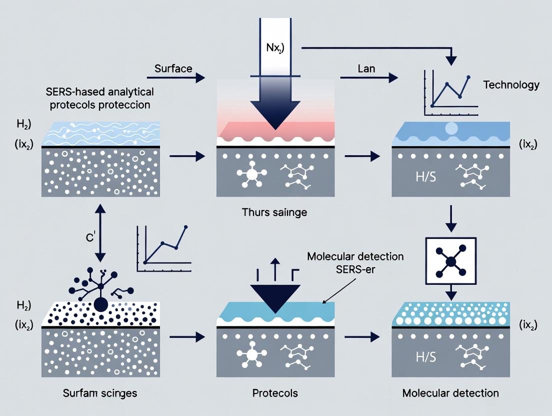
SERS-Based Analytical Protocols: Building Robust and Reproducible Detection Platforms for Biomedical Research
Surface-enhanced Raman scattering (SERS) offers exceptional sensitivity and molecular specificity, but translating this potential into reliable analytical protocols remains a significant challenge.
SERS-Based Analytical Protocols: Building Robust and Reproducible Detection Platforms for Biomedical Research
Abstract
Surface-enhanced Raman scattering (SERS) offers exceptional sensitivity and molecular specificity, but translating this potential into reliable analytical protocols remains a significant challenge. This article provides a comprehensive guide for researchers and drug development professionals, addressing the entire lifecycle of a SERS-based assay. We first explore the fundamental principles governing SERS signal generation and reproducibility. We then detail methodological strategies for substrate design, functionalization, and protocol standardization across diverse applications, from therapeutic drug monitoring to pathogen detection. A dedicated section tackles common troubleshooting and optimization hurdles, including signal variability, matrix effects, and quantification strategies. Finally, we present a framework for the rigorous validation and benchmarking of SERS protocols against established analytical techniques. This guide aims to equip scientists with the knowledge to develop SERS assays that are not only sensitive but also robust, reproducible, and ready for translational research.
The Foundations of Reliable SERS: Understanding Core Principles and Signal Generation Mechanisms
Application Notes
Surface-enhanced Raman scattering (SERS) is a powerful analytical technique that amplifies the inherently weak Raman scattering signal by several orders of magnitude, enabling single-molecule detection. The total SERS enhancement factor (EF) is a product of contributions from two distinct mechanisms: the electromagnetic enhancement mechanism (EM) and the chemical enhancement mechanism (CHEM). Understanding their interplay is critical for designing robust SERS-based analytical protocols.
Electromagnetic Enhancement (EM): This mechanism dominates the total SERS EF, typically contributing factors of 10^4 to 10^11. It arises from the localized surface plasmon resonance (LSPR) of metallic nanostructures (primarily Au, Ag, Cu). When laser light excites the LSPR, it generates intense, localized electromagnetic fields ("hot spots"). Molecules located within these hot spots experience dramatically enhanced incident and scattered electromagnetic fields. The EM mechanism is a long-range effect (effective over ~10 nm) and is largely non-specific to the analyte molecule.
Chemical Enhancement (CHEM): This mechanism provides a smaller, short-range contribution (10^1 to 10^3) that is molecule-specific. It involves charge transfer between the analyte molecule and the metal surface upon photoexcitation. This requires direct chemical interaction (physisorption or chemisorption) and is influenced by the electronic structure of both the molecule and the substrate. CHEM effects can alter the relative intensities of Raman bands, providing additional chemical information.
Quantitative Comparison of Enhancement Mechanisms
Table 1: Characteristics of SERS Enhancement Mechanisms
| Mechanism | Typical Enhancement Factor | Range | Substrate Dependence | Analyte Dependence | Key Physical Origin |
|---|---|---|---|---|---|
| Electromagnetic (EM) | 10^4 – 10^11 | Long-range (~10 nm) | Crucial (nanostructure geometry, material) | Weak (non-specific) | Localized Surface Plasmon Resonance |
| Chemical (CHEM) | 10^1 – 10^3 | Short-range (direct contact) | Moderate (surface chemistry) | Strong (specific adsorption/electronic states) | Charge-Transfer Resonance |
Table 2: Common SERS Substrates and Their Predominant Enhancement Mechanisms
| Substrate Type | Common Materials | Dominant Mechanism | Typical EF Range | Key Application |
|---|---|---|---|---|
| Colloidal Nanoparticles | Citrate-reduced Ag/Au spheres | EM | 10^6 – 10^8 | Solution-phase, bio-sensing |
| Nanostructured Films | Au/Ag films over nanospheres (FON) | EM | 10^7 – 10^9 | Solid-phase, environmental sensing |
| Tip-Enhanced (TERS) | Au/Ag-coated AFM tips | EM + CHEM | >10^9 | Nanoscale spatial resolution |
| Shell-Isolated Nanoparticles | Au core, SiO₂ shell | EM (CHEM minimized) | 10^5 – 10^7 | Inert, non-invasive analysis |
| Functionalized Substrates | Thiol-bound molecules on Au | EM + Strong CHEM | 10^6 – 10^10 | Targeted molecular detection |
Experimental Protocols
Protocol 1: Differentiating EM and CHEM Contributions via Distance-Dependence Study
This protocol isolates the EM contribution by systematically increasing the distance between the analyte and the metal surface.
Materials: See The Scientist's Toolkit below. Procedure:
- Substrate Preparation: Clean a silicon wafer (or glass slide) via sonication in acetone, isopropanol, and deionized water (DI H₂O) for 10 minutes each. Dry under N₂ stream.
- Spacer Layer Deposition: Use atomic layer deposition (ALD) to deposit an ultra-thin, uniform layer of alumina (Al₂O₃) onto the substrate. Perform separate depositions to create substrates with spacer thicknesses of 0.5 nm, 2 nm, 5 nm, and 10 nm.
- Metal Film Deposition: Deposit a 100 nm thick Ag film via electron-beam evaporation onto each spacer-coated substrate.
- Analyte Deposition: Prepare a 1 mM solution of a non-chemisorbing probe molecule (e.g., 1,2-bis(4-pyridyl)ethylene, BPE) in ethanol. Drop-cast 10 µL onto each substrate and allow to dry.
- SERS Measurement: Acquire Raman spectra using a 633 nm laser, 50x objective, 1 mW power, and 10 s integration time. Use a consistent spot size.
- Data Analysis: Plot the intensity of a key Raman band (e.g., BPE ~1200 cm⁻¹) versus spacer thickness. The exponential decay with distance confirms the EM mechanism's short-range nature. The signal at "zero" distance (on a bare Ag film) includes both EM and CHEM contributions.
Diagram Title: SERS Distance-Dependence Experiment Workflow
Protocol 2: Probing Chemical Enhancement via Adsorption Isotherm and Wavelength Dependence
This protocol investigates CHEM by studying adsorption behavior and excitation wavelength dependence.
Materials: See The Scientist's Toolkit below. Procedure:
- Substrate Functionalization: Immerse Au-coated substrates (e.g., Klarite) in a 1 mM ethanolic solution of 4-mercaptobenzoic acid (4-MBA) for 2 hours to form a self-assembled monolayer (SAM). Rinse thoroughly with ethanol and DI H₂O to remove physisorbed molecules. Dry under N₂.
- Adsorption Isotherm: Prepare a series of 4-MBA solutions in ethanol with concentrations from 1 µM to 10 mM. Incubate separate Au substrates in each concentration for 2 hours. After rinsing and drying, acquire SERS spectra using identical parameters (e.g., 785 nm laser, 5 mW, 5 s).
- Wavelength Dependence: Using the SAM substrate from step 1, acquire SERS spectra using multiple laser excitation lines (e.g., 532 nm, 633 nm, 785 nm). Keep laser power and integration time constant per unit area.
- Data Analysis:
- Plot SERS band intensity (e.g., ~1580 cm⁻¹ ring breathing) vs. solution concentration. A Langmuir-like isotherm suggests specific chemisorption.
- Compare the relative intensities of the ~1580 cm⁻¹ and ~1078 cm⁻¹ bands across different laser wavelengths. Significant changes indicate resonant charge-transfer contributions (CHEM), as the EM enhancement profile is relatively broad compared to specific molecule-metal charge-transfer resonances.
Diagram Title: Chemical Enhancement Investigation Pathways
The Scientist's Toolkit
Table 3: Essential Research Reagent Solutions for SERS Mechanism Studies
| Item / Reagent | Function / Purpose | Key Consideration |
|---|---|---|
| Gold/Silver Colloids | Provide tunable plasmonic nanoparticles for EM studies. | Size, shape, and aggregation state control LSPR wavelength. |
| Functional Thiols (e.g., 4-MBA) | Form self-assembled monolayers to study CHEM and create reproducible surfaces. | Purity is critical for monolayer formation. |
| Non-Interacting Spacers (Al₂O₃, SiO₂) | Create controlled nanoscale gaps to isolate EM distance dependence. | Uniformity and thickness control (e.g., via ALD) are essential. |
| Raman Probe Molecules (BPE, R6G, CV) | Standard analytes with known, strong Raman signatures for EF calculation. | Choice depends on laser wavelength to avoid fluorescence. |
| Plasmonic Substrates (Klarite, FON) | Standardized, reproducible SERS-active surfaces for controlled experiments. | Batch-to-batch consistency is vital for quantitative comparisons. |
| Atomic Layer Deposition (ALD) System | Deposits ultra-thin, conformal spacer or shell layers with atomic precision. | Enables precise distance-dependence experiments. |
| Raman Spectrometer with Multiple Lasers | Acquires SERS spectra; multiple wavelengths allow resonance/charge-transfer studies. | Stability, laser line options, and microscope integration are key. |
This application note, framed within a broader thesis on developing robust Surface-Enhanced Raman Spectroscopy (SERS)-based analytical protocols for reliable detection, details the critical substrate parameters governing SERS enhancement. The performance, reproducibility, and quantitative capability of a SERS protocol are fundamentally dictated by the engineered nanostructures that serve as the SERS-active substrate. This document provides detailed protocols and analyses for characterizing nanoparticle morphology and composition, and for engineering electromagnetic "hotspots," with a focus on applications in pharmaceutical and biomedical research.
Core Substrate Parameters and Quantitative Benchmarks
The efficacy of a SERS substrate is quantified by its Enhancement Factor (EF), which depends on the following interrelated parameters.
Table 1: Quantitative Metrics for SERS Substrate Characterization
| Parameter | Target Metric (Typical Range) | Measurement Technique | Impact on SERS Performance |
|---|---|---|---|
| Nanoparticle Size | 20-100 nm (spherical Au/Ag) | TEM, SEM | Determines plasmon resonance frequency; influences scattering cross-section. |
| Shape Uniformity | PDI < 0.1 (DLS); < 5% size deviation (TEM) | TEM, SEM, DLS | Critical for signal reproducibility and uniform analyte adsorption. |
| Interparticle Gap (Hotspot) | 1-5 nm for maximum EF | Cryo-TEM, SEM | Single most critical parameter; EF scales as ~(d/r)^12 for dimer gap d, particle radius r. |
| Composition Purity | > 95% elemental (Au or Ag) | EDX, ICP-MS | Affects plasmonic quality, oxidation resistance, and functionalization chemistry. |
| Aggregate Control | Defined cluster geometry (e.g., dimers, trimers) | TEM, Single-Particle Spectroscopy | Directs and localizes hotspot formation. |
| Surface Chemistry | Monolayer ligand density (~ 5 molecules/nm²) | TGA, NMR | Stabilizes morphology, prevents unspecific binding, enables bio-conjugation. |
| Batch-to-Batch EF Variance | < 15% RSD | SERS mapping with reporter molecule (e.g., 4-MBA) | Ultimate measure of substrate reliability for analytical protocols. |
Detailed Experimental Protocols
Protocol 3.1: Synthesis and Morphological Control of Anisotropic Au Nanostars for Hotspot Engineering
Objective: Synthesize gold nanostars (AuNS) with controlled tip sharpness and length to create multiple, high-intensity hotspots at their tips for superior SERS enhancement. Materials: Hydrogen tetrachloroaurate(III) trihydrate (HAuCl₄·3H₂O), silver nitrate (AgNO₃), L-ascorbic acid (AA), cetyltrimethylammonium bromide (CTAB), 4-mercaptobenzoic acid (4-MBA), ultrapure water (>18 MΩ·cm). Procedure:
- Seed Solution: In a 20 mL vial, mix 5 mL of 0.5 mM HAuCl₄ and 5 mL of 0.2 M CTAB. Gently stir.
- Reduction: Add 600 µL of ice-cold 10 mM NaBH₄. Stir vigorously for 2 min. Solution turns brownish-yellow. Age seeds at 27°C for 30 min before use.
- Growth Solution: In a 50 mL conical tube, combine 10 mL of 0.1 M CTAB, 500 µL of 10 mM HAuCl₄, 150 µL of 10 mM AgNO₃, and 85 µL of 100 mM AA. Gently mix (no vortex).
- Initiation: Add 12 µL of the aged seed solution. Swirl gently for 30 sec and let react undisturbed at 27°C for 1 hour.
- Purification: Centrifuge at 8000 RCF for 10 min. Discard supernatant and re-suspend pellet in 10 mL of 0.1 mM CTAB solution. Repeat once.
- Characterization: Analyze tip morphology via TEM. Measure plasmon resonance via UV-Vis-NIR spectroscopy (expect peaks ~650-900 nm).
Protocol 3.2: Quantitative SERS Enhancement Factor (EF) Calculation
Objective: Quantitatively determine the average EF of a synthesized substrate using a monolayer of 4-MBA as a Raman reporter. Materials: Purified nanoparticle colloid, 10 µM ethanolic 4-MBA solution, silicon wafer with 300 nm oxide layer (for drop-coated reference). Procedure:
- SERS Sample Preparation: Add 1 mL of purified nanoparticle colloid to 10 µL of 10 µM 4-MBA. Incubate for 30 min. Centrifuge and re-suspend in water to remove unbound reporter. Drop-cast 5 µL onto a clean substrate (e.g., glass slide) and air-dry.
- Non-SERS Reference Preparation: Drop-cast 5 µL of 1 mM 4-MBA in ethanol onto a Si/SiO₂ wafer and air-dry to form a monolayer.
- Raman Acquisition: Using a 785 nm laser with identical power and acquisition time:
- Acquire spectra from at least 20 random spots on the SERS sample.
- Acquire 5 spectra from the reference monolayer.
- Calculation: Use the formula: [ EF = (I{SERS} / N{SERS}) / (I{Ref} / N{Ref}) ] where I is the integrated intensity of the 4-MBA peak at ~1580 cm⁻¹, N_SERS is the number of molecules probed in the SERS hotspot volume, and N_Ref is the number of molecules probed in the reference. Estimate N_SERS from TEM-derived hotspot volume and monolayer packing density.
Protocol 3.3: Engineering Dimer Hotspots via DNA Origami
Objective: Assemble nanoparticle dimers with sub-5 nm gap precision using DNA origami scaffolds for uniform, quantifiable hotspot generation. Materials: DNA origami rectangle (e.g., p7308 scaffold), staple strands, thiolated capture strands, 40 nm Au nanoparticles (citrate-stabilized), TAE buffer with 12.5 mM Mg²⁺. Procedure:
- Origami Folding: Mix scaffold strand (10 nM) with 10x excess staple strands, including thiolated capture strands at two specific positions, in folding buffer. Thermally anneal from 80°C to 25°C over 2 hours.
- Purification: Use Amicon centrifugal filters (100 kDa MWCO) to remove excess staples. Confirm folding with agarose gel electrophoresis (2% gel, stained with SYBR Gold).
- Nanoparticle Conjugation: Incubate purified origami (1 nM) with 5 nM of AuNPs (pre-treated with TCEP to activate thiols) in conjugation buffer (TAE with 10 mM Mg²⁺) for 48 hours at 25°C.
- Separation: Use agarose gel electrophoresis (0.8% gel) to separate 1:1 origami-AuNP dimers from free AuNPs and larger aggregates. Extract dimer band.
- Validation: Image dimers using TEM. Perform single-particle SERS mapping to correlate dimer structure with EF.
Visualizing Protocols and Relationships
Diagram 1: SERS Substrate Development & Validation Workflow (83 chars)
Diagram 2: Nanoparticle Dimer & Electromagnetic Hotspot (72 chars)
The Scientist's Toolkit: Essential Research Reagent Solutions
Table 2: Key Reagents and Materials for SERS Substrate Development
| Item | Function in Protocol | Critical Parameters & Notes |
|---|---|---|
| HAuCl₄·3H₂O | Gold precursor for nanoparticle synthesis. | ≥99.9% trace metals basis; store desiccated, in amber vial. |
| CTAB (Cetyltrimethylammonium bromide) | Shape-directing surfactant for anisotropic growth. | Recrystallize from hot ethanol for high purity; critical for reproducibility. |
| DNA Origami Scaffold (e.g., p7308) | Precise template for nanoparticle positioning. | Commercial kits (from Tilibit, GattaQuant) ensure reliable folding. |
| Thiolated DNA/Oligo | Covalent attachment of nanoparticles to substrates or scaffolds. | Use HPLC-purified; treat with TCEP before use to reduce disulfides. |
| 4-Mercaptobenzoic Acid (4-MBA) | Standard Raman reporter for EF calculation and surface probing. | >95% purity; prepare fresh ethanolic solutions to avoid oxidation. |
| Ultra-Pure Water | Solvent for all aqueous syntheses and rinsing. | Resistivity >18 MΩ·cm (Milli-Q or equivalent) to avoid contamination. |
| TAE/Mg²⁺ Buffer | Folding and stability buffer for DNA origami structures. | Mg²⁺ concentration (typically 10-20 mM) is critical for structural integrity. |
| Silicon Wafer (with 300 nm SiO₂) | Ideal flat, reflective substrate for reference Raman measurements. | Low roughness (<1 nm RMS); clean with piranha solution before use (Caution: Highly corrosive). |
Surface-Enhanced Raman Spectroscopy (SERS) offers exceptional sensitivity for chemical and biological detection, making it highly attractive for analytical chemistry and drug development. However, its translation into reliable, standardized protocols is hindered by significant challenges in signal reproducibility. This document, framed within a broader thesis on developing robust SERS-based analytical protocols, details the key sources of variance and provides actionable application notes and protocols to mitigate them, enabling more reliable detection research.
The major contributors to SERS signal variance can be categorized and quantified. The following table summarizes empirical data on their relative impact.
Table 1: Quantified Primary Sources of Variance in SERS Measurements
| Source Category | Specific Factor | Typical Coefficient of Variation (CV) Range | Impact Level (H/M/L) |
|---|---|---|---|
| Substrate Heterogeneity | Nanoparticle Size Distribution | 15-25% | High |
| Hotspot Density & Distribution | 20-40% | High | |
| Batch-to-Batch Consistency | 10-30% | High | |
| Sample Preparation | Analyte Adsorption Uniformity | 10-20% | Medium |
| Aggregation State Control (for colloids) | 20-50% | High | |
| Substrate Cleaning Efficacy | 5-15% | Medium | |
| Instrumental Factors | Laser Power Stability | 1-5% | Low |
| Wavelength & Focus Point Drift | 2-8% | Medium | |
| Spectrometer Calibration | 3-10% | Medium | |
| Environmental & Operational | Measurement Spot Selection | 10-60% | High |
| Operator Technique | 5-25% | Medium | |
| Ambient Contamination | Variable | High |
Detailed Experimental Protocols
Protocol 2.1: Standardized Fabrication and QC of Citrate-Reduced Silver Colloids (AgNPs)
Objective: Produce a consistent batch of SERS-active colloidal nanoparticles with characterized size and aggregation state. Materials: See The Scientist's Toolkit (Section 4). Procedure:
- Preparation: Clean all glassware with aqua regia (3:1 HCl:HNO₃) for 30 minutes, followed by exhaustive rinsing with ultrapure water (18.2 MΩ·cm).
- Synthesis: In a 250 mL Erlenmeyer flask, heat 100 mL of 1 mM AgNO₃ solution to boiling under vigorous stirring.
- Reduction: Rapidly add 2 mL of 1% (w/v) trisodium citrate solution to the boiling AgNO₃.
- Reaction: Maintain boiling with stirring for 1 hour. Allow to cool to room temperature.
- Quality Control (QC):
- UV-Vis Spectroscopy: Measure the localized surface plasmon resonance (LSPR) peak. A primary peak at ~400 nm with FWHM < 80 nm indicates acceptable monodispersity.
- Dynamic Light Scattering (DLS): Determine the hydrodynamic diameter. Aim for a mean diameter of 50-60 nm with a polydispersity index (PDI) < 0.2.
- Baseline SERS Test: Using a standard probe like 1 µM benzenethiol, measure SERS intensity from 10 random 1 µL spots on a silicon wafer. Calculate the CV of the dominant peak (e.g., ~1075 cm⁻¹). Accept batches with CV < 20%.
Protocol 2.2: Controlled Aggregation for Signal Enhancement
Objective: Generate reproducible inter-particle "hotspots" using a controlled aggregating agent. Materials: AgNP colloid (from Protocol 2.1), 1 M MgSO₄ or 1 M KCl, analyte solution, vortex mixer. Procedure:
- Prepare an "analyte-NP" mixture by combining 450 µL of AgNP colloid, 50 µL of the analyte at 10x the desired final concentration, and vortexing for 10 seconds.
- Critical Step: Initiate aggregation by adding 5 µL of 1 M MgSO₄ (final conc. ~10 mM) directly into the solution while vortexing at medium speed.
- Immediately pipette 2 µL of the aggregating mixture onto a clean aluminum or silicon substrate.
- Allow the droplet to dry in a covered, dust-free environment at room temperature for 45 minutes.
- Note: The time between aggregation initiation and droplet deposition must be kept constant (±5 sec) across all replicates.
Protocol 2.3: Mapping Protocol for Substrate Homogeneity Assessment
Objective: Quantitatively map SERS activity across a substrate to evaluate hotspot distribution. Materials: Homogeneous SERS substrate, standard reporter molecule (e.g., 10 nM crystal violet), Raman microscope with motorized stage. Procedure:
- Immerse or spot-coat the substrate uniformly with the standard reporter molecule. Dry under nitrogen.
- Program the microscope stage to perform a raster scan over a defined area (e.g., 100 µm x 100 µm).
- Acquire a spectrum (e.g., 1 sec integration, 785 nm laser, 5 mW) at each point on a predefined grid (e.g., 5 µm step size).
- Extract the intensity of a key Raman band (e.g., 1175 cm⁻¹ for crystal violet) at each pixel.
- Generate a 2D intensity map and calculate the spatial CV across the mapped area. A CV > 25% indicates significant heterogeneity.
Visualization of Workflows and Relationships
Diagram 1: SERS Workflow & Key Variance Sources
Diagram 2: Mitigation Strategy for SERS Variance
The Scientist's Toolkit: Essential Research Reagent Solutions
Table 2: Key Materials and Reagents for Reproducible SERS
| Item Name | Function & Role in Reproducibility | Critical Specification / Note |
|---|---|---|
| High-Purity Metal Salts (AgNO₃, HAuCl₄) | Precursor for nanoparticle synthesis. Impurities affect nucleation/growth. | ≥99.99% trace metals basis. Use dedicated, unopened bottles for synthesis. |
| Chemical Reducers (Trisodium Citrate, NaBH₄) | Control reduction rate, determining NP size and morphology. | Freshly prepared solutions. Weigh accurately for molar ratio consistency. |
| Aggregation Agents (MgSO₄, KCl, HNO₃) | Induce controlled nanoparticle clustering to form hotspots. | Concentration is critical. Prepare stock solutions gravimetrically. |
| Internal Standard (IS) Molecules (e.g., 4-Mercaptobenzoic acid, Deuterated compounds) | Co-adsorb with analyte. IS signal normalizes for hotspot fluctuations. | Must have non-overlapping Raman bands and similar adsorption affinity. |
| Ultra-Pure Water | Solvent for all solutions. Ionic/organic contaminants alter aggregation. | 18.2 MΩ·cm resistivity, total organic carbon (TOC) < 5 ppb. |
| Standard Raman Probes (e.g., Benzenethiol, Crystal Violet) | For substrate quality control and inter-laboratory comparison. | Use analytical grade. Store in dark, anhydrous conditions. |
| Cleaned & Functionalized Substrates (Si wafers, ITO glass, Au films) | Provide a uniform, inert surface for droplet deposition or NP immobilization. | Clean with oxygen plasma or piranha solution immediately before use. (Caution: Hazardous) |
This protocol outlines the essential steps for performing reliable Surface-Enhanced Raman Spectroscopy (SERS) analysis, a cornerstone technique in the broader thesis on developing robust SERS-based analytical methods for detection in complex matrices. SERS amplifies the inherently weak Raman signal by several orders of magnitude through interactions with nanostructured metallic surfaces, enabling sensitive and specific molecular fingerprinting.
Sample Preparation Protocol
Objective: To prepare the analyte for optimal interaction with the SERS-active substrate.
Liquid Sample Preparation
- Materials: Analyte solution, appropriate solvent (e.g., deionized water, ethanol), aggregating agent (e.g., NaCl, MgSO₄, HNO₃).
- Protocol:
- Prepare a stock solution of the target analyte at a known concentration.
- If using colloidal nanoparticles (e.g., citrate-reduced Ag or Au NPs), mix the analyte solution with the colloid at a defined volume ratio (typical range 1:1 to 1:10 v/v).
- Add a controlled volume of an aggregating agent (e.g., 1-10 µL of 1M NaCl to 1 mL of colloid-analyte mix) to induce nanoparticle aggregation and form "hot spots". Vortex briefly.
- Allow the mixture to incubate for a standardized time (2-10 minutes) to ensure adsorption equilibrium.
- Transfer an aliquot (e.g., 20-50 µL) to a well plate or onto a solid substrate for measurement.
Solid/Surface-Based Sample Preparation
- Materials: Solid SERS substrate (e.g., Si wafer with Au nanorods, commercial SERS chips), analyte solution.
- Protocol:
- Clean the solid substrate according to manufacturer or lab protocol (often plasma cleaning or solvent rinse).
- Apply a precise volume (e.g., 2-5 µL) of the analyte solution onto the active area of the substrate.
- Allow the droplet to dry under controlled conditions (ambient temperature, in a desiccator, or under nitrogen flow).
- For quantitative analysis, use a consistent drying method and spot size.
Substrate Selection & Handling
The choice of substrate is critical. The following table summarizes common options.
Table 1: Common SERS Substrates and Their Characteristics
| Substrate Type | Typical Enhancement Factor (EF) Range | Key Advantages | Key Limitations |
|---|---|---|---|
| Citrate-Ag/Au Colloids | 10⁶ - 10⁸ | Easy synthesis, solution-phase mixing, low cost. | Batch-to-batch variability, temporal instability. |
| Lithographically Patterned Chips | 10⁷ - 10⁹ | Excellent reproducibility, spatially uniform signal. | High cost, limited active area. |
| Electrochemically Roughened Electrodes | 10⁵ - 10⁷ | In-situ tunability, good for electrochemical SERS. | Lower EF, complex preparation. |
| Commercial SERS Chips | 10⁶ - 10⁸ | User-friendly, consistent, often functionalized. | Higher per-sample cost, proprietary surfaces. |
Note: Enhancement Factors (EF) are approximate and highly dependent on specific morphology and analyte.
Spectral Acquisition Protocol
Objective: To obtain high-quality, reproducible SERS spectra.
Instrument Setup
- Materials: Raman spectrometer (with microscope), laser source (e.g., 532 nm, 633 nm, 785 nm), calibration standard (e.g., silicon wafer for 520.7 cm⁻¹ peak).
- Protocol:
- Power on the spectrometer and laser. Allow a minimum 30-minute warm-up for stability.
- Calibrate the spectrometer using the designated standard.
- Set acquisition parameters:
- Laser Power: Optimize to avoid photodecomposition. Start low (e.g., 0.1-1 mW at sample) and increase.
- Integration Time: Typically 1-10 seconds per accumulation.
- Number of Accumulations: 5-50 to improve signal-to-noise ratio.
- Grating & Spectral Range: Select appropriate for target analyte fingerprints.
- For mapping, define the spatial step size (e.g., 0.5-2 µm) and number of points.
Data Collection
- Focus the laser beam on the sample. For colloids in a well plate, focus just inside the liquid droplet. For solid substrates, focus on the dried film.
- Acquire a background spectrum from a clean area or blank solution.
- Acquire spectra from the sample. For heterogeneous samples, collect multiple spectra from random points (n ≥ 10) or perform a 2D map.
- Save spectra in a standardized format (e.g., .txt, .spc).
Critical Experimental Workflow
Title: Core SERS Experimental Workflow
Data Pre-processing Steps
Raw SERS spectra require pre-processing before analysis. A typical sequence is shown below.
Title: SERS Data Pre-processing Sequence
The Scientist's Toolkit: Key Research Reagent Solutions
Table 2: Essential Materials for a SERS Protocol
| Item | Function & Rationale |
|---|---|
| Gold or Silver Colloids | The most common SERS-active media. Provide plasmonic enhancement. Citrate-reduced are standard; can be synthesized or purchased. |
| Aggregating Agent (e.g., 1M NaCl) | Induces controlled aggregation of colloidal nanoparticles, creating inter-particle gaps ("hot spots") for extreme signal enhancement. |
| Internal Standard (e.g., 4-Mercaptobenzoic acid, KNO₃) | A compound with a known, sharp Raman band added at constant concentration to normalize signal and correct for instrumental/ preparation variance. |
| Raman Calibration Standard (Silicon Wafer) | Used to calibrate the spectrometer's wavelength axis before measurement, ensuring spectral accuracy and reproducibility. |
| Functionalized SERS Substrates (e.g., aptamer-coated chips) | Substrates with immobilized capture probes (antibodies, aptamers) for selective detection of target analytes from complex mixtures. |
| Quartz Cuvettes/ Well Plates | For holding liquid samples during measurement. Have low intrinsic Raman background. |
Designing and Implementing SERS Protocols: Step-by-Step Strategies for Diverse Applications
Within the broader thesis on SERS-based analytical protocols for reliable detection, systematic development of the sensing platform is paramount. This involves three interdependent pillars: the selection of an optimal plasmonic substrate, its chemical functionalization for target capture, and the design of an effective SERS probe. This application note provides detailed protocols for each stage, aimed at creating robust assays for pharmaceutical and biomedical research.
Substrate Selection: Materials and Characterization
The substrate forms the foundation of the SERS effect. Selection criteria include enhancement factor (EF), reproducibility, cost, and compatibility with the target matrix.
Table 1: Comparison of Common SERS Substrates
| Substrate Type | Typical EF | Relative Cost | Reproducibility (RSD%) | Key Advantage | Primary Use Case |
|---|---|---|---|---|---|
| Colloidal Au Nanoparticles | 10^6 - 10^8 | Low | 10-20% (batch) | Solution-phase, flexible functionalization | Homogeneous assays, in-vitro sensing |
| Colloidal Ag Nanoparticles | 10^8 - 10^10 | Low | 15-25% (batch) | Highest intrinsic enhancement | High sensitivity detection in clean matrices |
| Lithographic Nanoarrays (Au) | 10^7 - 10^9 | Very High | <5% (spot-to-spot) | Exceptional uniformity | Quantitative, multiplexed assays |
| Commercial SERS Chips | 10^6 - 10^8 | Medium-High | <8% (chip-to-chip) | User-friendly, ready-to-use | Diagnostic prototype development |
| In-situ Grown Nanostructures | 10^5 - 10^7 | Low | 20-30% | On-demand fabrication | Custom research applications |
Protocol 2.1: Synthesis and Optimization of Citrate-Reduced Gold Nanoparticles (AuNPs) for SERS
- Objective: To synthesize reproducible 60nm AuNP colloids as a versatile SERS substrate.
- Materials: Hydrogen tetrachloroaurate(III) trihydrate (HAuCl₄·3H₂O), Trisodium citrate dihydrate, ultrapure water (18.2 MΩ·cm).
- Procedure:
- Clean all glassware with aqua regia (3:1 HCl:HNO₃) and rinse thoroughly with ultrapure water.
- Bring 100 mL of 0.25 mM HAuCl₄ solution to a rolling boil in a round-bottom flask under vigorous stirring.
- Rapidly add 2.5 mL of 1% (w/v) trisodium citrate solution.
- Continue heating and stirring until the solution color changes from pale yellow to deep red (~10 mins).
- Reflux for an additional 15 minutes, then cool to room temperature while stirring.
- Characterize by UV-Vis spectroscopy (λ_max ~535 nm for 60 nm) and dynamic light scattering (DLS) for size distribution.
- Critical Notes: Batch-to-batch reproducibility is enhanced by strict control of temperature, stirring speed, and injection rate. Store at 4°C in dark.
Substrate Functionalization for Target Capture
Functionalization anchors capture molecules (e.g., antibodies, aptamers) to the metallic surface, conferring specificity.
Protocol 3.1: Thiol-Based Functionalization of AuNPs with a Mixed Self-Assembled Monolayer (SAM)
- Objective: To create a stable, bio-inert, and reactive surface on AuNPs for subsequent probe attachment.
- Materials: Polyethylene glycol (PEG) thiol (HS-PEG, MW 2000), carboxyl-terminated thiol (HS-C11-EG6-COOH), ethanol, phosphate buffered saline (PBS, 0.01 M, pH 7.4).
- Procedure:
- Concentrate 10 mL of as-synthesized AuNPs (Protocol 2.1) by centrifugation at 8,000 x g for 15 min. Discard supernatant and resuspend in 1 mL PBS.
- Prepare a 1 mM mixed thiol solution in ethanol with a 100:1 molar ratio of HS-PEG:HS-C11-EG6-COOH. The PEG provides antifouling properties; the carboxyl enables covalent coupling.
- Add 100 µL of the mixed thiol solution to 1 mL of concentrated AuNPs under gentle vortexing.
- Incubate overnight at room temperature in the dark.
- Purify functionalized AuNPs by three cycles of centrifugation/resuspension in PBS (pH 7.4).
- Critical Notes: The ratio of functional to passivating thiols controls probe density. Use fresh thiol solutions and degas solvents to prevent oxidation.
SERS Probe Design and Conjugation
The SERS probe consists of a reporter molecule (Raman dye) and a targeting moiety attached to a plasmonic nanoparticle.
Protocol 4.1: Conjugation of a Raman Reporter and Antibody to Functionalized AuNPs
- Objective: To create an immunologically active SERS tag targeting a specific analyte.
- Materials: Functionalized AuNPs (from Protocol 3.1), Raman reporter (e.g., Malachite Green Isothiocyanate, MGITC), N-(3-Dimethylaminopropyl)-N′-ethylcarbodiimide hydrochloride (EDC), N-Hydroxysuccinimide (NHS), anti-target monoclonal antibody (mAb), borate buffer (0.01 M, pH 8.5).
- Procedure:
Part A: Reporter Loading
- Add 10 µL of 1 mM MGITC in DMSO to 1 mL of functionalized AuNPs.
- Incubate for 2 hours at room temperature in the dark with mild agitation.
- Centrifuge to remove excess dye. Resuspend in 1 mL of borate buffer. Part B: Antibody Conjugation via EDC/NHS Chemistry
- Activate the carboxyl groups on the probe surface by adding 50 µL of fresh 40 mM EDC and 50 µL of 100 mM NHS in water. React for 20 mins.
- Purify probes via centrifugation to remove excess EDC/NHS.
- Resuspend activated probes in 1 mL borate buffer.
- Add 20 µg of the target mAb. Incubate for 4 hours at room temperature.
- Quench the reaction by adding 100 µL of 1M ethanolamine (pH 8.5) for 15 mins.
- Block remaining active sites with 1% (w/v) bovine serum albumin (BSA) for 30 mins.
- Purify the final SERS probes by centrifugation (3x) and resuspend in 1 mL PBS with 0.1% BSA. Store at 4°C.
- Critical Notes: Optimize the dye-to-antibody ratio to maximize signal while maintaining immunoreactivity. Avoid over-conjugation, which can cause nanoparticle aggregation.
Integrated Workflow for a SERS Assay
The following diagram illustrates the logical and experimental pathway from substrate to result.
Diagram 1: SERS Assay Development and Execution Workflow
The Scientist's Toolkit: Key Research Reagent Solutions
Table 2: Essential Materials for SERS Protocol Development
| Item | Function & Role in Protocol | Example Product/Chemical |
|---|---|---|
| Chloroauric Acid (HAuCl₄) | Gold precursor for synthesizing plasmonic AuNP substrates. | Hydrogen tetrachloroaurate(III) trihydrate |
| Citrate Stabilizer | Reducing agent and colloidal stabilizer for AuNP synthesis. | Trisodium citrate dihydrate |
| Functional Thiols | Form self-assembled monolayers (SAMs) on Au for surface chemistry. | HS-PEG-OH, HS-C11-EG6-COOH |
| Raman Reporter Dye | Provides the intense, characteristic SERS signal for detection. | Malachite Green Isothiocyanate (MGITC), Cyanine dyes |
| Crosslinker (EDC/NHS) | Activates carboxyl groups for covalent coupling of proteins/ligands. | N-(3-Dimethylaminopropyl)-N′-ethylcarbodiimide hydrochloride |
| Capture Bioreceptor | Confers specificity by binding the target analyte. | Monoclonal antibodies, DNA aptamers |
| Blocking Agent | Reduces non-specific adsorption on sensor surface. | Bovine Serum Albumin (BSA), casein |
| Wash Buffer | Removes unbound reagents while maintaining assay integrity. | Phosphate Buffered Saline (PBS) with Tween-20 (PBST) |
Standardizing Sample Preparation and Handling for Complex Matrices (Serum, Cell Lysate, etc.)
Within the thesis framework for developing robust Surface-Enhanced Raman Spectroscopy (SERS) analytical protocols, standardized sample preparation is the critical first step. Variability in handling complex biological matrices directly compromises the reproducibility, sensitivity, and quantitative accuracy of SERS detection. This document details standardized protocols and application notes for serum and cell lysates to ensure reliable analyte detection for research and drug development.
Table 1: Effect of Sample Preparation Variables on SERS Signal Reproducibility (Relative Standard Deviation, % RSD)
| Preparation Variable | Non-Standardized Protocol (RSD%) | Standardized Protocol (RSD%) | Key Observation |
|---|---|---|---|
| Serum Deproteinization | 25-40% | 8-12% | Acid/Organic solvent type & incubation time critical. |
| Cell Lysis (Duration) | 30-35% | 10-15% | Sonication pulse consistency reduces biomolecular degradation. |
| Analyte Extraction Efficiency | 60-75% | 85-95% | Use of internal standard (e.g., deuterated analog) is essential. |
| Matrix Effect on Nanoparticle Aggregation | High (Subjective) | Low (Quantified) | Controlled salt/buffer addition stabilizes SERS substrate. |
Table 2: Recommended Benchmarks for SERS-Ready Sample Quality
| Matrix | Acceptable Protein Content | Acceptable Salt Concentration | Optimal pH for Nanoparticle Mixing | Maximum Viscosity (cP) |
|---|---|---|---|---|
| Human Serum | < 0.5 mg/mL | 10-150 mM | 7.2 - 7.6 | < 1.5 |
| Cell Lysate (Mammalian) | 1-2 mg/mL | 50-200 mM | 7.0 - 7.4 | < 2.0 |
Detailed Experimental Protocols
Protocol 3.1: Standardized Deproteinization of Serum for Small Molecule SERS Analysis
Objective: To reproducibly remove interfering proteins and lipids from serum prior to SERS detection of low molecular weight analytes (e.g., drugs, metabolites).
Materials:
- Fresh or properly thawed serum sample (100 µL).
- Internal Standard Solution (e.g., 10 µM deuterated analyte in methanol).
- Cold Acetonitrile (ACN, LC-MS grade).
- 0.1% Formic Acid in water.
- Centrifugal filters (10 kDa MWCO).
- Vacuum concentrator.
- Reconstitution Buffer (10 mM phosphate, pH 7.4).
Procedure:
- Spike & Denature: To 100 µL of serum, add 10 µL of Internal Standard Solution. Vortex for 10 sec.
- Protein Precipitation: Add 300 µL of cold ACN dropwise while vortexing. Vortex vigorously for 1 min.
- Incubate & Pellet: Incubate at -20°C for 20 min. Centrifuge at 14,000 x g for 15 min at 4°C.
- Clarify: Transfer the supernatant to a 10 kDa MWCO centrifugal filter. Centrifuge at 12,000 x g for 20 min at 4°C.
- Dry & Reconstitute: Transfer the filtrate to a clean tube. Dry under vacuum at 30°C for 45-60 min. Reconstitute the pellet in 50 µL of Reconstitution Buffer by vortexing for 2 min and brief sonication (30 sec).
- SERS Mixing: Combine 10 µL of processed sample with 90 µL of citrate-reduced gold nanoparticle colloid. Mix by gentle pipetting. Incubate for 5 min at room temperature in the dark before SERS measurement.
Protocol 3.2: Standardized Preparation of SERS-Ready Cell Lysate
Objective: To generate a homogeneous, nanoparticle-compatible lysate for intracellular SERS mapping or metabolite detection.
Materials:
- Cell pellet (1 x 10^6 cells).
- Ice-cold Lysis Buffer (e.g., 25 mM Tris, 150 mM NaCl, 1% NP-40, pH 7.4, supplemented with protease/phosphatase inhibitors).
- Nuclease (e.g., Benzonase).
- Probe sonicator with microtip.
- Bradford or BCA protein assay kit.
Procedure:
- Wash: Resuspend cell pellet in 1 mL of ice-cold PBS. Centrifuge at 500 x g for 5 min at 4°C. Discard supernatant.
- Lysate: Resuspend the washed pellet in 100 µL of Ice-cold Lysis Buffer. Incubate on ice for 15 min.
- Nuclease Treatment: Add 2 µL of nuclease (250 U/µL). Invert to mix. Incubate on ice for 10 min.
- Standardized Sonication: Using a probe sonicator on ice, apply three pulses of 5 sec each at 20% amplitude, with a 30 sec rest on ice between pulses. This minimizes heat-induced degradation.
- Clarify: Centrifuge the lysate at 12,000 x g for 15 min at 4°C.
- Quantify & Normalize: Transfer the clear supernatant to a new tube. Determine protein concentration using a BCA assay. Dilute all samples to a standardized concentration of 1.0 mg/mL using Lysis Buffer.
- SERS Preparation: For direct mixing, dilute 10 µL of normalized lysate with 80 µL of 10 mM phosphate buffer (pH 7.2). Add 10 µL of aggregated nanoparticle solution. For centrifugal cleaning, process through a 3 kDa MWCO filter (2 min, 10,000 x g) before mixing with nanoparticles.
Diagrams: Workflows and Relationships
SERS Sample Preparation Workflow
Matrix Effect vs. Standardization on SERS Outcome
The Scientist's Toolkit: Key Research Reagent Solutions
Table 3: Essential Materials for Standardized SERS Sample Preparation
| Item | Function & Rationale | Example Product/Chemical |
|---|---|---|
| Internal Standard (IS) | Corrects for losses during preparation; enables quantification. Isotopically labeled version of the target analyte is ideal. | Deuterated or 13C-labeled drug standard. |
| Protein Precipitation Solvent | Removes bulk proteins to reduce viscosity and nonspecific binding to nanoparticles. | Cold Acetonitrile (ACN) or Methanol. |
| Centrifugal Filters (MWCO) | Rapid buffer exchange, desalting, and removal of residual large biomolecules. | 3kDa or 10kDa Molecular Weight Cut-Off (MWCO) filters. |
| SERS-Compatible Buffer | Maintains nanoparticle stability and analyte integrity during mixing. Low chloride content. | 10 mM Phosphate or HEPES buffer, pH 7.2-7.4. |
| Aggregating/Stabilizing Agent | Consistently induces the nanoparticle plasmonic coupling required for strong SERS enhancement. | Controlled concentrations of NaCl, MgSO4, or poly-L-lysine. |
| Protease/Phosphatase Inhibitors | Preserves the native state of phosphoproteins or protein targets in cell lysates. | Commercial cocktail tablets in lysis buffer. |
| Standardized Nanoparticle Colloid | The core SERS substrate. Batch-to-batch consistency is paramount. | Citrate-reduced gold nanospheres (60nm), OD~1. |
Within the broader thesis on establishing robust Surface-Enhanced Raman Spectroscopy (SERS)-based analytical protocols for reliable detection in pharmaceutical and clinical research, consistent spectral collection is paramount. The inherent sensitivity of SERS to instrumental parameters and environmental conditions necessitates rigorous standardization of data acquisition practices. This document outlines best practices and detailed protocols to ensure reproducibility and reliability in SERS spectral data, aimed at supporting method validation in drug development and diagnostic research.
Foundational Principles of SERS Instrumentation Calibration
Reliable SERS detection begins with a fully characterized and calibrated instrument. Daily verification of key performance parameters is essential.
Wavelength Calibration Protocol
Objective: To ensure the accuracy of the reported Raman shift (cm⁻¹) axis. Materials: Neon or argon calibration lamp, NIST-traceable polystyrene film. Procedure:
- Install the calibration lamp in the sample position.
- Acquire an emission spectrum with the laser off, using the same grating and center wavelength used for SERS experiments.
- Match the observed emission peaks to their known wavelengths (e.g., Ne: 540.06 nm, 585.25 nm).
- Apply a pixel-to-wavelength correction within the instrument software.
- Validate calibration using a standard polystyrene film. Acquire its Raman spectrum and verify that key peaks (e.g., 1001.4 cm⁻¹, 1602.4 cm⁻¹) are within ±1 cm⁻¹ of their certified positions. Frequency: Daily, or at the start of each experimental session.
Intensity Calibration (Radiometric Response)
Objective: To correct for the wavelength-dependent efficiency of the spectrometer and detector. Materials: NIST-traceable tungsten-halogen irradiance standard lamp. Procedure:
- Warm up the standard lamp as per manufacturer instructions (typically 15-30 minutes).
- Place the lamp at the prescribed distance from the spectrometer entrance.
- Acquire a spectrum of the lamp's output.
- Divide the certified spectral output of the lamp (in µW/cm²/nm/sr) by the acquired raw counts to generate a correction array.
- Apply this correction array to all subsequent Raman/SERS spectra. Frequency: Quarterly, or after any major optical realignment.
Laser Power Stability and Focus
Objective: To ensure consistent excitation power and optimal spatial alignment. Protocol:
- Measure laser power at the sample plane using a calibrated photodiode power meter before and after a series of measurements.
- Record fluctuations; accept variations of <2% over a 4-hour period.
- Using a standard Si wafer or a SERS substrate with a uniform coating (e.g., evaporated Au), optimize the focus by maximizing the signal from a known peak while minimizing the laser power. The goal is to find the focus that gives the highest signal-to-noise ratio, not merely the highest intensity.
Systematic Data Acquisition Workflow for SERS
The following workflow must be followed for every sample batch to ensure internal consistency and allow for meaningful comparison.
Diagram Title: Systematic SERS Data Acquisition Workflow
Key Experimental Protocols for Method Validation
Protocol: Establishing a SERS Analytical Calibration Curve
Objective: To quantify an analyte (e.g., an antibiotic or a metabolite) using SERS. Reagents: See "The Scientist's Toolkit" below. Procedure:
- Substrate Preparation: Activate commercially purchased or fabricated Au nanoparticle-coated slides in a UV-ozone cleaner for 15 minutes.
- Standard Preparation: Prepare a dilution series of the target analyte in a suitable buffer (e.g., 10 mM phosphate, pH 7.4) covering 3 orders of magnitude (e.g., 1 µM to 1 nM). Include a blank (buffer only).
- Spotting and Binding: Apply 2 µL of each standard solution to distinct, marked locations on the SERS substrate. Allow to incubate in a humidity chamber (80% RH) for 30 minutes to prevent evaporation artifacts.
- Rinsing and Drying: Gently rinse each spot with 50 µL of deionized water to remove unbound analyte and salts. Dry under a gentle stream of nitrogen.
- Spectral Acquisition: Using a 785 nm laser, 10 mW power, 10-second integration time, acquire 20 spectra from random points within each spotted area.
- Data Processing: For each concentration, average the 20 spectra. Subtract the system blank spectrum. Vector-normalize the averaged spectra. Integrate the area under the characteristic analyte peak.
- Curve Fitting: Plot integrated peak area vs. logarithmic analyte concentration. Fit with a logistic or linear model as appropriate.
Protocol: Intra- and Inter-day Reproducibility Assessment
Objective: To validate the precision of the SERS method. Procedure:
- Prepare three separate batches of QC samples (low, mid, high concentration) on three different days.
- Each day, prepare fresh substrates and solutions from the same stock.
- For each batch, acquire 50 spectra from across the substrate.
- Calculate the Relative Standard Deviation (RSD) of the characteristic peak intensity within a single substrate (intra-assay) and between the three different days (inter-assay).
- Acceptance Criterion: For bioanalytical applications, inter-day RSD should typically be <15-20%.
Table 1: Typical SERS Instrument Calibration Parameters and Tolerances
| Parameter | Calibration Standard | Target Value | Acceptable Tolerance | Validation Frequency |
|---|---|---|---|---|
| Wavelength Accuracy | Polystyrene (1001.4 cm⁻¹) | 1001.4 cm⁻¹ | ± 1.0 cm⁻¹ | Daily |
| Laser Power Stability | Calibrated Photodiode | As set (e.g., 10 mW) | ± 2% over 4 hrs | Pre-/Post-run |
| Spectrometer Response | NIST Irradiance Lamp | Manufacturer Cert. | Curve Applied | Quarterly |
| System Noise (RMS) | Dark Spectrum | -- | < 5 counts | Daily |
Table 2: Example QC Metrics for a 1 mM BPE (Bis-pyridyl ethylene) Standard
| QC Peak (cm⁻¹) | Expected Intensity (a.u.) | Intra-batch RSD (n=50) | Inter-day RSD (n=3 days) | Action Limit (Deviation) |
|---|---|---|---|---|
| 1200 | 15,000 ± 1500 | ≤ 8% | ≤ 12% | ± 5% Intensity |
| 1600 | 22,000 ± 2200 | ≤ 7% | ≤ 15% | ± 5% Intensity |
The Scientist's Toolkit: Essential Research Reagent Solutions
Table 3: Key Materials for Reliable SERS-Based Detection Protocols
| Item | Function & Rationale | Example Product/Specification |
|---|---|---|
| NIST-Traceable Polystyrene Film | Provides absolute Raman shift calibration for instrument validation. Peaks are certified. | e.g., National Institute of Standards and Technology SRM 2241 |
| Irradiance Standard Lamp | Enables intensity/radiometric calibration of the full optical path. | e.g., Tungsten-Halogen lamp, 300-1100 nm range, calibrated traceable to NIST |
| Reference SERS Substrate | A stable, homogeneous plasmonic substrate for system performance monitoring. | e.g., Commercial Au nanoparticle arrays on Si, with specified enhancement factor (EF) |
| Internal Standard Molecule | Co-spiked with analyte to correct for point-to-point signal variation on substrate. | e.g., Deuterated compounds (d8-thiophenol), or isotopically labeled target analyte |
| Quality Control (QC) Analyte | A stable molecule with strong, known SERS peaks to monitor day-to-day system performance. | e.g., 1,2-Bis(4-pyridyl)ethylene (BPE) at 1 mM in ethanol |
| UV-Ozone Cleaner | For consistent, chemical-free cleaning and activation of metal SERS substrates prior to use. | -- |
| Humidity Chamber | Controls evaporation rate during sample incubation, critical for reproducible analyte deposition. | Custom or commercial, maintaining 75-85% RH |
Advanced Considerations: Signal Correction Pathway
For high-precision quantitative SERS, a multi-stage correction must be applied to raw spectral data.
Diagram Title: SERS Spectral Data Pre-processing Pathway
Adherence to these instrumentation and data acquisition best practices forms the bedrock of any thesis aiming to develop reliable SERS-based analytical protocols. Consistent calibration, a rigorous acquisition workflow, and standardized experimental protocols are non-negotiable for producing spectral data that is comparable across instruments, laboratories, and time. This disciplined approach directly addresses major challenges in reproducibility, ultimately accelerating the translation of SERS detection from research into validated applications in drug development and clinical diagnostics.
Troubleshooting SERS Assays: Solving Common Problems and Optimizing for Sensitivity & Reproducibility
Diagnosing and Mitigating Signal Inconsistency and Background Noise
Within Surface-Enhanced Raman Spectroscopy (SERS)-based analytical protocols for reliable detection, signal inconsistency and background noise are primary obstacles to quantitative reproducibility and low-concentration analyte detection. This application note details diagnostic procedures and mitigation strategies to enhance the robustness of SERS assays in pharmaceutical and biomedical research.
Table 1: Primary Sources of SERS Signal Inconsistency and Reported Impact
| Source of Variability | Typical Coefficient of Variation (CV%) | Primary Impact on Signal |
|---|---|---|
| Nanoparticle Aggregation State | 25-60% | Peak intensity & wavelength |
| Laser Power Fluctuation | 5-15% | Overall signal intensity |
| Substrate "Hot Spot" Heterogeneity | 30-70% | Spatial intensity mapping |
| Analyte Adsorption Kinetics | 20-50% | Temporal signal drift |
| Fluorescence Background | N/A (Signal-to-Background Ratio) | Obscures Raman peaks |
Table 2: Mitigation Strategies and Efficacy
| Strategy | Target Variability | Typical CV Reduction Achieved |
|---|---|---|
| Internal Standardization (e.g., deuterated compounds) | Aggregation, laser power | 25% to <10% |
| Shell-Isolated Nanoparticle Enhancement (SHINERS) | Substrate heterogeneity, contamination | 40% to ~15% |
| Spectral Baseline Correction Algorithms | Fluorescence background | Improves SBR by 5-10x |
| Microfluidic Sample Delivery | Analyte adsorption, aggregation | 30% to ~12% |
| Controlled Laser Power Monitoring | Laser fluctuation | 15% to <5% |
Experimental Protocols
Protocol 1: Diagnosing Signal Inconsistency with Internal Standard Calibration
Objective: To distinguish between signal variation due to nanoparticle enhancement field fluctuations and analyte concentration changes. Materials: SERS substrate (e.g., citrate-reduced Au nanoparticles), target analyte (e.g., drug candidate), internal standard (e.g., 4-mercaptobenzonitrile, 1 mM in ethanol). Procedure:
- Prepare a mixture of the target analyte and internal standard at a fixed molar ratio (e.g., 1:1).
- Functionalize the SERS substrate with 100 µL of the mixture for 1 hour.
- Rinse gently with deionized water and air-dry under nitrogen.
- Acquire SERS spectra from 10 random spots on the substrate using a 785 nm laser, 1s integration time, 5 mW power.
- Calculate the peak intensity ratio (Analyte Peak / IS Peak) for each spot. Diagnosis: A high CV (>20%) in the raw analyte peak intensity with a low CV (<10%) in the intensity ratio indicates variability is largely from enhancement factor inhomogeneity, not analyte distribution.
Protocol 2: Mitigating Fluorescence Background via Photobleaching and Wavelength Optimization
Objective: To suppress fluorescent background that obscures Raman signals. Materials: SERS substrate, fluorescent sample (e.g., cell lysate containing drug). Procedure:
- Place the sample on the substrate and mount on the Raman stage.
- Pre-bleaching Step: Expose the measurement area to the full laser power (e.g., 50 mW at 785 nm) for 30-60 seconds.
- Reduce laser power to analytical level (e.g., 5 mW).
- Acquire spectrum immediately after bleaching.
- Alternative Wavelength Screening: Repeat acquisition on a separate sample spot using lasers at 638 nm and 830 nm, comparing the signal-to-background ratio. Analysis: Fluorescence typically decays faster than Raman signal under laser exposure. The optimal wavelength is often longer (830 nm) for highly fluorescent biological samples.
Protocol 3: Protocol for Consistent Substrate Fabrication via Microfluidic Mixing
Objective: To produce homogeneous nanoparticle aggregates for reproducible SERS signals. Materials: Gold nanoparticle colloid (60 nm), aggregating agent (e.g., NaCl, 10 mM), syringe pump, Y-shaped microfluidic chip. Procedure:
- Load one syringe with Au colloid and another with NaCl solution.
- Connect both to inlets of a Y-shaped microfluidic chip using PTFE tubing.
- Using a dual-syringe pump, set equal flow rates (e.g., 10 µL/min) for both streams to achieve 1:1 mixing.
- Collect the mixed, aggregated colloid from the outlet directly onto a clean slide or into a measurement cuvette.
- Immediately introduce analyte and acquire spectra. Result: Continuous flow mixing provides temporally consistent aggregant-to-nanoparticle ratios, reducing CV compared to bulk vortex mixing.
Visualizations
Diagnosis and Mitigation Workflow for SERS
Noise Sources and Mitigation Pathways
The Scientist's Toolkit: Essential Research Reagent Solutions
Table 3: Key Reagents and Materials for Reliable SERS Protocols
| Item | Function & Rationale |
|---|---|
| 4-Mercaptobenzonitrile (4-MBN) | An ideal internal standard. Its nitrile peak (~2230 cm⁻¹) appears in a quiet region of the Raman spectrum, avoiding overlap with most analytes. |
| Shell-Isolated Nanoparticles (SHINs) | Gold nanoparticles coated with an ultra-thin, inert silica or alumina shell (2-4 nm). They provide uniform enhancement while preventing direct analyte-substrate chemical interaction and reducing heterogeneity. |
| Hydroxylamine-reduced Gold Nanoparticles | Provides more monodisperse and reproducible colloidal substrates compared to citrate reduction, leading to lower batch-to-batch variability. |
| Deuterated Solvents (e.g., D₂O) | Used for preparing analyte solutions to shift or eliminate solvent O-H stretching bands that can overlap with analyte signals. |
| Raman-Calibrated Bandpass Filters | Optical filters placed before the detector to rigorously exclude elastically scattered laser light (Rayleigh scatter), a major source of background. |
| Microfluidic Mixing Chips | For controlled, reproducible mixing of analytes with colloidal nanoparticles, ensuring consistent aggregation state across experiments. |
| Silanized Glass Slides or Wafers | Provide a hydrophobic and uniform surface for depositing nanoparticle colloids, promoting even coffee-ring-free drying and sample distribution. |
Within the development of robust Surface-Enhanced Raman Spectroscopy (SERS) analytical protocols for reliable detection, matrix interference and non-specific binding (NSB) represent the foremost barriers to clinical and research translation. Biological samples (serum, plasma, urine, tissue homogenates) contain a complex milieu of proteins, lipids, salts, and metabolites that can foul plasmonic nanostructures, quench signal, and generate spurious background. This Application Note details validated protocols to overcome these challenges, ensuring specificity, sensitivity, and quantitative accuracy in SERS-based assays.
Key Interference Mechanisms and Mitigation Strategies
Table 1: Common Interferents in Biological Matrices and Their Impact on SERS
| Interferent Class | Primary Impact on SERS | Consequence |
|---|---|---|
| Albumin & High-Abundance Proteins | Non-specific adsorption to nanoparticles; formation of a protein corona; physical blocking of hot spots. | Reduced enhancement factor; signal suppression; poor reproducibility. |
| Lipids & Lipoproteins | Adsorption on hydrophobic surfaces; aggregation of nanoparticles; increased light scattering. | Background fluorescence; altered plasmon resonance; nanoparticle instability. |
| Salts & Ionic Species | Induced aggregation of citrate-reduced nanoparticles; destabilization of colloids. | Inconsistent SERS enhancement; precipitation. |
| Endogenous Fluorophores | Generation of broad-band fluorescence. | Swamping of the sharper Raman signal. |
| Non-Target Biomolecules | Competitive binding to capture ligands (e.g., antibodies, aptamers). | Reduced assay sensitivity; false positives/negatives. |
Core Experimental Protocols
Protocol 1: Synthesis and Functionalization of Zwitterion-Coated SERS Nanotags for NSB Reduction
Objective: To create stable, low-fouling SERS nanotags for direct detection in complex media. Materials: Gold nanorods (AuNRs, 50nm x 15nm), Raman reporter (DTTC iodide), Zwitterionic sulfobetaine silane (SB), ethanol, phosphate buffer saline (PBS). Procedure:
- Reporter Labeling: Concentrate AuNRs to 1 nM. Add DTTC from a 1 mM stock to a final concentration of 10 µM. Incubate in the dark for 30 min.
- Zwitterionic Capping: Add SB silane to the mixture at a 1 mM final concentration. Adjust pH to ~8.0. React for 4 hours at 40°C with gentle stirring.
- Purification: Centrifuge at 8000 x g for 10 min. Decant supernatant and resuspend pellet in 1X PBS. Repeat 3x.
- Validation: Incubate tags in 50% fetal bovine serum (FBS) for 2 hours. Centrifuge and measure SERS intensity of the pellet vs. a control. >90% signal retention indicates effective anti-fouling.
Protocol 2: Magnetic Solid-Phase Extraction (MSPE) for Matrix Cleanup Pre-SERS
Objective: To isolate and preconcentrate analytes from bulk biological matrix. Materials: Carboxyl-functionalized magnetic beads (100 nm), EDC/NHS coupling reagents, target-specific capture probe (antibody/aptamer), washing buffers (PBS with 0.1% Tween-20, 0.5M NaCl), elution buffer (low-pH glycine or formamide). Procedure:
- Bead Functionalization: Activate 1 mg of beads with 200 µL of 50 mg/mL EDC and 50 mg/mL NHS in MES buffer for 30 min. Wash.
- Probe Immobilization: Incubate activated beads with 10 µg of capture probe for 2 hours. Block with 1% BSA for 1 hour.
- Sample Processing: Mix 100 µL of processed sample (e.g., diluted serum) with 20 µL of probe-bead suspension. Incubate 30 min.
- Washing: Use a magnetic rack. Wash twice with 0.1% PBST and once with 0.5M NaCl buffer.
- SERS Detection: Resuspend bead-analyte complex in 20 µL of buffer. Mix with 5 µL of reporter nanotags (from Protocol 1) or add directly to a SERS substrate for measurement.
The Scientist's Toolkit: Essential Research Reagent Solutions
Table 2: Key Reagents for Mitigating Interference in SERS Assays
| Reagent / Material | Function & Role in Overcoming Interference |
|---|---|
| Zwitterionic Polymers (e.g., SB, CB) | Form a hydration layer on nanoparticles, dramatically reducing non-specific protein adsorption (fouling). |
| PEGylated Thiols (e.g., mPEG-SH) | Common passivating agent to block vacant sites on gold surfaces, minimizing NSB. |
| Carboxylated Magnetic Beads | Enable MSPE for physical separation of analyte from interfering matrix components. |
| Blocking Agents (Casein, BSA, SuperBlock) | Saturate non-specific binding sites on substrates and nanoparticles before assay. |
| Chaotropic Salt Washes (e.g., NaCl, MgCl2) | High-ionic strength buffers disrupt weak, non-specific interactions during washing steps. |
| Surface Regenerable SERS Substrates (e.g., Si/Au) | Allow for harsh cleaning (piranha) between experiments to remove fouling layers. |
| Internal Standard (IS) Nanotags (e.g., 4-MBA on AuNPs) | Co-delivered with target-specific tags; IS signal normalizes for instrumental and matrix variance. |
Data Presentation: Quantitative Performance Metrics
Table 3: Performance Comparison of SERS Protocols with and without Interference Mitigation
| Assay Format | Matrix | Limit of Detection (LOD) | Signal-to-Background Ratio | %CV (Reproducibility) |
|---|---|---|---|---|
| Bare AuNPs, Direct Mixing | 10% Serum | 100 nM | 2.5 | 25% |
| PEGylated AuNRs, Direct Mixing | 10% Serum | 50 nM | 5.8 | 18% |
| Zwitterionic AuNTags (Protocol 1) | 50% Serum | 10 nM | 12.3 | 9% |
| MSPE + Bare SERS Tags | Whole Serum | 5 nM | 8.7 | 15% |
| MSPE + Zwitterionic Tags | Whole Serum | 1 nM | 25.1 | 7% |
Visualization of Workflows and Mechanisms
Title: Integrated MSPE-SERS Assay Workflow
Title: Mechanism of Specific vs. Non-Specific Binding
Within the development of robust SERS-based analytical protocols for reliable detection, the optimization of instrumental and chemical parameters is critical. This application note details the systematic tuning of three interdependent levers: laser power, spectral integration time, and metallic nanoparticle (substrate) concentration. Proper balancing of these factors is essential to maximize the signal-to-noise ratio (SNR), enhance sensitivity, and ensure reproducibility in applications ranging from pharmaceutical contaminant screening to biomolecular sensing.
Quantitative Parameter Interplay & Optimization Data
The following table summarizes the effects and recommended optimization ranges for key parameters, based on current literature and standard protocols in SERS-based drug development research.
Table 1: Optimization Parameters for SERS-Based Detection Protocols
| Parameter | Typical Optimization Range | Primary Effect on SERS Signal | Risk of Excessive Increase | Recommended Starting Point for Assay Development |
|---|---|---|---|---|
| Laser Power (mW) | 0.1 - 10 mW (at sample) | Linear increase in signal intensity. | Photothermal damage, sample degradation, substrate melting, increased background. | 1.0 mW (633 nm laser) |
| Integration Time (s) | 0.1 - 10 s | Linear increase in total collected photons. | Increased detector noise, saturation, prolonged data acquisition, potential for sample drift. | 1 s |
| Nanoparticle (Au/Ag) Concentration | 0.05 - 1.0 nM (for ~60 nm spheres) | Increases number of "hot spots"; signal increases to a plateau. | Excessive scattering/absorption, increased aggregation leading to irreproducibility, high cost. | 0.2 nM (colloidal gold) |
| Resulting Key Metric: Signal-to-Noise Ratio (SNR) | Maximize | SNR ∝ (Laser Power × Integration Time) / (Background + System Noise) | Diminishing returns, potential signal loss. | Target SNR > 10 for LOD determination |
Detailed Experimental Protocols
Protocol 3.1: Systematic Calibration Using a Raman Reporter
Objective: To establish the optimal combination of laser power and integration time for a given SERS substrate and instrument.
- Reagent Preparation:
- Prepare a 1 µM solution of a stable Raman reporter (e.g., 4-aminothiophenol, 4-ATP) in ethanol.
- Mix 10 µL of 4-ATP solution with 990 µL of citrate-stabilized 60 nm gold nanoparticle colloid (0.2 nM). Incubate for 15 minutes for thiol-gold binding.
- Substrate Deposition:
- Pipette 5 µL of the functionalized colloid onto a clean aluminum-coated slide or silicon wafer. Allow to air dry under ambient conditions.
- Instrumental Parameter Sweep:
- Using a Raman microscope with a 633 nm laser, focus on a homogeneous spot of the dried substrate.
- Fix integration time at 1 s. Collect spectra at laser powers of 0.1, 0.5, 1.0, 2.5, 5.0, and 7.5 mW (measured at sample).
- Fix laser power at the level yielding the highest SNR from the previous step. Collect spectra at integration times of 0.1, 0.5, 1, 2, 5, and 10 s.
- Data Analysis:
- For each spectrum, plot the intensity of the characteristic 4-ATP peak (~1078 cm⁻¹) against the baseline noise (standard deviation of signal from 1200-1250 cm⁻¹).
- Calculate SNR. The combination yielding the highest SNR without spectral broadening or baseline distortion is optimal.
Protocol 3.2: Optimizing Substrate Concentration for Analytic Detection
Objective: To determine the minimal effective nanoparticle concentration for reliable detection of a target analyte in solution-phase assay format.
- Serial Dilution of Substrate:
- Prepare a stock solution of citrate-reduced silver nanoparticles (AgNPs, ~50 nm). Determine approximate concentration via UV-Vis absorbance (λmax ~400 nm).
- Perform serial dilutions in purified water to create concentrations of 1.0, 0.5, 0.25, 0.1, 0.05, and 0.01 nM.
- Assay Assembly & Measurement:
- Prepare a constant, low-concentration solution of the target analyte (e.g., 10 µM antibiotic).
- In a microtube, combine 50 µL of analyte solution with 50 µL of each AgNP concentration. Include a negative control (water + AgNPs).
- Incubate for 5 minutes to allow for analyte adsorption to the NP surface.
- Load each mixture into a quartz capillary cell or a well plate suitable for Raman measurement.
- Using the optimized laser power and integration time from Protocol 3.1, acquire 10 spectra from random positions for each sample.
- Analysis:
- Calculate the mean intensity of the primary analyte Raman peak and its relative standard deviation (RSD%) for each AgNP concentration.
- Plot mean intensity and RSD% versus AgNP concentration. The optimal concentration is at the beginning of the signal plateau region where the RSD is minimized (<15%), ensuring high signal and reproducibility.
Visualization of Optimization Workflow & Relationships
Diagram 1: SERS Protocol Optimization Decision Workflow
Diagram 2: Parameter Effects on Final SERS Performance
The Scientist's Toolkit: Essential Research Reagent Solutions
Table 2: Key Reagents and Materials for SERS Protocol Optimization
| Item | Typical Specification/Example | Function in Optimization |
|---|---|---|
| Metallic Nanoparticles | Citrate-stabilized gold nanospheres, 60 nm diameter, OD~1. | The foundational SERS substrate. Concentration is a primary optimization lever. Size and shape define plasmonic properties. |
| Raman Reporter Molecule | 4-Aminothiophenol (4-ATP), 99% purity. | A model analyte with a strong, well-characterized Raman fingerprint. Used for initial instrument and power/time calibration. |
| Target Analytic Standards | Pharmaceutical compounds (e.g., antibiotics, APIs), biomarkers, contaminants. | High-purity standards are required to build calibration curves and determine LOD/LOQ for the optimized protocol. |
| Functionalization Ligands | Polyethylene glycol thiol (mPEG-SH), specific antibodies, aptamers. | For converting non-specific substrates into targeted SERS assays. Can affect optimal nanoparticle concentration. |
| Aggregation/Enhancer Agents | Magnesium sulfate (MgSO₄), poly-L-lysine, sodium chloride (NaCl). | Used to controllably induce nanoparticle aggregation, amplifying signal. Concentration must be optimized to balance signal vs. reproducibility. |
| Reference Material | Silicon wafer with 520 cm⁻¹ Raman peak. | For daily instrumental calibration and verification of laser power and wavelength accuracy. |
| Specialized Substrates | Commercially available SERS substrates (e.g., Klarite, SERSitive slides). | Used as standardized platforms to compare performance across labs or to bypass colloidal optimization steps. |
This application note, framed within a thesis on SERS-based analytical protocols for reliable detection, details the practical strategies and experimental protocols required to transition Surface-Enhanced Raman Scattering (SERS) from a qualitative fingerprinting technique to a robust quantitative analytical method. The core challenge lies in overcoming signal variability stemming from nanoparticle heterogeneity, substrate reproducibility, and molecular adsorption dynamics. The following sections provide a structured guide for researchers, scientists, and drug development professionals to implement reliable quantitative SERS (qSERS).
Core Principles & Critical Strategies for qSERS
Quantitative SERS requires rigorous control over experimental parameters and data analysis. The foundational strategies are summarized below.
Table 1: Key Strategies for Implementing Quantitative SERS
| Strategy | Description | Primary Challenge Addressed |
|---|---|---|
| Internal Standardization | Co-adsorption or encapsulation of a known, stable Raman reporter molecule with the analyte. | Signal fluctuation due to laser power, focus drift, and substrate hot-spot density. |
| Isotope-Edited Standards | Use of deuterated or ¹³C-labeled analogs of the target analyte as an internal standard. | Matrix effects and differential adsorption competition between analyte and a foreign internal standard. |
| Substrate Engineering | Use of highly uniform, reproducible substrates (e.g., templated nanoparticle arrays, calibrated colloidal aggregates). | Spatial heterogeneity of enhancement factors across the substrate. |
| Standard Addition Method | Spiking known concentrations of analyte into the sample matrix and measuring the SERS response. | Matrix-induced suppression or enhancement of the SERS signal (matrix effects). |
| Advanced Statistical Calibration | Employing partial least squares (PLS) regression or machine learning models on full spectral datasets. | Non-linear response and multivariate interference from complex samples. |
| Controlled Aggregation | Using aggregating agents (e.g., salts, polymers) at fixed, optimized concentrations to ensure consistent nanoparticle clustering. | Inconsistent formation of "hot spots" in colloidal solutions. |
Detailed Experimental Protocols
Protocol 2.1: qSERS Using a Co-Adsorbed Internal Standard in Colloidal Suspension
Objective: To quantify an unknown concentration of a target molecule (e.g., drug compound) using a mixed monolayer approach with 4-mercaptobenzoic acid (4-MBA) as an internal standard.
Materials:
- Citrate-reduced gold nanoparticles (e.g., 60 nm diameter).
- Target analyte stock solution.
- Internal standard stock solution (e.g., 1 mM 4-MBA in ethanol).
- Aggregation agent (e.g., 1 M MgSO₄ or KCl).
- Deionized water, vortex mixer, microcentrifuge tubes, Raman spectrometer.
Procedure:
- Sample Preparation: For each calibration point and unknown sample, prepare a mixture containing a fixed, final concentration of the internal standard (e.g., 10 µM 4-MBA) and varying/unknown concentrations of the target analyte in a total volume of 900 µL.
- Nanoparticle Addition: Add 100 µL of the stock AuNP suspension to each mixture. Vortex briefly.
- Controlled Aggregation: Add 10 µL of 1 M MgSO₄ to each tube. Vortex immediately and thoroughly for 10 seconds.
- Incubation: Allow the mixture to incubate for exactly 2 minutes at room temperature to ensure consistent aggregate formation.
- SERS Measurement: Pipette the aggregated suspension into a well plate or capillary tube. Acquire SERS spectra using fixed instrument parameters (laser power, integration time, objective).
- Data Analysis: Identify a unique peak for the analyte (IA) and the internal standard (IIS). Calculate the intensity ratio (IA / IIS) for each standard. Plot this ratio against the known analyte concentration to generate a calibration curve. Use the curve to determine the unknown concentration.
Title: qSERS Workflow with Internal Standard
Protocol 2.2: qSERS on Planar Substrates Using Standard Addition
Objective: To quantify an analyte in a complex matrix (e.g., biological fluid) where matrix effects are significant.
Materials:
- Commercial or fabricated planar SERS substrate (e.g., Si wafer with Au nanodomes).
- Sample containing unknown analyte concentration.
- Analyte standard solution for spiking.
- Matrix-matched blank (e.g., pure serum, buffer).
- Automatic dispensing/spotting system or precision pipettes.
Procedure:
- Substrate Segmentation: Divide a single, homogeneous planar substrate into multiple measurement zones (e.g., 6-8 zones).
- Sample Spiking: Prepare a constant volume of the unknown sample. To successive aliquots, spike increasing, known amounts of the pure analyte standard. One aliquot remains unspiked.
- Deposition: Precisely deposit a fixed volume (e.g., 0.5 µL) of each spiked sample onto distinct zones of the substrate. Allow to dry uniformly under controlled conditions.
- SERS Mapping: Perform SERS mapping (e.g., 10x10 points) on each zone using consistent parameters. Average the spectra from each zone to obtain a representative spectrum for that spike level.
- Data Analysis: For each zone, measure the intensity of a key analyte peak. Plot this intensity against the spiked analyte concentration. Perform a linear regression. The absolute value of the x-intercept of this line corresponds to the original concentration of the analyte in the unknown sample.
Title: Standard Addition Method for qSERS
The Scientist's Toolkit: Essential Reagents & Materials
Table 2: Key Research Reagent Solutions for qSERS
| Item | Function/Description | Example Product/Note |
|---|---|---|
| Calibrated Colloidal Nanoparticles | Provide consistent, batch-to-batch SERS enhancement. Monodisperse Au or Ag NPs with characterized size and shape. | citrate-AuNPs (60nm), PVP-Ag nanocubes. Stability and concentration certification is key. |
| Planar SERS Substrates | Offer high spatial reproducibility for mapping and multiplexing. | Klarite, Silmeco, or in-house fabricated nanopillar/nanodome arrays. |
| Raman Internal Standards | Molecules with strong, distinct Raman bands used for signal normalization. | 4-mercaptobenzoic acid (4-MBA), 4-aminothiophenol (4-ATP), deuterated solvents (D₂O). |
| Aggregating Agents | Induce controlled nanoparticle clustering to generate reproducible "hot spots". | Salts (MgSO₄, KCl, NaCl), polymers (poly-L-lysine). Concentration must be optimized. |
| Surface Passivators | Reduce non-specific adsorption in complex matrices (e.g., serum, urine). | Alkanethiols (e.g., mercaptohexanol), surfactant mixtures (Tween-20). |
| Calibration Standards | Certified reference materials (CRMs) of the target analyte for building primary calibration curves. | Pharmaceutical grade API, accredited reference material from NIST or similar body. |
| Matrix-Matching Buffers | Simulate the sample environment to correct for matrix effects during calibration. | Artificial saliva, simulated body fluid, defined serum protein mixtures. |
Data Analysis & Validation
Table 3: Typical qSERS Performance Metrics from Recent Studies
| Analyte | Matrix | qSERS Strategy | Linear Range | Limit of Detection (LOD) | Reference (Type) |
|---|---|---|---|---|---|
| Cocaine | Oral Fluid | Isotope-edited internal standard (D₃-cocaine) | 0.1 - 50 µg/mL | 0.03 µg/mL | Anal. Chem. 2023 |
| Antibiotic (Ciprofloxacin) | River Water | Internal Standard (4-MBA) on AgNPs | 0.5 - 100 nM | 0.15 nM | Sensors & Actuators B 2024 |
| Cancer Biomarker (miRNA-21) | Buffer | DNA-capture on ordered nanopillar array | 10 fM - 1 nM | 3.2 fM | Nature Comm. 2023 |
| Glucose | Serum | Standard addition on hydrophobic plasmonic paper | 0.1 - 25 mM | 0.05 mM | ACS Sensors 2024 |
| Pesticide (Thiram) | Apple Peel | Direct PLS regression on colloidal Au | 0.1 - 100 ppm | 0.04 ppm | Food Chem. 2023 |
Validation Protocol: For any qSERS method, validation against a gold-standard technique (e.g., LC-MS/MS) is mandatory. Perform spike-recovery experiments in the relevant matrix at low, medium, and high concentration levels. Acceptable recovery is typically 80-120%. Assess intra-day and inter-day precision (relative standard deviation, RSD), aiming for RSD < 15-20%.
Reliable quantitative SERS is achievable through a systematic approach combining engineered substrates, intelligent internal referencing, robust calibration methodologies, and rigorous statistical analysis. By adhering to the protocols and strategies outlined in this application note, researchers can develop SERS-based assays that provide not only molecular identification but also accurate, precise, and validated concentration data, thereby fulfilling its potential in drug development, diagnostics, and environmental monitoring.
Validating SERS Performance: Benchmarking Protocols and Establishing Analytical Figures of Merit
Within the development of Surface-Enhanced Raman Spectroscopy (SERS)-based analytical protocols for reliable detection, establishing rigorous validation criteria is paramount. This document outlines the essential validation parameters—Limit of Detection (LOD), Limit of Quantification (LOQ), Linearity, Precision, and Accuracy—providing detailed application notes and experimental protocols to ensure data reliability for research and drug development.
Validation Parameters: Definitions and Protocols
Limit of Detection (LOD) & Limit of Quantification (LOQ)
Definition: LOD is the lowest analyte concentration that can be detected but not necessarily quantified. LOQ is the lowest concentration that can be quantified with acceptable precision and accuracy. Protocol for Determination (Based on Signal-to-Noise and Calibration Curve):
- Prepare a series of standard solutions at concentrations near the expected detection limit.
- Analyze each solution (n≥10 for the blank/lowest standard).
- Method A (Signal-to-Noise): LOD = 3 × (S/N), LOQ = 10 × (S/N), where S/N is the ratio of the analyte signal to the background noise.
- Method B (Calibration Standard Deviation): LOD = 3.3σ/S, LOQ = 10σ/S, where σ is the standard deviation of the response (y-intercept) and S is the slope of the calibration curve.
Linearity
Definition: The ability of the method to obtain test results proportional to the analyte concentration within a given range. Protocol for Evaluation:
- Prepare at least 5 standard solutions across the claimed range (e.g., 50%, 75%, 100%, 125%, 150% of target).
- Analyze each concentration in triplicate.
- Plot mean response vs. concentration.
- Perform linear regression (y = mx + c). Calculate the correlation coefficient (r), coefficient of determination (R²), y-intercept, and slope.
- Assess via residual plots.
Precision
Definition: The closeness of agreement between a series of measurements from multiple sampling under prescribed conditions. Includes repeatability (intra-day) and intermediate precision (inter-day, inter-analyst). Protocol for Evaluation (Repeatability):
- Prepare a homogeneous sample at three concentration levels (low, mid, high) within the linear range.
- For each level, perform at least 6 independent sample preparations and analyses in one day by one analyst using the same equipment.
- Calculate the mean, standard deviation (SD), and relative standard deviation (%RSD) for each level.
Accuracy
Definition: The closeness of agreement between the measured value and a known reference value (true value). Often assessed as %Recovery. Protocol for Evaluation (Spike/Recovery):
- Select a blank matrix (e.g., buffer, serum) known to be free of the analyte.
- Spike the matrix with known quantities of the analyte at three levels (low, mid, high) across the linear range (n≥3 per level).
- Analyze the spiked samples using the validated SERS protocol.
- Calculate %Recovery = (Measured Concentration / Spiked Concentration) × 100.
Summarized Quantitative Data from Validation Studies
Table 1: Example Validation Summary for a Hypothetical SERS-based Drug Detection Assay
| Parameter | Criteria | Result | Acceptance Criteria |
|---|---|---|---|
| Linear Range | 0.1 – 100 µM | 0.1 – 100 µM | R² ≥ 0.990 |
| Slope | Sensitivity | 1250 ± 50 cps/µM | N/A |
| Y-Intercept | 85 cps | Not statistically different from zero | |
| LOD (3.3σ/S) | 0.03 µM | S/N ≥ 3 | |
| LOQ (10σ/S) | 0.1 µM | %RSD < 20%, Recovery 80-120% | |
| Precision (Repeatability, %RSD, n=6) | Low QC (0.3 µM) | 4.5% | ≤ 15% |
| Mid QC (10 µM) | 3.1% | ≤ 10% | |
| High QC (80 µM) | 2.8% | ≤ 10% | |
| Accuracy (%Recovery, n=3) | Low QC (0.3 µM) | 98.5% | 85-115% |
| Mid QC (10 µM) | 101.2% | 90-110% | |
| High QC (80 µM) | 99.8% | 90-110% |
The Scientist's Toolkit: Key Research Reagent Solutions
Table 2: Essential Materials for SERS-based Analytical Validation
| Item | Function in SERS Validation |
|---|---|
| Gold or Silver Nanoparticles | Provide the plasmonic enhancement for Raman signal amplification. Critical for sensitivity. |
| Analyte-specific Capture Probe | Functionalized ligand (e.g., antibody, aptamer) for selective target binding on the SERS substrate. |
| Raman Reporter Molecule | A compound with a strong, unique Raman signature used as a label for indirect detection. |
| Internal Standard (IS) | A compound with a distinct Raman peak added uniformly to samples to correct for signal variability. |
| Standard Reference Material | High-purity analyte for preparing calibration standards to establish the quantitative curve. |
| Matrix-matching Blank | A sample identical to the test matrix but without the analyte, for preparing calibration standards. |
Visualized Workflows and Relationships
SERS Validation Parameter Workflow
SERS Accuracy Assessment Protocol
Within the broader thesis on establishing robust Surface-Enhanced Raman Spectroscopy (SERS)-based analytical protocols, this document provides critical application notes and protocols for benchmarking SERS performance against established gold-standard techniques: High-Performance Liquid Chromatography (HPLC), Enzyme-Linked Immunosorbent Assay (ELISA), and Polymerase Chain Reaction (PCR). The objective is to provide a standardized framework for validating SERS assays in terms of sensitivity, specificity, reproducibility, and operational efficiency for applications in therapeutic drug monitoring, pathogen detection, and biomarker analysis.
The following table summarizes key performance metrics for each technique, based on recent literature and application-specific data.
Table 1: Comparative Performance Metrics of Analytical Techniques
| Parameter | SERS | HPLC | ELISA | PCR (qPCR) |
|---|---|---|---|---|
| Typical LOD | pM-fM (10⁻¹² - 10⁻¹⁵ M) | nM (10⁻⁹ M) | pM (10⁻¹² M) | aM-single copy (10⁻¹⁸ M) |
| Assay Time | 5 - 30 minutes | 20 - 60 minutes | 2 - 5 hours | 1 - 3 hours |
| Sample Volume | µL - nL (1-10 µL typical) | µL - mL (10-100 µL typical) | µL - mL (50-100 µL typical) | µL - nL (1-10 µL typical) |
| Multiplexing Capacity | High (spectral fingerprinting) | Low-Moderate (chromatographic separation) | Low (typically 1-2 analytes/well) | Moderate (multiplex up to 4-6 targets) |
| Quantification | Yes (calibration curve) | Yes (gold standard) | Yes (calibration curve) | Yes (standard curve, Ct value) |
| Key Strength | Ultra-sensitive, rapid, multiplex, fingerprint | Excellent separation, quantitative, broad compound range | High-throughput, specific, established | Ultimate sensitivity, genetic specificity |
| Key Limitation | Substrate reproducibility, matrix effects | Low sensitivity, bulky equipment | Long protocol, limited dynamic range | Inhibitors, requires nucleic acid extraction |
Detailed Experimental Protocols for Benchmarking
Protocol 1: Benchmarking SERS vs. HPLC for Small Molecule Quantification
Objective: To compare the accuracy and sensitivity of SERS and HPLC in quantifying a model drug (e.g., Doxorubicin) in serum.
Materials: Doxorubicin hydrochloride, human serum, methanol (HPLC grade), colloidal gold nanoparticles (60 nm), aggregating agent (e.g., MgSO₄ or KCl), C18 reverse-phase HPLC column, Raman spectrometer with 785 nm laser, HPLC system with UV/Vis detector.
Procedure:
- Sample Preparation: Spike doxorubicin into drug-free human serum at concentrations from 1 nM to 100 µM. For SERS, deproteinize 100 µL serum with 300 µL methanol, vortex, centrifuge (10,000 rpm, 10 min). For HPLC, process similarly but dilute supernatant 1:1 with mobile phase.
- SERS Assay: Mix 10 µL of pretreated sample with 90 µL of Au colloid and 10 µL of 0.1 M MgSO₄. Incubate for 2 min. Pipette 5 µL onto an aluminum slide and air dry. Acquire Raman spectra (785 nm, 5 mW, 10 sec integration). Use the peak at 1258 cm⁻¹ (ring stretch) for quantification.
- HPLC Assay: Inject 20 µL of pretreated sample onto a C18 column. Use an isocratic mobile phase of 40:60 (v/v) 10 mM ammonium acetate:acetonitrile at 1 mL/min. Detect at 480 nm.
- Data Analysis: Generate calibration curves for both methods. Calculate LOD (3σ/slope), LOQ (10σ/slope), and compare recovery rates (%) at low, mid, and high concentrations.
Protocol 2: Benchmarking SERS vs. ELISA for Protein Detection
Objective: To compare the sensitivity and specificity of a SERS immunoassay vs. ELISA for detecting a target antigen (e.g., Prostate-Specific Antigen, PSA).
Materials: Recombinant PSA, mouse anti-PSA capture antibody, Raman reporter-labeled gold nanostar conjugates (e.g., DTNB@AuNS), 96-well microplate (for ELISA), nitrocellulose membrane or functionalized SERS substrate (for SERS), TMB substrate, plate reader.
Procedure:
- SERS Immunoassay: Immobilize capture antibody on a functionalized SERS substrate. Block with BSA. Incubate with PSA samples (0.1 pg/mL - 100 ng/mL) for 1 hour. Wash. Incubate with DTNB@AuNS detection conjugate for 45 min. Wash, dry, and map SERS signals (638 nm laser, 1 sec/point). Use DTNB peak at 1330 cm⁻¹ for quantification.
- ELISA (Sandwich): Coat plate with capture antibody overnight at 4°C. Block with BSA. Incubate with PSA samples for 2 hours. Wash. Incubate with HRP-conjugated detection antibody for 1 hour. Wash. Add TMB substrate for 15 min, stop with H₂SO₄. Read absorbance at 450 nm.
- Data Analysis: Plot standard curves for both methods. Perform statistical analysis (t-test) on triplicate measurements at critical concentration levels (e.g., clinical cut-off of 4 ng/mL). Compare inter-assay CV%.
Protocol 3: Benchmarking SERS vs. PCR for Nucleic Acid Detection
Objective: To compare the LOD and specificity of a SERS-based hybridization assay vs. qPCR for a target DNA sequence (e.g., a fragment of the SARS-CoV-2 N gene).
Materials: Synthetic target DNA, complementary Raman reporter-tagged probe DNA (Cy5), capture DNA immobilized on magnetic beads, TaqMan probe/master mix, qPCR instrument.
Procedure:
- SERS DNA Assay: Hybridize target DNA with capture DNA on beads and Cy5-tagged probe for 30 min at 37°C. Wash beads twice. Resuspend in buffer and place on a slide for SERS measurement (633 nm laser). Monitor Cy5 peak at ~1485 cm⁻¹.
- qPCR Assay: Prepare 25 µL reactions with TaqMan master mix, primers/probe, and the same DNA samples (10⁰ - 10⁶ copies/µL). Run in a real-time PCR cycler: 95°C for 10 min, followed by 40 cycles of 95°C for 15 sec and 60°C for 1 min. Record Ct values.
- Data Analysis: Determine LOD for each method via probit analysis or 3σ method. Assess cross-reactivity with non-target sequences. Compare total hands-on and assay-to-result time.
Visualization of Methodologies and Relationships
Diagram 1: SERS Benchmarking Workflow Logic
Diagram 2: SERS Immunoassay Protocol Steps
The Scientist's Toolkit: Essential Research Reagent Solutions
Table 2: Key Reagents and Materials for SERS Benchmarking Experiments
| Item Name | Function & Role in Benchmarking |
|---|---|
| Gold Nanoparticles (Colloidal, 60nm) | Core SERS substrate. Provides plasmonic enhancement. Reproducibility of synthesis is critical for assay consistency. |
| Raman Reporter Molecules (e.g., DTNB, Cy5) | Molecules adsorbed on nanoparticles providing unique Raman fingerprints. Enable indirect detection of targets. |
| Functionalized SERS Substrates (e.g., SiO₂@Ag chips) | Solid-phase platforms for assay immobilization. Offer enhanced stability and signal uniformity vs. colloids. |
| Antibody Pairs (Matched, high affinity) | Essential for immunoassays (SERS & ELISA). Specificity of these reagents directly determines assay specificity. |
| TaqMan Probe/Primer Sets | For qPCR benchmarking. High-quality, validated sets are the gold standard for nucleic acid detection. |
| Magnetic Beads (Streptavidin-coated) | Used for separation in SERS DNA assays. Allow for efficient washing to reduce background signal. |
| Reference Standard (Analytical Grade) | Pure analyte for generating calibration curves. Accuracy of this standard underpins quantification in ALL methods (SERS, HPLC, ELISA). |
| Matrix-Matched Blank | Drug-free serum, buffer, etc. Used to prepare standards and assess matrix effects, a key validation parameter. |
Assessing Inter-laboratory Reproducibility and Protocol Transferability
1. Introduction and Context Within the broader thesis on developing robust SERS-based analytical protocols for reliable detection in clinical diagnostics and drug development, assessing inter-laboratory reproducibility is paramount. The successful transfer of a protocol from a developing lab to external validation sites is a critical bottleneck. This document outlines application notes and detailed protocols for a model assay—the SERS-based detection of a kinase activity biomarker—to systematically evaluate transferability.
2. Quantitative Data Summary: Inter-laboratory Ring Trial Three independent laboratories (Lab A: developer, Lab B & C: external) performed the identical SERS assay for p38α MAP kinase activity using a peptide substrate. Key results are summarized below.
Table 1: Inter-laboratory Comparison of SERS Assay Key Metrics
| Performance Metric | Lab A (Developer) | Lab B | Lab C | Acceptance Criterion |
|---|---|---|---|---|
| Limit of Detection (LOD) | 0.15 nM | 0.18 nM | 0.32 nM | ≤ 0.35 nM |
| Signal CV (Intra-assay, n=12) | 4.8% | 5.5% | 7.2% | ≤ 10% |
| Signal CV (Inter-assay, n=3 days) | 7.2% | 9.1% | 11.5% | ≤ 15% |
| Z'-Factor (High Control) | 0.72 | 0.68 | 0.61 | ≥ 0.5 |
| Mean SERS Intensity (1 nM Kinase) | 12540 ± 950 | 11870 ± 1020 | 9650 ± 1350 | Within 30% of Lab A |
Table 2: Protocol Transferability Success Checklist
| Transfer Component | Status (Pass/Fail) | Notes |
|---|---|---|
| Reagent Preparation | Pass | Centralized reagent kit reduced variability. |
| Substrate Functionalization | Pass | Visual QC (color change) was effective. |
| Instrument Calibration | Conditional | Lab C required spectrometer recalibration. |
| Data Processing Script | Pass | Standardized Python script yielded consistent results. |
| Final Result Interpretation | Pass | All labs correctly identified blind samples. |
3. Detailed Experimental Protocols
3.1. Core SERS Substrate Preparation (Gold Nanourchins)
- Materials: Hydrogen tetrachloroaurate(III) trihydrate (HAuCl₄·3H₂O), hydrochloric acid (HCl), ascorbic acid, silver nitrate (AgNO₃), cyclohexane, cetyltrimethylammonium bromide (CTAB).
- Procedure:
- Seed Solution: In a clean vial, mix 0.5 mL of 1% HAuCl₄, 0.5 mL of 0.1M HCl, and 9 mL of cyclohexane with 0.1 g of CTAB. Vortex until homogeneous.
- Reduction: Add 0.5 mL of fresh 0.1M ascorbic acid. Vortex vigorously for 30 sec. Solution turns from yellow to colorless, indicating Au(III) to Au(I) reduction.
- Growth Solution: In a separate tube, prepare 9.75 mL of 0.1M CTAB, 0.2 mL of 10mM HAuCl₄, and 0.05 mL of 10mM AgNO₃.
- Nanourchin Synthesis: Add 0.15 mL of the seed solution to the growth solution. Mix gently and let it sit undisturbed at 30°C for 3 hours.
- Washing: Centrifuge at 8500 x g for 10 min. Decant supernatant and resuspend pellet in deionized water. Repeat twice.
- Characterization: Validate via UV-Vis (peak ~650 nm) and SEM (diameter 80-100 nm, spiky morphology).
3.2. SERS Assay for p38α MAP Kinase Activity
- Materials: Functionalized Au nanourchins, biotinylated kinase substrate peptide (KKLNRTLSFAEPG), active p38α kinase, ATP, SERS reporter tag (Cy5-azide), streptavidin-coated 96-well plate, wash buffer (TBST).
- Procedure:
- Kinase Reaction: In a low-binding microtube, combine 10 µL of peptide substrate (1 µM), 5 µL of kinase (serially diluted in reaction buffer), and 5 µL of 1 mM ATP. Incubate at 30°C for 60 min. Include no-kinase and no-ATP controls.
- Capture: Transfer 15 µL of each reaction mixture to a streptavidin-coated well. Incubate for 30 min at RT to capture biotinylated peptides.
- Click Labeling: Wash wells 3x with TBST. Add 100 µL of click reaction mixture (containing Cy5-azide) to each well. Incubate in the dark for 20 min.
- SERS Measurement: Wash wells 5x with TBST. Add 100 µL of functionalized Au nanourchin solution. Incubate for 15 min. Wash 3x. Measure SERS signal using a 633 nm laser, 10 sec integration time, across 5 random spots per well.
4. Diagrams
SERS Assay Workflow for Kinase Detection
Protocol Development and Transferability Assessment Logic
5. The Scientist's Toolkit: Essential Research Reagent Solutions
| Item | Function & Importance |
|---|---|
| Functionalized Au Nano-Urchins | Core SERS substrate. High enhancement factor and reproducible morphology are critical for signal uniformity across labs. |
| Centralized Assay Kit | Pre-aliquoted, QC-tested reagents (peptide, Cy5-azide, buffers) to minimize preparation variability during transfer. |
| Reference SERS Standard (e.g., 4-Mercaptobenzoic acid) | Daily instrument performance check and inter-instrument signal normalization. |
| Standardized Data Processing Script | Python/R script for consistent baseline correction, peak fitting, and intensity calculation, shared via version control (Git). |
| Calibrated Spectrophotometer | For verifying nanoparticle concentration (UV-Vis) and ensuring consistent substrate dosing across labs. |
| Positive/Negative Control Slides | Fixed SERS signal slides for daily validation of spectrometer focus, alignment, and laser power. |
Critical Analysis of SERS Strengths and Limitations for Specific Biomedical Use Cases
This document serves as a critical analysis of Surface-Enhanced Raman Spectroscopy (SERS) for specific biomedical applications, framed within a broader thesis on developing robust SERS-based analytical protocols for reliable detection in research and clinical settings. SERS leverages plasmonic nanostructures to amplify the inherently weak Raman signal of molecules by factors exceeding 10⁸, enabling sensitive, fingerprint-specific detection. The following sections dissect its core strengths and limitations through the lens of concrete use cases, supported by current data and detailed protocols.
Quantitative Comparison of SERS Strengths and Limitations
Table 1: SWOT Analysis of SERS for Biomedical Detection
| Category | Aspect | Impact & Quantitative Context |
|---|---|---|
| Strength | Sensitivity | Single-molecule detection reported; practical limits in biofluids: ~pM–fM for optimized assays. |
| Strength | Specificity | Narrow vibrational bands (< 2 nm FWHM) allow multiplexing; 5–10 unique tags can be distinguished simultaneously. |
| Strength | Robustness | Minimal photobleaching vs. fluorescence; signals stable over hours of continuous excitation. |
| Strength | Aqueous Compatibility | Water is a weak Raman scatterer, ideal for analysis in physiological buffers and biofluids. |
| Limitation | Reproducibility | Signal variance (RSD) 10-30% common due to hotspot heterogeneity; requires stringent nanostructure QC. |
| Limitation | Quantification | Nonlinear signal dependence on hotspot localization complicates standard curve generation. |
| Limitation | Substrate Cost & Complexity | High-performance plasmonic substrates (e.g., tailored Au/Ag nanoshells) are expensive to fabricate. |
| Limitation | Matrix Interference | Non-specific protein adsorption in serum can attenuate signal by up to 40-60% without passivation. |
Table 2: SERS Performance in Key Biomedical Use Cases
| Use Case | Key SERS Strength Exploited | Primary Limitation Encountered | Reported LOD (Current) |
|---|---|---|---|
| Circulating Tumor DNA (ctDNA) Detection | Multiplexing for mutation panels; direct label-free reading of nucleobases. | Low analyte concentration in vast background of native DNA; requires pre-concentration. | ~0.1% mutant allele frequency (with pre-enrichment). |
| Therapeutic Drug Monitoring (e.g., Antibiotics) | Fingerprint identification of drug and metabolites in serum without separation. | Competitive protein binding alters free drug SERS signal, leading to overestimation. | ~50 ng/mL for Vancomycin in spiked serum. |
| Intracellular pH Sensing | Ratiometric measurement using pH-sensitive dye on nanoparticle; spatial mapping. | Nanoparticle endocytosis variability and lysosomal localization skewing readings. | pH resolution of ±0.2 units in live cells. |
| Lateral Flow Assay (LFA) Readout | Extreme sensitivity for low-abundance biomarkers (cardiac troponin, pathogens). | Inhomogeneous flow causing uneven "hotspot" formation on test line, affecting precision. | 10-100x lower LOD vs. conventional colorimetric LFA for SARS-CoV-2 nucleocapsid. |
Detailed Application Notes & Protocols
Application Note 1: SERS-Based Multiplexed Detection of MicroRNAs in Serum for Cancer Profiling
Objective: To simultaneously quantify three distinct microRNA (miR-21, miR-155, miR-10b) cancer biomarkers from 100 µL of human serum.
Core Strengths Leveraged: Multiplexing capability and high sensitivity in a complex matrix. Key Limitations Mitigated: Use of internal standard (IS) probes for quantification and ultrathin silica shell coating to reduce matrix interference.
Protocol: SERS-Tag Synthesis and Assay Workflow
Step 1: SERS Nanotag Fabrication.
- Synthesize 60nm Au nanoparticles (AuNPs) via citrate reduction.
- Partition AuNPs into three batches. Incubate each with a unique Raman reporter molecule (DTNB, 4-MBA, 4-ATP) at 10 µM for 30 min.
- Add thiolated DNA probe complementary to target miRNA. Incubate 1 hour.
- Passivate with mercaptohexanol. Add silica precursor (TEOS) to form a ~2nm shell. Centrifuge and resuspend in PBS.
Step 2: Sample Preparation and Hybridization.
- Extract total small RNA from 100 µL serum using magnetic bead-based kit.
- Mix extracted RNA with a combined solution containing the three SERS nanotags and an IS nanotag (with a non-human sequence). Denature at 95°C for 2 min, then hybridize at 37°C for 60 min.
Step 3: Magnetic Capture and Washing.
- Add magnetic beads conjugated with capture sequences. Incubate 30 min.
- Use a magnetic rack to separate bead-complexed SERS tags from unbound ones. Wash twice with hybridization buffer.
Step 4: SERS Measurement and Analysis.
- Resuspend beads in 20 µL DI water and spot onto a hydrophobic slide.
- Acquire SERS spectra using a 785 nm laser, 10 mW power, 5 sec integration.
- Quantify each miRNA by taking the ratio of its reporter's characteristic peak intensity to the IS reporter's peak intensity. Use a calibration curve from synthetic miRNA spiked into healthy serum.
Visualization: SERS Multiplex miRNA Assay Workflow
Title: Workflow for multiplexed miRNA detection using SERS nanotags.
Application Note 2: In Situ SERS Monitoring of Drug Release from Polymeric Nanoparticles
Objective: To monitor the pH-triggered release of Doxorubicin (Dox) from a poly(lactic-co-glycolic acid) (PLGA) nanoparticle carrier within a live cell model.
Core Strengths Leveraged: Molecular specificity and suitability for in situ, aqueous environments. Key Limitations Mitigated: Use of enhanced nanostructures (Au nanostars) for consistent signal and time-course mapping to account for heterogeneity.
Protocol: Intracellular Drug Release Tracking
Step 1: Preparation of SERS-Active Drug Carrier.
- Synthesize Au nanostars via seed-mediated growth.
- Co-load Dox and 4-MBA (as a stable SERS reporter for particle localization) into PLGA nanoparticles using a double emulsion method.
- Conjugate the drug-loaded PLGA nanoparticles to the Au nanostars via EDC/NHS chemistry.
Step 2: Cell Culture and Incubation.
- Culture MCF-7 breast cancer cells in a glass-bottom dish.
- Incubate cells with the SERS-nanocarrier construct (equivalent to 10 µg/mL Dox) for 4 hours.
Step 3: Time-Course SERS Mapping.
- Replace medium with fresh, phenol-free imaging medium.
- Using a confocal Raman microscope with a 633 nm laser, define a region of interest over several cells.
- Acquire a spectral map (1 µm step size) at time zero (t=0). Incubate the dish at 37°C/5% CO2.
- Repeat mapping of the same cellular coordinates every 30 minutes for 6 hours.
Step 4: Data Processing.
- For each spectrum, identify peaks: 4-MBA (~1585 cm⁻¹) for particle location; Dox (~1250 cm⁻¹, 1618 cm⁻¹) for drug.
- Calculate the ratio of Dox peak intensity to 4-MBA peak intensity (IDox/I4MBA) at each time point and location.
- A decrease in the ratio indicates drug release from the particle.
Visualization: Intracellular SERS Drug Release Monitoring Logic
Title: Logic flow for SERS monitoring of intracellular drug release.
The Scientist's Toolkit: Essential Research Reagents & Materials
Table 3: Key Reagent Solutions for Featured SERS Experiments
| Item Name | Function / Role in Protocol | Critical Specification / Note |
|---|---|---|
| Citrate-Reduced Gold Nanoparticles (60 nm) | Plasmonic core for SERS nanotag fabrication. Provides signal enhancement. | Preferable OD~1 at λmax; low polydispersity (PDI < 0.1) for reproducibility. |
| Raman Reporter Molecules (DTNB, 4-MBA, 4-ATP) | Generate unique, intense SERS fingerprints for multiplexing or as internal standards. | Must have strong affinity for metal (thiol, carboxyl) and distinct, non-overlapping peaks. |
| Thiolated Polyethylene Glycol (SH-PEG) | Passivation agent to prevent non-specific adsorption and stabilize nanoparticles in biofluids. | Use heterobifunctional (e.g., SH-PEG-COOH) for subsequent conjugation. |
| (3-Aminopropyl)triethoxysilane (APTES) | Coupling agent for silica coating or functionalizing solid SERS substrates with amine groups. | High purity to avoid uncontrolled multilayer formation. |
| Magnetic Beads (Streptavidin-Coated) | Solid-phase capture support for separation and washing in solution-phase assays. | Uniform size (~1 µm) and high magnetic responsiveness are key. |
| Hydrophobic Slide / Well Plate | Substrate for droplet evaporation SERS measurements, concentrates nanoparticles into a tight spot. | Ensures consistent and enhanced laser sampling from a defined area. |
| Raman Internal Standard Solution (e.g., KNO₃) | Added directly to sample for signal normalization, correcting for instrumental fluctuation. | Should have a sharp peak in a silent region of the sample's spectrum (e.g., ~1045 cm⁻¹). |
| Nuclease-Free Water & Buffers | Preparation of all DNA/RNA-related SERS assays to prevent degradation of biomolecular probes. | Essential for maintaining probe integrity and hybridization efficiency. |
Conclusion
The development of reliable SERS-based analytical protocols hinges on a meticulous, multi-faceted approach that integrates foundational understanding, rigorous methodology, systematic troubleshooting, and comprehensive validation. Moving beyond proof-of-concept demonstrations requires a shift in focus from maximizing sensitivity alone to ensuring robustness, reproducibility, and quantitative accuracy. By adhering to the principles outlined—standardizing substrate fabrication and functionalization, controlling experimental variables, implementing robust data analysis, and rigorously benchmarking performance—researchers can transform SERS from a powerful research tool into a validated analytical platform. The future of SERS in biomedical and clinical research lies in the creation of standardized, kit-based protocols and automated systems that minimize user variance, paving the way for its adoption in point-of-care diagnostics, high-throughput drug screening, and real-time bioanalytical monitoring. This evolution will be critical for translating the exceptional molecular fingerprinting capability of SERS into tangible impacts on healthcare and therapeutic development.