SPR Regeneration Strategy Selection Guide: A Comprehensive Framework for Drug Development Researchers
This article provides a systematic guide for researchers, scientists, and drug development professionals on selecting and optimizing surface plasmon resonance (SPR) regeneration solutions.
SPR Regeneration Strategy Selection Guide: A Comprehensive Framework for Drug Development Researchers
Abstract
This article provides a systematic guide for researchers, scientists, and drug development professionals on selecting and optimizing surface plasmon resonance (SPR) regeneration solutions. Covering foundational principles to advanced validation, it details the critical role of regeneration in reliable kinetics and affinity measurements. The content explores the chemical mechanisms of common regenerants, method development for diverse biomolecular interactions, troubleshooting for common assay failures, and rigorous validation strategies. This framework enables the development of robust, reproducible SPR assays crucial for characterizing therapeutic candidates like monoclonal antibodies, small molecules, and other biologics.
Understanding SPR Regeneration: Principles, Pitfalls, and Chemical Foundations for Robust Assays
The Critical Role of Regeneration in SPR Assay Reliability and Throughput
Technical Support Center
Troubleshooting Guides & FAQs
Q1: My sensorgrams show a continuous baseline drift upward after multiple regeneration cycles. What is the cause and how can I fix it? A: This is typically caused by incomplete regeneration, leading to analyte accumulation. First, verify that your regeneration solution is appropriately stringent. A two-step regeneration protocol (e.g., 10 mM Glycine-HCl pH 2.0 followed by 0.05% SDS for 30 seconds each) is often more effective than a single step for challenging ligands. Ensure the regeneration contact time is sufficient; increasing from 30 to 60 seconds can help. If the problem persists, the ligand may be denaturing or degrading over cycles; consider a more stable immobilization chemistry or a different ligand orientation.
Q2: After regeneration, my ligand activity (binding capacity) drops significantly over 10 cycles. How can I improve ligand stability? A: Ligand decay is a common throughput limiter. Key factors are regeneration solution pH and ionic strength. The following table summarizes optimized conditions for common ligand types:
Table 1: Regeneration Solution Optimization for Ligand Stability
| Ligand Type | Analyte | Recommended Regeneration Solution | Avg. Cycles to 80% Activity | Key Consideration |
|---|---|---|---|---|
| Protein A/G | Human IgG | 10 mM Glycine, pH 2.5 | 100+ | Gentle acid works well; avoid chaotropes. |
| Anti-His mAb | His-tagged Protein | 350 mM Imidazole, pH 8.0 | 40-50 | Mild alkaline imidazole is effective. |
| Streptavidin | Biotinylated Peptide | 50 mM NaOH, 1M NaCl | 70+ | High pH and salt; monitor SA stability. |
| Target Receptor | Small Molecule | 0.5% (v/v) Phosphoric Acid | 20-30 | Harsh conditions often needed; ligand decay is expected. |
Protocol for Ligand Stability Testing:
- Immobilize ligand to a baseline response of ~100 RU.
- Perform a complete binding cycle (analyte injection → dissociation → regeneration) 10 times.
- After cycle 5 and 10, inject a reference concentration of analyte and record the maximum binding response (Rmax).
- Calculate % Activity = (Rmax at cycle N / Rmax at cycle 1) * 100.
- If activity falls below 80% before 50 cycles, systematically test less harsh regeneration solutions (e.g., higher pH acids, low concentration chaotropes like 1-2 M MgCl2).
Q3: I get inconsistent binding responses in subsequent cycles even with the same analyte concentration. What could be wrong? A: Inconsistent responses often point to unreliable regeneration. First, ensure your regeneration solution is freshly prepared and properly degassed to prevent microbubbles in the flow system. Check that the regeneration time is consistent and allows for complete baseline return before the next injection. Implement a "conditioning" procedure: run 5-10 initial regeneration cycles on a new sensor chip before collecting data to stabilize the surface. If using an automated system, verify the liquid handler's precision in dispensing the regeneration solution.
Q4: How do I select a regeneration solution for a novel protein-protein interaction? A: Adopt a systematic screening approach. The thesis context emphasizes that selection is empirical and must balance complete analyte removal with ligand integrity.
Experimental Protocol: Regeneration Solution Screening
- Immobilize your ligand using standard amine coupling.
- Bind your analyte at a moderate concentration to achieve a robust signal.
- Screen a panel of regeneration solutions in order of increasing stringency:
- Low Stringency: pH shift (e.g., 10 mM Glycine pH 2.0, 2.0, 2.5; 10 mM NaOH).
- Medium Stringency: Salt solutions (e.g., 1-3 M MgCl2, 2 M NaCl).
- High Stringency: Chaotropes (e.g., 4 M Guanidine-HCl), surfactants (e.g., 0.05% SDS).
- Inject each candidate for 30-60 seconds and monitor baseline return.
- Assess by injecting a control analyte concentration after each regeneration. The ideal solution returns the baseline to within 1-2 RU of the original and maintains consistent control binding for at least 5 cycles.
- Document the percentage baseline shift and ligand activity in a comparison table.
Table 2: Example Screening Results for a Novel Antibody-Antigen Pair
| Solution | Baseline Return (RU) | Ligand Activity Cycle 5 | Throughput Rating |
|---|---|---|---|
| 10 mM Glycine pH 2.0 | +15 | 40% | Poor |
| 10 mM Glycine pH 1.5 | +2 | 15% | Poor |
| 0.1 M Phosphoric Acid | +1 | 85% | Excellent |
| 1 M MgCl2 | +45 | 95% | Unusable |
| 0.05% SDS | 0 | 78% | Good |
Q5: Can I use the same regeneration solution for different ligands on a multi-channel SPR instrument? A: It is possible but not always advisable. The thesis research highlights that cross-talk or carryover between flow cells can occur. If you must use a common solution, design your experiment so that ligands requiring the mildest regeneration are in the upstream flow cells. Always include a "blank" reference cell regenerated with the same solution to monitor nonspecific carryover effects. For highest throughput and reliability, dedicate series of cycles to each ligand/regeneration condition separately.
The Scientist's Toolkit: Research Reagent Solutions
Table 3: Essential Materials for SPR Regeneration Research
| Item | Function | Key Consideration |
|---|---|---|
| Glycine-HCl Buffer (pH 1.5-3.0) | Mild acid for disrupting electrostatic & hydrophobic interactions. | Workhorse solution; test in 0.5 pH unit increments. |
| Phosphoric Acid (0.1-0.5%) | Strong acid for challenging protein-protein complexes. | Highly effective but can degrade sensitive ligands. |
| Sodium Hydroxide (1-50 mM) | High pH for removing acidic analytes or charged molecules. | Useful for DNA/RNA interactions; can hydrolyze ligands. |
| Magnesium Chloride (1-3 M) | High salt to disrupt ionic interactions. | Gentle on ligand activity; may not fully regenerate. |
| Guanidine-HCl (4-6 M) | Chaotrope to denature and remove tightly bound analytes. | "Last resort" option; often destroys ligand activity. |
| SDS (0.01-0.1%) | Ionic surfactant to solubilize hydrophobic aggregates. | Excellent for removing lipids; requires thorough washout. |
| Ethylene Glycol (10-25%) | Reduces hydrophobic interactions by altering solvent polarity. | Mild option for peptide or small molecule interactions. |
| HBS-EP+ or PBS-P Buffer | Running buffer for re-equilibration post-regeneration. | Critical for restoring stable baseline before next cycle. |
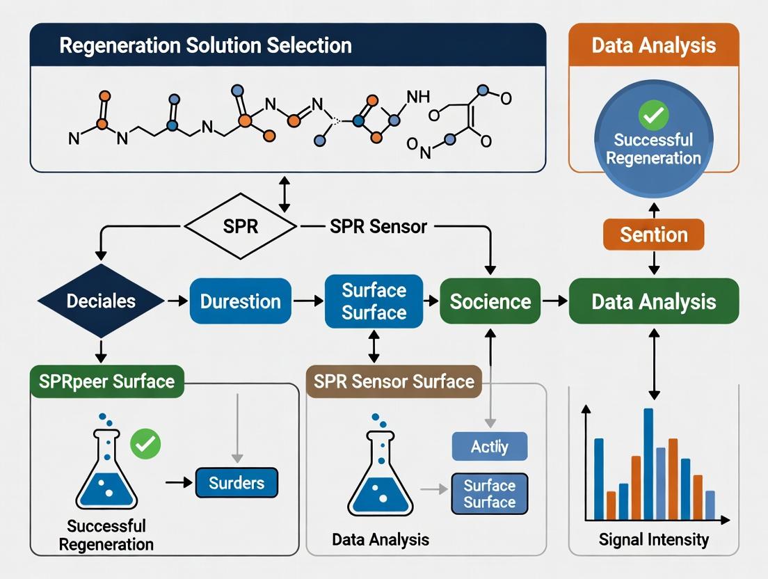
Experimental Workflow & Pathway Diagrams
Title: SPR Regeneration Cycle Decision Workflow
Title: SPR Regeneration Troubleshooting Logic Tree
Technical Support Center
Troubleshooting Guide: Common Regeneration Failures
Issue: Incomplete Regeneration (High Residual Response After Regeneration)
- Cause: Regenerant is too mild, exposure time is insufficient, or the complex is extremely high-affinity.
- Solution: Perform a scouting experiment. Increase regenerant concentration in a stepwise manner (e.g., 10 mM to 100 mM glycine-HCl) or switch to a harsher solution type (e.g., from low pH to chaotropic agent). Increase contact time from 30s to 60-90s.
- SPR Thesis Context: This highlights the non-universality of regeneration conditions and underscores the core thesis that optimal regenerant selection is target- and complex-specific, requiring empirical determination.
Issue: Loss of Ligand Activity (Steady Decline in Binding Capacity Over Cycles)
- Cause: Regenerant is too harsh, denaturing the immobilized ligand.
- Solution: Immediately switch to a milder regenerant. Test neutral pH options like 1-5 mM NaOH or 3-5 mM MgCl₂ for sensitive ligands. Ensure the regenerant is not left in contact with the sensor surface for extended periods.
- SPR Thesis Context: This trade-off between regeneration efficiency and ligand stability is a central challenge in regeneration solution research, driving the search for novel, targeted regenerants.
Issue: High Non-Specific Binding During Regeneration Phase
- Cause: Regenerant composition or pH causes aggregation or non-specific adsorption of analytes or contaminants.
- Solution: Include a mild detergent (e.g., 0.05% Tween 20) in the regenerant or running buffer. Filter all solutions. Test different pH regimes.
- SPR Thesis Context: Non-specific interactions during regeneration confound data, emphasizing the need for regenerants that promote specific complex disruption without introducing new artifacts.
Issue: Unstable Baseline Post-Regeneration
- Cause: Slow re-equilibration of the system to running buffer pH/ionic strength, or micro-bubbles.
- Solution: Extend the post-regeneration stabilization period. Degas all buffers thoroughly. Include a "buffer-only" injection after regeneration before the next analyte cycle.
- SPR Thesis Context: Baseline instability reduces data quality and throughput, a practical problem that regeneration optimization protocols within the thesis aim to minimize.
Frequently Asked Questions (FAQs)
Q1: What are the primary chemical mechanisms by which common regenerants work? A: The mechanisms are: 1) pH Disruption (Low/High pH): Alters ionization states of amino acid side chains, disrupting hydrogen bonds and electrostatic interactions (e.g., 10-100 mM Glycine-HCl pH 1.5-3.0, 1-50 mM NaOH). 2) Chaotropic Agents: Disrupts hydrophobic interactions and hydrogen bonding by destabilizing water structure (e.g., 1-6 M Guanidine-HCl, 2-8 M Urea). 3) High Salt: Shields electrostatic interactions (e.g., 1-4 M MgCl₂, NaCl). 4) Surfactants: Disrupts hydrophobic interfaces (e.g., 0.05-0.5% SDS). 5) Competitive Elution: Uses a high concentration of soluble ligand or analogue to competitively displace the bound analyte.
Q2: How do I systematically select a starting regenerant for a new protein-ligand pair? A: Follow a scouting protocol. Start with mild conditions and escalate: 1) Mild acid (10 mM Glycine, pH 2.5). 2) Mild base (5 mM NaOH). 3) High salt (2 M NaCl). 4) Chaotrope (2 M MgCl₂). 5) Combination (e.g., 1 M NaCl in Glycine pH 2.0). Inject each for 30-60s and monitor both regeneration efficiency (% recovery) and ligand stability over 5-10 cycles. Choose the mildest condition that achieves >95% removal of analyte.
Q3: Why does a regenerant that works for one antigen-antibody pair fail for another? A: The exact epitope-paratope interface is unique. A regenerant targeting ionic bonds may fail if the primary interactions are hydrophobic. The stability of the antibody's own structure to the regenerant also varies. This specificity is a core rationale for the ongoing research into developing prediction tools for regenerant selection based on interface properties.
Q4: Can I use the same regenerant for capture-based and direct immobilization assays? A: Extreme caution is needed. Capture systems (e.g., anti-His, streptavidin) often use a mild regenerant to remove the analyte while leaving the captured ligand intact. A second, harsher regeneration is then used to clear the capture ligand itself. Directly immobilized ligands may withstand slightly harsher conditions, but the capture molecule's stability is the limiting factor.
Q5: What are the key quantitative metrics to evaluate regeneration success? A: The two critical metrics are: 1) Regeneration Efficiency (%): [(Response after regeneration) / (Initial binding response)] * 100. Target >95% removal. 2) Ligand Stability (% Residual Activity): [(Binding response at cycle N) / (Binding response at cycle 1)] * 100. A drop of >5% over 10 cycles indicates ligand degradation.
Data Presentation: Common Regenerants & Performance
Table 1: Efficacy and Typical Use Cases of Common Regeneration Solutions
| Regenerant Solution | Typical Concentration | Primary Mechanism | Typical Use Case | Advantage | Risk |
|---|---|---|---|---|---|
| Glycine-HCl | 10 - 100 mM, pH 1.5-3.0 | pH Disruption (Acidic) | Antigen-Antibody complexes | Highly effective for many mAbs | Can denature sensitive proteins |
| NaOH | 1 - 50 mM | pH Disruption (Basic) | Robust antibodies, some peptides | Low cost, effective | High risk of ligand denaturation |
| HCl | 10 - 100 mM | pH Disruption (Acidic) | Strong ionic complexes | Very strong | Highly destructive to ligand |
| MgCl₂ | 1 - 4 M | High Salt / Mild Chaotrope | Disrupting ionic interactions | Gentler than low pH | May be ineffective for high-affinity complexes |
| Guanidine-HCl | 1 - 6 M | Chaotropic Agent | Very stable protein complexes | Powerful disruption | High risk of permanent ligand denaturation |
| SDS | 0.05 - 0.5% (w/v) | Surfactant | Hydrophobic interactions, last resort | Extremely effective | Very difficult to wash out, ruins surface |
Table 2: Scouting Experiment Results (Hypothetical Data for an IgG-Antigen Pair)
| Regenerant | Conc. | Contact Time | % Regeneration (Cycle 5) | % Ligand Activity (Cycle 10 vs. 1) | Pass/Fail |
|---|---|---|---|---|---|
| Glycine-HCl | 10 mM, pH 2.0 | 30s | 78% | 99% | Fail (Incomplete) |
| Glycine-HCl | 50 mM, pH 2.0 | 30s | 97% | 98% | Pass |
| Glycine-HCl | 50 mM, pH 2.0 | 60s | 99% | 95% | Pass (Optimal) |
| NaOH | 10 mM | 30s | 99% | 82% | Fail (Ligand Loss) |
| MgCl₂ | 3 M | 60s | 85% | 100% | Fail (Incomplete) |
Experimental Protocols
Protocol 1: Basic Regeneration Scouting Experiment Objective: To empirically determine the optimal regenerant for a specific immobilized ligand. Materials: SPR instrument, sensor chip with immobilized ligand, analyte sample, running buffer (e.g., HBS-EP+), series of regeneration solutions. Method:
- Establish Binding: Inject a saturating concentration of analyte over the ligand surface for 2-3 minutes. Allow dissociation in running buffer for 3-5 minutes.
- First Regeneration Test: Inject Regenerant A (e.g., 10 mM Glycine pH 2.0) for 30-60 seconds at a standard flow rate (e.g., 30 µL/min).
- Stabilize: Allow the baseline to stabilize in running buffer for 1-2 minutes.
- Assess: The response after stabilization should return to near the original baseline. Calculate % Regeneration.
- Verify Ligand Activity: Re-inject the same analyte sample. The binding response should be nearly identical to the first injection. Calculate % Ligand Activity.
- Cycle: Repeat steps 1-5 for 5-10 cycles to assess cumulative effects.
- Scout Next Condition: Switch to a new flow cell or fresh ligand surface. Repeat steps 1-6 with Regenerant B (e.g., 50 mM Glycine pH 2.0), then C (e.g., 10 mM NaOH), etc.
- Analysis: Plot % Regeneration and % Ligand Activity vs. cycle number for each condition. The optimal condition maintains >95% regeneration and >95% activity over all cycles.
Protocol 2: Two-Step Regeneration for Capture Systems Objective: To fully regenerate a capture sensor surface (e.g., Protein A or Streptavidin) without damaging the capture molecule. Materials: SPR instrument, capture sensor chip, captured ligand, analyte, running buffer. Method:
- Capture & Bind: Capture the ligand, then inject analyte to form the complex.
- Step 1 - Mild Regeneration: Inject a mild, specific regenerant (e.g., 10 mM Glycine pH 1.7 for Protein A) to dissociate the analyte and/or the captured ligand. This clears the complex but may leave the capture layer intact.
- Step 2 - Baseline Check: If the response returns to the post-capture baseline, the cycle can continue. If a drift or residual response is observed, proceed to Step 3.
- Step 3 - Harsher Regeneration (Occasional): Periodically (e.g., every 3-5 cycles), inject a harsher solution (e.g., 0.5% SDS, 50 mM NaOH) designed to fully strip the capture surface. Warning: This may degrade the capture surface over many cycles.
- Re-condition: After a harsh regeneration, the capture surface must be re-equilibrated with running buffer thoroughly before the next capture step.
Mandatory Visualization
The Scientist's Toolkit: Research Reagent Solutions
Table 3: Essential Reagents for SPR Regeneration Studies
| Item | Function & Role in Research |
|---|---|
| Glycine-HCl Buffer (pH 1.5-3.0) | The workhorse acidic regenerant. Used to systematically test pH disruption. Different concentrations and pHs are key variables in scouting experiments. |
| Sodium Hydroxide (NaOH) Solution (1-50 mM) | The standard basic regenerant. Tests ligand stability to high pH and is often used for cleaning surfaces. |
| Magnesium Chloride (MgCl₂) Solution (1-4 M) | A high-salt, mild chaotrope. Used to disrupt electrostatic interactions with lower denaturation risk, providing a gentler alternative. |
| Guanidine Hydrochloride (Gdn-HCl) Solution (1-6 M) | A strong chaotropic agent. Used to disrupt very stable complexes involving extensive hydrophobic interactions. Serves as a "last resort" benchmark. |
| Sodium Dodecyl Sulfate (SDS) Solution (0.05-0.5%) | An ionic surfactant. Used to disrupt hydrophobic interfaces and for deep cleaning of sensor surfaces between experiments. |
| HBS-EP+ Buffer | The standard running buffer (10 mM HEPES, 150 mM NaCl, 3 mM EDTA, 0.05% Surfactant P20). Provides a stable, low non-specific binding baseline for all regeneration cycles. |
| Sensor Chips (CM5, SA, Protein A) | The solid support. Different chip chemistries (carboxylated dextran, streptavidin, Protein A) influence ligand orientation and stability, affecting regenerant choice. |
| High-Precision pH Meter & Calibrated Buffers | Critical for accurate and reproducible preparation of all regenerant solutions, especially pH-based ones. |
| 0.22 µm Syringe Filters | Essential for removing particulates and microbes from all solutions before introduction to the sensitive fluidics of the SPR instrument. |
Technical Support Center: Troubleshooting & FAQs for SPR Regeneration
Q1: After multiple cycles, my baseline drifts upward, and binding response decreases. What is the likely cause, and how can I fix it? A: This indicates incomplete regeneration and progressive ligand/surface fouling. The incomplete removal of analyte leads to cumulative occupancy. First, increase the contact time of your current regenerant by 50-100%. If ineffective, implement a two-step regeneration: 1) a 30-second pulse of 10 mM Glycine-HCl (pH 1.7), followed by 2) a 60-second pulse of 0.05% (w/v) SDS. Rinse extensively with running buffer between steps. This combination often removes stubborn, non-specifically bound material.
Q2: My immobilized ligand appears to lose activity after just 2-3 regeneration cycles with 50 mM NaOH. What alternatives should I test? A: NaOH, while effective for many antibodies, can denature sensitive proteins. Consider a screening approach using a 96-well plate format before committing to the chip. Test these regenerants in order of increasing stringency:
- High-Salt Buffer: 2 M MgCl₂ for 30-60 seconds (disrupts ionic interactions).
- Mild Acid: 10 mM Glycine-HCl, pH 2.0-2.5, for 30 seconds.
- Chaotropic Agent: 4 M Guanidine-HCl for 30 seconds.
- Surfactant: 0.5% (v/v) Tween-20 or 0.1% SDS for 60 seconds. Monitor ligand activity via a reference analyte injection after each test regeneration. The gentlest effective solution should be selected.
Q3: I am working with a small molecule inhibitor binding to a kinase. What regeneration strategy is most suitable for this low-affinity (µM range), non-covalent interaction? A: For low-affinity interactions, mild conditions are often sufficient and preferred to maintain kinase conformation. A brief (30-second) pulse of a solution that alters ionic strength or pH is typically effective. Recommended starting protocol: Use 1 M NaCl in HBS-EP buffer (pH 7.4) for 30 seconds. If incomplete, switch to 10 mM Glycine-HCl pH 2.0 for 20 seconds. The mild acid often disrupts weak hydrophobic and ionic bonds without denaturing the enzyme.
Q4: How do I systematically select a regeneration solution for a novel protein-protein interaction? A: Follow this validated screening and optimization workflow.
Experimental Protocol: Regenerant Screening Cascade
- Immobilization: Immobilize the ligand to a CM5 chip via standard amine coupling to achieve ~5,000 RU.
- Baseline & Binding: Establish a stable baseline in running buffer. Inject a single concentration of analyte to achieve a robust binding signal (~100 RU).
- Regenerant Screening: Inject each candidate regenerant for 30-60 seconds at a flow rate of 30 µL/min. Use the following order, pausing if activity loss is observed:
- Step 1: Running buffer (negative control).
- Step 2: 10 mM Glycine, pH 2.0.
- Step 3: 10 mM Glycine, pH 1.5.
- Step 4: 10 mM NaOH (or 50 mM HCl).
- Step 5: 0.5% (v/v) Tween-20 or 0.05% SDS.
- Step 6: 4 M Guanidine-HCl.
- Assessment: After each regenerant pulse, inject the analyte again. Calculate the percent recovery: (Response Post-Regeneration / Initial Response) x 100%.
- Optimization: For the mildest effective regenerant, optimize contact time (15-120 sec) and concentration (e.g., pH gradient for glycine) to achieve >95% recovery for at least 5 cycles.
Table 1: Quantitative Performance of Common Regenerants
| Regenerant Solution | Typical Concentration | Contact Time (sec) | Effective Against | Risk of Ligand Denaturation | % Recovery (Typical Range)* |
|---|---|---|---|---|---|
| Glycine-HCl (low pH) | 10 mM, pH 1.5-2.5 | 30-60 | Ionic, hydrophobic bonds | Low-Moderate | 85-99% |
| NaOH / HCl | 10-50 mM | 30-60 | Ionic, some hydrophobic | High | 70-98% |
| High Salt (MgCl₂) | 1-3 M | 60-120 | Purely ionic interactions | Very Low | 60-90% |
| Chaotropic (Gdn-HCl) | 2-4 M | 30-90 | Hydrophobic, some H-bonds | Moderate-High | 80-95% |
| Ionic Detergent (SDS) | 0.01-0.1% (w/v) | 60-120 | Hydrophobic, aggregates | Moderate-High | 90-99% |
| Non-Ionic Detergent (Tween-20) | 0.5-1% (v/v) | 60-120 | Weak hydrophobic, lipid layers | Low | 75-95% |
*Recovery is highly system-dependent. Values represent common observations from published screening studies.
Q5: Can I mix different regenerant types, and what are the advantages? A: Yes, sequential or mixed regenerants are a powerful strategy. A common combination is a brief acid step (to disrupt specific bonds) followed by a mild surfactant (to remove aggregated or lipid-bound material). This can be more effective and gentler than using a single, harsher agent. Example Protocol for Challenging Systems: 1) 30-second pulse of 10 mM Glycine pH 1.7, 2) 45-second pulse of 0.025% SDS, 3) 2-minute stabilization in running buffer.
The Scientist's Toolkit: Key Research Reagent Solutions
| Item | Function in SPR Regeneration Research |
|---|---|
| Glycine-HCl Buffer (pH 1.5-3.0) | Mild acidic regenerant; protonates carboxylates and amines, disrupting ionic and hydrogen bonds. The workhorse for many antibody-antigen systems. |
| NaOH Solution (10-100 mM) | Strong base; effective for removing tightly bound analytes and sanitizing surfaces. High denaturation risk for sensitive ligands. |
| Guanidine-HCl (2-6 M) | Chaotropic agent; disrupts hydrophobic interactions and unfolds proteins by altering water structure. Used for stubborn, non-specific binding. |
| Sodium Dodecyl Sulfate (SDS) (0.01-0.5%) | Ionic detergent; solubilizes membranes and denatures proteins by disrupting hydrophobic forces. Excellent for removing aggregates. Use at lowest effective concentration. |
| Tween-20 or Triton X-100 (0.1-1%) | Non-ionic detergents; disrupt weak hydrophobic and lipid-based interactions with minimal denaturation. Good for mild washing steps. |
| Magnesium Chloride (MgCl₂, 1-3 M) | High-salt solution; disrupts electrostatic interactions via charge shielding. The gentlest option for salt-sensitive interactions. |
| HBS-EP Running Buffer | Standard SPR buffer (HEPES, NaCl, EDTA, Surfactant P20). The baseline for all experiments and a negative control regenerant. |
| Piezo Fluidic Valve & Multi-Channel Kit | Hardware for automated, sequential injection of multiple regenerants during screening without manual intervention. |
Visualization: Regenerant Selection Workflow
Title: SPR Regenerant Selection Decision Tree
Title: Regeneration Screening Experimental Protocol
Technical Support Center: Troubleshooting & FAQs
Frequently Asked Questions
Q1: Our immobilized ligand shows a significant drop in binding capacity after regeneration cycles. What is the likely cause and how can we address it? A: This is commonly caused by ligand degradation or conformational change due to harsh regeneration conditions. First, verify ligand stability by screening a panel of regeneration solutions with varying pH and ionic strength. For protein ligands, use a solution that deviates minimally from its optimal pH and pI. For small molecule or nucleic acid ligands, consider the chemical stability of functional groups. Implement a stability test protocol (see Experimental Protocol 1). Switch to a gentler regeneration solution, such as a mild acidic (pH 4.0-5.0) or basic (pH 8.5-9.0) buffer, or consider a high-salt solution (e.g., 1-2 M NaCl) if the binding is ionic.
Q2: Our analyte (a large protein complex) shows poor binding response and slow kinetics. How do analyte properties guide regeneration strategy? A: Large, multi-domain analytes are susceptible to denaturation. Aggressive regeneration can disrupt their native structure, preventing rebinding in subsequent cycles. Key properties to consider are the analyte's pI, known chemical sensitivities, and structural domains. Use regeneration solutions with pH values at least 1.5 units away from the analyte's pI to avoid precipitation. Prefer shorter contact times (15-30 seconds) with solutions like 10 mM Glycine-HCl (pH 2.5-3.0) or 3 M MgCl₂. Always perform a post-regeneration stability injection of a control analyte to confirm the analyte's structure remains intact.
Q3: How do we select a regeneration solution for a low-affinity interaction (KD in the micromolar range) without dissociating the ligand-analyte complex during the association phase? A: For low-affinity interactions, the binding interface is often small or involves weak forces. Regeneration must be very mild to avoid stripping the ligand. First, try solutions that weaken specific non-covalent bonds. For hydrophobic interfaces, use low concentrations of surfactants (e.g., 0.05% SDS) or chaotropic agents (1 M urea). For ionic interactions, use a high-salt buffer (e.g., 2 M NaCl). The key is to find a solution that weakens the interface just enough for dissociation without causing irreversible ligand damage. A stepwise screening protocol is essential (see Experimental Protocol 2).
Q4: We cannot find a solution that fully regenerates the surface without damaging the ligand. What are our options? A: When no single solution provides complete regeneration and stability, consider a multi-step approach. A two-step regeneration using two different solutions (e.g., high salt followed by mild acid) can be effective for complex interfaces. Alternatively, consider switching to a single-cycle kinetics (SCK) or multi-cycle kinetics (MCK) approach that does not require regeneration between analyte concentrations. Lastly, evaluate if a capture-based immobilization method (e.g., capturing a His-tagged ligand via an anti-His antibody surface) is more suitable, as the captured ligand can be replenished each cycle.
Q5: How many regeneration cycles should we test to confirm ligand stability? A: For a robustness test, a minimum of 50-100 regeneration cycles is recommended for publication-quality data. For initial screening, 10-20 cycles per candidate regeneration solution can identify clear failures. Monitor the baseline shift (indicating ligand loss) and the binding response to a reference analyte over these cycles. A decline in binding response >10% typically indicates unacceptable ligand instability.
Experimental Protocols
Experimental Protocol 1: Ligand Stability Screening
Objective: To assess the stability of an immobilized ligand against a panel of regeneration candidates.
- Immobilization: Immobilize the ligand to the desired response level (typically 50-100 RU for kinetics) on a suitable sensor chip using standard amine, thiol, or capture coupling.
- Baseline: Establish a stable baseline in running buffer.
- Binding Cycle: Inject a middle concentration of analyte (from a kinetic series) for 3-5 minutes to achieve near-saturation.
- Dissociation: Allow dissociation in running buffer for 5-10 minutes.
- Regeneration Test: Inject the candidate regeneration solution for 30-60 seconds.
- Stability Check: Re-inject the same analyte concentration. Record the binding response.
- Repetition: Repeat steps 3-6 for 10-20 cycles per regeneration solution.
- Analysis: Plot binding response versus cycle number. The solution causing the smallest decline in response and baseline drift is the most suitable.
Experimental Protocol 2: Stepwise Regeneration Solution Screening
Objective: To systematically identify the mildest effective regeneration condition.
- Prepare Solution Matrix: Create a matrix of solutions varying in pH (1.5-3.0 for acid; 8.5-10 for base), chaotrope concentration (0.5-4 M MgCl₂, 0.5-2 M GuHCl), and surfactant concentration (0.01-0.1% SDS).
- Initial Test: Start with the mildest condition (e.g., pH 5.0, 1 M NaCl). Perform 3-5 regeneration cycles as in Protocol 1.
- Efficacy Check: Calculate the percentage of residual analyte after regeneration. Aim for <5% residual binding.
- Escalation: If regeneration is insufficient (<95% removal), move to the next slightly harsher condition in the matrix.
- Ligand Activity Check: After each successful regeneration step, perform a control analyte injection to confirm ligand activity remains >90% of initial.
- Documentation: Record the exact condition that first achieves >95% regeneration with >90% ligand activity retention.
Data Presentation
Table 1: Regeneration Solution Efficacy vs. Ligand Type Stability
| Ligand Type | Optimal Regeneration Solution | Typical Contact Time | Max Cycles (Response Loss <10%) | Key Risk Factor |
|---|---|---|---|---|
| IgG Antibody | 10 mM Glycine, pH 2.0-2.5 | 30-60 sec | 100-200 | Acid-induced aggregation |
| Streptavidin | 1 M NaCl, 50 mM NaOH | 30 sec | >200 | High pH denaturation |
| His-Tagged Protein | 350 mM EDTA, pH 8.0 | 60 sec | 50-100 | Metal chelation, ligand leaching |
| Membrane Protein | 0.05% SDS, 40 mM Octyl Glucoside | 60-120 sec | 20-50 | Detergent denaturation |
| DNA Oligo | 50 mM NaOH, 1 M NaCl | 60 sec | >200 | Strand separation at high pH |
Table 2: Analyte Properties & Regeneration Harshness Guide
| Analyte Property | Regeneration Sensitivity | Recommended Solution Class | Solution to Avoid |
|---|---|---|---|
| pI < 4.5 | Low sensitivity to acid, high to base | Mild acidic (pH 3.0-4.0) | High pH (>9.0) |
| pI > 9.5 | Low sensitivity to base, high to acid | Mild basic (pH 8.5-9.5) | Low pH (<3.0) |
| Hydrophobic Interface | Sensitive to surfactants | High salt (>2 M NaCl) or mild pH | Harsh detergents (SDS >0.1%) |
| Multi-subunit Complex | High sensitivity to chaotropes | Short pulse of mild acid/base | High [Chaotropes] (>2 M) |
| Low Affinity (µM KD) | Very high sensitivity | Very mild (pH shift 1-2 units, 0.5 M NaCl) | Standard glycine pH 2.0 |
Diagrams
Title: SPR Regeneration Solution Selection Logic Flow
Title: Ligand Stability Screening Experimental Workflow
The Scientist's Toolkit: Research Reagent Solutions
| Reagent / Material | Primary Function in Regeneration Studies |
|---|---|
| Glycine-HCl Buffer (pH 1.5-3.5) | Mild acidic regeneration; disrupts ionic and hydrogen bonds. Common first-line screen. |
| Sodium Acetate Buffer (pH 4.0-5.5) | Weaker acid for sensitive ligands/analytes near neutral pI. |
| Tris/NaOH Buffer (pH 8.5-10) | Mild basic regeneration; effective for acidic ligands or hydrophobic interactions. |
| High-Salt Solutions (1-4 M NaCl/MgCl₂) | Disrupts electrostatic interactions. Often gentler on ligand conformation. |
| Chaotropic Agents (1-4 M MgCl₂, GuHCl) | Disrupts hydrogen bonding and hydrophobic packing; for strong complexes. |
| Surfactants (0.01-0.5% SDS, Tween 20) | Disrupts hydrophobic interfaces and prevents non-specific binding. |
| Chelators (10-350 mM EDTA/EGTA) | Regeneration for immobilized metal affinity (e.g., His-tag) surfaces. |
| CM5 or Series S Sensor Chips (Gold) | Standard carboxymethylated dextran chips for amine coupling of ligands. |
| Pioneer Chip J2 | Low nonspecific binding, high stability chip for challenging samples like membrane proteins. |
| HBS-EP+ Running Buffer | Standard SPR running buffer (HEPES, NaCl, EDTA, surfactant). Provides stable baseline. |
Impact of Regeneration on Baseline Stability, Signal Drift, and Sensorgram Quality Over Time
Troubleshooting Guides & FAQs
Q1: Why does the baseline not return to the original level after multiple regeneration cycles? A: This is typically caused by incomplete regeneration or cumulative, non-specific binding. Incomplete regeneration leaves residual analyte, causing a progressive baseline shift. Aggressive regeneration can sometimes damage the ligand, reducing binding capacity and lowering the baseline. Ensure your regeneration solution is strong enough to fully dissociate the complex but not degrade the immobilized ligand. Perform a "blank" injection (buffer only) to check for bulk refractive index shifts.
Q2: What causes significant signal drift over a series of analyte injections? A: Signal drift often stems from two main sources: 1) Instrumental/Temperature Instability: Ensure the instrument and all solutions are fully thermally equilibrated. 2) Ligand Decay or Surface Fouling: Repeated regeneration can gradually denature the ligand or promote non-specific deposition of contaminants. This leads to a changing baseline. Using a reference flow cell and incorporating regular "conditioning" injections with a weak acid/base can help stabilize the surface.
Q3: How can I improve poor sensorgram quality (high noise, unstable binding curves) over time? A: Degrading sensorgram quality is frequently linked to a deteriorating sensor chip surface. Noisy baselines can result from micro-bubbles in the fluidics; thoroughly degas all buffers. Unstable curves may indicate an uneven or dirty surface. Implement more stringent filtering and regular maintenance cycles. If using covalent coupling, ensure the surface is not over-activated, as this can lead to heterogeneous ligand attachment and unstable signals upon regeneration.
Q4: How do I select the optimal regeneration solution for my specific interaction? A: Selection is empirical and critical for long-term stability. The solution must break the specific interaction without harming the ligand. Start with a scouting experiment using a matrix of conditions (pH, ionic strength, additives). The goal is to find the mildest solution that achieves >95% dissociation of the analyte. Harsh conditions (e.g., low pH, chaotropes) give high efficiency but accelerate baseline drift due to ligand damage. Gentle conditions preserve the ligand but risk incomplete regeneration.
Data Presentation: Regeneration Solution Impact Study
Table 1: Comparison of Regeneration Solutions on SPR Performance Over 100 Cycles
| Regeneration Solution (pH) | Baseline Shift (RU) after 100 Cycles | Signal Drift (RU/min) | % Ligand Activity Remaining | Recommended for Ligand Type |
|---|---|---|---|---|
| Glycine-HCl, pH 2.0 | +125 | 0.8 | 65% | High-stability antibodies |
| Glycine-HCl, pH 2.5 | +45 | 0.3 | 85% | Most antibodies |
| NaOH, 10 mM | +200 | 1.2 | 40% | Robust protein A/G captures |
| NaCl, 3.0 M | +15 | 0.1 | 95% | Ionic interactions |
| EDTA, 10 mM (for metal chelate) | +30 | 0.2 | 90% | His-tagged proteins |
Table 2: Sensorgram Quality Metrics vs. Regeneration Count
| Regeneration Cycle # | Baseline Noise (RU, RMS) | Binding Response (RU) | Chi² Value (Goodness of Fit) |
|---|---|---|---|
| 1-10 | 0.5 | 100.0 | 0.9 |
| 11-30 | 0.6 | 98.5 | 1.2 |
| 31-60 | 0.8 | 95.2 | 2.8 |
| 61-100 | 1.5 | 88.7 | 5.6 |
Experimental Protocols
Protocol 1: Regeneration Solution Scouting for Antibody-Antigen Interactions
- Immobilize the antibody ligand on a CMS sensor chip using standard amine coupling to a level of ~10,000 RU.
- Inject a saturating concentration of antigen to achieve maximum binding.
- Inject a series of candidate regeneration solutions (e.g., Glycine-HCl pH 2.0-3.0, Phosphoric acid, NaOH) for 30-60 seconds.
- Monitor the immediate drop in response unit (RU). The ideal solution returns the signal to within ±5 RU of the original baseline.
- Repeat the binding-regeneration cycle 5-10 times with the promising candidates. Select the solution that maintains consistent binding response and stable baseline over these cycles.
Protocol 2: Long-Term Stability Assessment of Regeneration Conditions
- Prepare a sensor chip with ligand immobilized in flow cell 2; use flow cell 1 as a reference.
- Establish a stable baseline in running buffer for at least 10 minutes.
- Program an automated cycle: Inject analyte (2-5 min) -> Dissociation in buffer (2-5 min) -> Inject regeneration solution (30-60 sec) -> Re-equilibration in buffer (2-3 min).
- Repeat this cycle 100+ times, recording the baseline RU at a fixed point before each analyte injection.
- Periodically (e.g., every 20 cycles) perform a "binding capacity check" with a standard analyte concentration.
- Plot baseline RU and binding response RU versus cycle number to quantify drift and decay.
The Scientist's Toolkit: Key Research Reagent Solutions
| Item | Function in SPR Regeneration Research |
|---|---|
| CM5/CM7 Sensor Chip | Gold surface with a carboxymethylated dextran matrix for covalent ligand immobilization. The stability of this matrix under regeneration is key. |
| Glycine-HCl Buffer (pH 1.5-3.0) | Mild acid commonly used to disrupt protein-protein interactions by protonating carboxylates and histidines. |
| Sodium Hydroxide (NaOH, 10-100 mM) | Strong base used for stringent regeneration; can hydrolyze esters or denature proteins. |
| High-Salt Solutions (e.g., 3M NaCl) | Disrupts electrostatic interactions. Gentle on ligand structure but only effective for certain binding modes. |
| Chaotropic Agents (e.g., Guanidine HCl) | Disrupts hydrogen bonding and hydrophobic interactions. Very effective but highly denaturing. |
| Surfactants (e.g., SDS, 0.01-0.1%) | Solubilizes hydrophobic interactions and cleans non-specific deposits. Can be difficult to wash off completely. |
Visualizations
Developing Your SPR Regeneration Protocol: A Step-by-Step Method for Diverse Molecular Interactions
Technical Support Center: Troubleshooting Guides & FAQs
FAQ 1: Why is my post-regeneration baseline signal unstable or drifting?
- Answer: Unstable baselines are often caused by incomplete regeneration or carryover of analyte or regeneration reagent. First, ensure your regeneration solution is strong enough to fully dissociate the ligand-analyte complex. A stepped or multi-pulse injection of regenerant can help. Second, extend the dissociation and post-regeneration stabilization time in your method. Third, consider incorporating a "conditioning" or "wash" step with running buffer between the regenerant injection and the next analyte cycle to flush residual regenerant from the system.
FAQ 2: How do I choose between acidic, basic, and chaotropic regeneration solutions for my protein target?
- Answer: The choice is empirical and target-dependent. Follow a systematic scouting approach:
- Start with broad screening: Test a standard panel of solutions (see Table 1) at moderate concentrations (e.g., 10 mM Glycine pH 2.0-3.0, 10 mM NaOH, 0.5-1.0 M NaCl, 0.05% SDS).
- Assess activity: After each regeneration, inject the analyte again. A stable, reproducible response indicates the ligand remains active.
- Rank by efficiency: Calculate the % regeneration (response after regen / initial response * 100). Aim for >95%.
- Optimize the lead: For the best 1-2 candidates, perform a concentration or pH gradient to find the mildest condition that gives complete regeneration, minimizing ligand denaturation over multiple cycles.
FAQ 3: My ligand activity decays rapidly over multiple binding cycles. What can I do?
- Answer: This indicates the regeneration condition is too harsh. You must balance regeneration efficiency with ligand stability.
- Short-Term Fix: Reduce the contact time of the regenerant (inject for 5-15 seconds instead of 30-60).
- Systematic Approach: Re-scout using milder alternatives. If you used 10 mM Glycine pH 2.0, try pH 2.5 or 3.0. Replace ionic detergents (SDS) with non-ionic ones (e.g., 0.1% Triton X-100) or chaotropic salts (MgCl₂). Consider using a combination of two mild reagents (e.g., low pH followed by high salt) in sequence for synergistic effects.
FAQ 4: How many regeneration cycles should I test to confirm stability?
- Answer: For initial scouting, 3-5 regeneration cycles per condition are sufficient to identify promising candidates. For final validation of a selected regenerant, you must demonstrate ligand stability over a number of cycles at least 2-3 times the number you plan to use in your actual assay. For typical kinetic characterization (≈100 cycles), validate over 10-20 cycles initially and monitor for gradual decay.
Experimental Protocols
Protocol 1: Initial Regenerant Scouting Panel Objective: To rapidly identify candidate regeneration solutions that fully dissociate a high-affinity protein-protein complex without damaging the immobilized ligand. Method:
- Immobilize the ligand to a CMS sensor chip using standard amine coupling to achieve an appropriate response level (e.g., 50-100 RU for kinetics).
- Establish a binding cycle: Inject analyte at a single concentration to achieve near-saturation (e.g., 5x KD) for 2-3 minutes, followed by a dissociation phase in running buffer for 3-5 minutes.
- Inject each candidate regenerant from Table 1 for 30-60 seconds at a flow rate of 30 µL/min.
- Allow a 2-5 minute stabilization period in running buffer.
- Repeat steps 2-4 for 3-5 cycles per regenerant.
- Analyze sensorgrams for complete return to baseline and reproducible analyte binding response.
Protocol 2: Concentration Gradient Optimization for a Lead Regenerant Objective: To find the minimum effective concentration/pH of a promising regenerant. Method:
- Prepare a series of the lead regenerant at varying strengths (e.g., Glycine HCl at pH 2.0, 2.2, 2.5, 2.7, 3.0).
- Using a fresh ligand surface, bind and dissociate analyte as in Protocol 1.
- Inject each regenerant strength in sequence from mildest to harshest, performing 2-3 binding cycles per strength.
- Plot the normalized ligand activity (Response Cycle n / Response Cycle 1) and % Regeneration against regenerant strength.
- Select the condition that maintains >95% activity and >98% regeneration over the tested cycles.
Data Presentation
Table 1: Standard Regenerant Scouting Panel & Typical Results Data framed within SPR regeneration solution selection research. Responses are hypothetical averages over 5 cycles.
| Regenerant Solution | Typical Concentration/ pH | % Regeneration (Mean ± SD) | Ligand Activity Remaining (Cycle 5/Cycle 1) | Recommended Use Case |
|---|---|---|---|---|
| Glycine HCl | 10 mM, pH 2.0 | 99.5 ± 0.3% | 85% | High-affinity antibody-antigen pairs. Can be harsh. |
| Phosphoric Acid | 10 mM, pH 2.0 | 98.7 ± 0.5% | 88% | Alternative to glycine for some targets. |
| Sodium Acetate | 10 mM, pH 4.0-5.0 | 65.2 ± 5.1% | 99% | Very mild; for low-affinity or pH-sensitive complexes. |
| Sodium Hydroxide | 10 mM - 50 mM | 99.8 ± 0.1% | 45% | Very harsh; for robust ligands or removing non-specific binds. |
| NaCl (High Salt) | 1.0 - 2.0 M | 70.1 ± 3.2% | 98% | Disrupts electrostatic interactions. |
| MgCl₂ (Chaotropic) | 1.0 - 3.0 M | 92.3 ± 1.8% | 95% | Disrupts water structure; good for hydrophobic interfaces. |
| SDS (Ionic Detergent) | 0.01% - 0.1% | 99.0 ± 0.4% | 60%* | Removes strongly aggregated or denatured material. |
| Guanidine HCl | 0.5 - 2.0 M | 99.5 ± 0.2% | 30% | Extreme denaturant; last-resort for stubborn complexes. |
*Activity can sometimes be restored with a gentle wash after SDS.
Visualizations
Title: Systematic Regenerant Scouting Decision Workflow
Title: Regenerant Scouting in Thesis Research Context
The Scientist's Toolkit: Key Research Reagent Solutions
| Item | Function in Regenerant Scouting |
|---|---|
| Glycine HCl Buffer (pH 1.5-3.0) | Acidic regenerant; protonates carboxylates and histidines, disrupting salt bridges and hydrogen bonds. A first-line scouting reagent. |
| Sodium Hydroxide (10-50 mM) | Basic regenerant; deprotonates amines and tyrosine, disrupting hydrogen bonds and causing conformational change. Powerful but often denaturing. |
| High-Salt Solutions (1-3 M NaCl) | Disrupts electrostatic (ionic) interactions by shielding opposite charges. A mild starting point for suspected charge-based complexes. |
| Chaotropic Salts (MgCl₂, GuHCl) | Disrupts hydrogen bonding in water, weakening hydrophobic effect and promoting solubilization of hydrophobic interfaces. |
| Ionic Detergent (SDS, 0.01-0.1%) | Binds to and solubilizes denatured protein aggregates; useful for removing non-specifically bound material. Often damages the ligand. |
| Non-Ionic Detergent (Triton X-100) | Milder surfactant for disrupting hydrophobic and some non-covalent interactions with less denaturation risk than SDS. |
| Ethylene Glycol (10-50%) | Reduces solution polarity, weakening hydrophobic interactions. Useful for optimizing mild conditions. |
| Running Buffer (e.g., HBS-EP+) | Critical for post-regeneration stabilization and washing. Must be pH-stable and compatible with the ligand. |
Technical Support Center: Troubleshooting Guides & FAQs
Q1: Our SPR sensorgram shows a high, drifting baseline during buffer injection after regeneration. What's wrong? A: This typically indicates incomplete regeneration or carryover. First, verify that your regeneration solution's contact time is sufficient. For harsh solutions (e.g., 10 mM Glycine-HCl, pH 2.0), 30-60 seconds is often enough, but for stable interactions, you may need 2-3 minutes. Increase contact time incrementally. Second, check injection volume; ensure at least a 3x flow cell volume (typically 60-120 µL) to fully displace the previous solution. A final "conditioning" injection of running buffer can stabilize the baseline.
Q2: After multiple cycles, ligand activity drops significantly (>20% Rmax loss). How can I prevent this? A: This is ligand degradation due to harsh regeneration conditions. Optimize pH and concentration. Try a milder pH first. If using acid, test pH 2.5, 3.0, and 3.5. For alkali, test pH 8.5 vs. 9.0. Reduce the concentration of chaotropic agents (e.g., try 0.5 M MgCl₂ before 1 M). See the table below for a systematic comparison. Always use the minimal effective condition.
Q3: We get variable binding responses in sequential cycles, but regeneration seems complete. What should we check? A: Focus on injection volume precision and pH stability. Ensure your regeneration solution is freshly prepared and pH-checked. Variable volumes from an autosampler can cause inconsistent contact times. Program a "draw speed" and "inject speed" that are consistent and avoid air bubbles. Also, include a 1-minute stabilization period post-regeneration before the next analyte injection.
Q4: No regeneration solution we've tried works for our antibody-antigen pair. What's the next step? A: Consider a multi-step or pulsed regeneration protocol. Inject a short pulse (5-10 µL) of a harsh solution (e.g., pH 1.5), immediately followed by a longer pulse of a milder, stabilizing solution (e.g., pH 8.0). This can dissociate the complex while quickly returning the ligand to a native pH. Diagram 1 illustrates this logical optimization workflow.
Q5: How many regeneration cycles should an SPR method withstand to be considered robust? A: For publication or assay validation, aim for a minimum of 100 cycles with <10% loss in initial ligand activity (Rmax) and a consistent baseline (RU drift <5 RU). Document the response for a mid-level analyte concentration at cycles 1, 10, 50, and 100.
Data Presentation: Regeneration Solution Efficacy
Table 1: Comparison of Common Regeneration Solutions & Optimal Conditions
| Regeneration Solution | Typical Concentration Range | Optimal pH | Contact Time (s) | Effective Against | Ligand Stability Risk |
|---|---|---|---|---|---|
| Glycine-HCl | 10-100 mM | 1.5 - 3.0 | 30-120 | Antibody-Antigen | Medium-High |
| NaOH | 1-100 mM | 11.0 - 13.0 | 30-60 | High-affinity, multivalent | High |
| HCl | 1-10 mM | 1.0 - 2.0 | 30-90 | Generic acidic | High |
| MgCl₂ | 0.5 - 2 M | N/A | 60-180 | Ionic interactions | Low |
| SDS | 0.01 - 0.1% (w/v) | N/A | 60-120 | Hydrophobic | Medium |
| Guanidine HCl | 0.5 - 6 M | N/A | 30-90 | Strong complexes | Very High |
| Phosphoric Acid | 10-50 mM | ~1.5 | 30-60 | His-tag/NTA | Medium |
Table 2: Impact of Injection Volume on Regeneration Consistency (Example for a 30 nL Flow Cell)
| Injection Volume (µL) | Flow Cell Volumes | Baseline Stability (RU SD over 10 cycles) | Observation |
|---|---|---|---|
| 30 | 1x | >15 RU | Poor, inconsistent regeneration |
| 60 | 2x | 8 RU | Moderate, occasional drift |
| 90 | 3x | <3 RU | Good, stable baseline |
| 120 | 4x | <2 RU | Excellent, but uses more sample |
Experimental Protocols
Protocol 1: Systematic Screening of Regeneration Conditions
- Ligand Immobilization: Immobilize your ligand (e.g., antibody) on a CM5 chip using standard amine coupling to achieve ~5000 RU.
- Single-Cycle Test: Inject a saturating concentration of analyte (10x KD) for 2 minutes. Dissociate in running buffer for 3 minutes.
- Regeneration Test: Inject a candidate regeneration solution for 60 seconds at 30 µL/min.
- Baseline Check: Monitor the baseline for 2 minutes post-injection.
- Efficacy Assessment: Inject analyte again. Calculate % Regeneration = (Response post-regeneration / Initial response) x 100. Target >95%.
- Stability Assessment: Repeat steps 3-5 for 10 cycles. Calculate % Rmax remaining.
- Vary Parameters: Repeat entire protocol altering one parameter at a time (pH, contact time, concentration).
Protocol 2: Determination of Minimal Effective Contact Time
- Following ligand immobilization and analyte binding, inject the chosen regeneration solution.
- Use the instrument's "pulse" or "contact time series" mode to inject the same solution for 5, 15, 30, 45, and 60 seconds in sequential cycles (with re-binding steps in between).
- Plot contact time vs. % Regeneration. The minimal effective time is the point where the curve plateaus at >95% regeneration.
Mandatory Visualization
Title: SPR Regeneration Condition Optimization Workflow
Title: Basic SPR Regeneration Cycle Steps
The Scientist's Toolkit: Key Research Reagent Solutions
Table 3: Essential Materials for SPR Regeneration Studies
| Item | Function & Importance in Regeneration Studies |
|---|---|
| CM5 Sensor Chip (or equivalent) | Gold surface with a carboxymethylated dextran matrix. The standard substrate for ligand immobilization via amine coupling, testing regeneration stress on the chip surface. |
| HBS-EP+ Buffer (10x) | (10 mM HEPES, 150 mM NaCl, 3 mM EDTA, 0.05% v/v Surfactant P20). The standard running buffer for many SPR systems. Provides a stable, non-interacting baseline. Surfactant prevents non-specific binding. |
| Glycine-HCl Stock (1 M, pH 2.0) | A versatile acidic regeneration stock solution. Can be diluted to various concentrations and pH levels to fine-tune stringency. |
| NaOH (50 mM) | Common basic regeneration solution. Effective for disrupting many high-affinity interactions but can damage sensitive ligands. |
| High-Salt Solution (e.g., 2 M MgCl₂) | Disrupts interactions heavily dependent on ionic or electrostatic forces. Generally gentler on ligand structure than extreme pH. |
| Chaotropic Agent (e.g., 4 M Guanidine HCl) | Disrupts hydrogen bonding and hydrophobic interactions. A "last resort" solution for very strong complexes. |
| pH Meter & Calibration Buffers | Critical for accurate preparation and reproducibility of regeneration solutions. Small pH changes (0.2 units) can significantly impact efficacy and ligand stability. |
| Automated Liquid Handler | Ensures precise and repeatable injection volumes and contact times, critical for robust, high-throughput condition screening. |
Application-Specific Strategies for mAbs, Bispecifics, Fc-Fusion Proteins, and Small Molecules
Troubleshooting Guide & FAQs: SPR Regeneration Solution Selection
Thesis Context: This technical support content is framed within ongoing research to develop a rational framework for selecting optimal Surface Plasmon Resonance (SPR) regeneration solutions based on the biochemical characteristics of the analyte-ligand complex. The goal is to minimize activity loss while achieving complete complex dissociation for reusable sensor chips.
Frequently Asked Questions
Q1: After injecting my monoclonal antibody (mAb) analyte, I cannot regenerate the protein A/G surface without significant loss of ligand binding capacity. What are my options?
A: Protein A/G surfaces present a common challenge due to the high-affinity, multi-domain binding of mAbs. Standard glycine pH 1.5-2.5 often causes irreversible denaturation.
- Strategy: Implement a multi-step or mild acidic regeneration approach.
- Protocol: Try a two-step regeneration: 1) 10-30 mM HCl for 30-60 seconds, followed by 2) 10 mM Glycine-HCl, pH 2.0. Monitor ligand activity over 5 cycles. If loss >10%, switch to a milder solution like 3 mM NaOH or 0.5% SDS for 60 seconds.
- Data Summary:
| Ligand Type | Recommended Regeneration Solution | Exposure Time | Typical Cycle Life (Rmax loss <10%) |
|---|---|---|---|
| Protein A/G + mAb | 10-30 mM HCl | 30-60 sec | 50-70 cycles |
| Protein A/G + mAb | 3 mM NaOH | 60 sec | 80-100 cycles |
| Anti-Fc Capture + mAb | 3 M MgCl2 | 30 sec | 100-150 cycles |
Q2: My bispecific antibody with a low-affinity arm shows selective loss of function for one target after regeneration on an anti-capture surface. How can I preserve functionality?
A: This indicates the regeneration solution is disrupting the structure of the more sensitive binding arm.
- Strategy: Screen solutions that dissociate the antibody from the capture reagent without affecting the antibody's native conformation.
- Protocol: Perform a regeneration screen using a high-throughput microfluidic SPR system (if available). Test these solutions in order: 1) 10 mM Glycine, pH 2.0, 2) 10 mM Glycine, pH 2.5, 3) 1-3 M MgCl2, 4) 0.5-1 M NaCl, 5) 0.1% (v/v) Tween 20. Use a multi-cycle kinetics experiment with both target analytes sequentially to assess function after each regeneration.
- Key Reagent: HBS-EP+ buffer (10 mM HEPES, 150 mM NaCl, 3 mM EDTA, 0.05% v/v Surfactant P20) for stable baseline.
Q3: For Fc-fusion proteins, what regeneration strategy balances complete dissociation from an anti-Fc capture antibody with maintaining ligand stability?
A: Fc-fusion proteins can be sensitive to low pH due to their non-antibody fusion component.
- Strategy: Prioritize high-salt or chelating agent solutions before resorting to low pH.
- Protocol: Immobilize an anti-Fc antibody (e.g., anti-human IgG Fc) via amine coupling. Perform binding/regeneration cycles. Start with 3 M MgCl2 or 4 M NaCl for 60 seconds. If dissociation is incomplete (carryover >5% RUs), test a milder acid like 10 mM Glycine, pH 2.5. The fusion partner (e.g., receptor, enzyme) dictates sensitivity.
Q4: Small molecule inhibitors often show non-specific binding to the dextran matrix, complicating kinetics and regeneration. How is this addressed?
A: Small molecules require a different capture strategy to avoid matrix interactions.
- Strategy: Use a direct, high-density immobilization of the protein target or a stable, high-affinity capture system (e.g., His-tag capture for a His-tagged kinase).
- Protocol: Direct amine coupling of the target protein at high density (>10,000 RUs) can create a defined binding surface. For regeneration, use 100% DMSO for 30-60 seconds, followed by a quick transition to running buffer. This is highly effective for dissociating small molecules without damaging the protein target if it is properly immobilized. Always include a solvent compensation channel.
- Data Summary:
| Analytic Class | Preferred Immobilization | Optimal Regeneration | Key Consideration |
|---|---|---|---|
| Small Molecule | High-density target protein | 50-100% DMSO, 30-60 sec | Solvent correction required |
| mAb (for Kinetics) | Anti-Fc capture | 3 M MgCl2 or 10 mM Gly pH 2.0 | Preserves mAb activity |
| Bispecific | Anti-Fab or target antigen capture | pH gradient (2.5 to 2.0) | Screen for arm-specific damage |
| Fc-Fusion | Anti-Fc capture | High salt (3-4 M) first | Fusion partner stability |
Experimental Protocol: Systematic Regeneration Solution Screening
Objective: To empirically determine the optimal regeneration solution for a given ligand-analyte pair.
- Ligand Immobilization: Immobilize your ligand (e.g., target antigen) to a CM5 sensor chip via standard amine coupling to achieve ~5-10,000 RU.
- Analyte Binding: Inject a single concentration of analyte (e.g., mAb) sufficient to achieve ~75-100 RU binding.
- Regeneration Screening: In sequential cycles on the same spot, inject a series of regeneration candidates for 30-60 seconds each. Common candidates in order: 10 mM Glycine pH 2.5, pH 2.0, pH 1.5; 10 mM HCl; 3 M MgCl2; 3 M NaCl; 0.5% SDS; 10 mM NaOH.
- Stability Test: After each regeneration candidate, re-inject the analyte. Calculate the percentage of initial binding response remaining.
- Selection Criteria: The optimal solution is the one that returns the baseline to within ±2 RU of the original and maintains ≥95% of the initial analyte binding response for at least three consecutive cycles.
The Scientist's Toolkit: Key Research Reagent Solutions
| Item | Function in SPR Regeneration Research |
|---|---|
| CM5 Series S Sensor Chip | Gold sensor surface with a carboxymethylated dextran matrix for ligand immobilization. |
| HBS-EP+ Buffer (10x) | Standard running buffer for most experiments; provides ionic strength and reduces non-specific binding. |
| 1-Ethyl-3-(3-dimethylaminopropyl)carbodiimide (EDC) | Crosslinker for activating carboxyl groups on the sensor chip during amine coupling. |
| N-Hydroxysuccinimide (NHS) | Stabilizes the amine-reactive intermediate formed by EDC activation. |
| 1 M Ethanolamine-HCl, pH 8.5 | Quenches unreacted NHS-esters after ligand immobilization, blocking remaining active sites. |
| Regeneration Scouting Kit | Commercial kit containing vials of common regeneration solutions (acids, bases, salts, solvents). |
| Glycine-HCl Buffer (pH 1.5-3.0) | Most common acidic regeneration solutions for disrupting protein-protein interactions. |
| 4-6 M Solutions of MgCl₂ or NaCl | High-ionic strength solutions for disrupting electrostatic interactions. |
| 0.1-0.5% Sodium Dodecyl Sulfate (SDS) | Ionic detergent for stripping strongly bound or denatured proteins (can damage some ligands). |
| 50-100% Dimethyl Sulfoxide (DMSO) | Organic solvent for dissociating small molecule analytes from protein targets. |
Diagrams
SPR Regeneration Selection Logic Flow
SPR Regeneration Screening Workflow
Troubleshooting Guides & FAQs
Q1: After repeated regeneration cycles, my baseline signal increases significantly. What is the cause and how can I resolve it? A: A rising baseline is often caused by the incomplete removal of tightly bound analyte or ligand denaturation/degradation. For pM-affinity pairs, standard acidic or basic regeneration may be insufficient.
- Troubleshooting Steps:
- Verify Regeneration Efficacy: Perform a "blank injection" (running buffer only) after regeneration. A stable baseline indicates successful removal of non-specifically bound material.
- Assess Ligand Stability: Immobilize a fresh ligand surface and subject it to your regeneration buffer without any analyte binding cycles. A stable baseline confirms ligand stability; a drifting baseline indicates the buffer itself is damaging the ligand.
- Optimize Solution: For resistant complexes, consider a two-step regeneration (e.g., brief high pH glycine followed by low pH glycine) or the inclusion of mild chaotropes (e.g., 0.5-1 M MgCl₂). Always test on a separate sensor chip first.
Q2: My immobilized antibody loses binding capacity after 5-10 regeneration cycles. How can I improve ligand stability? A: This indicates ligand degradation. The goal is to find the mildest solution that disrupts the high-affinity interaction.
- Troubleshooting Steps:
- Reduce Exposure Time: Shorten the regeneration injection contact time from 60s to 30s or even 15s while maintaining flow rate.
- Lower Concentration: Titrate down the concentration of harsh additives (e.g., SDS, chaotropes) to the minimum effective dose.
- Alternative Chemistry: If using amine coupling, consider switching to a capture method (e.g., anti-Fc or Protein A). The captured antibody can be replenished each cycle, avoiding repeated stress on the same molecule. See the protocol below.
Q3: I cannot find a regeneration condition that fully dissociates my pM complex without damaging the ligand. What are my options? A: When no single solution works, a multi-pronged strategy is required.
- Troubleshooting Steps:
- Scouting in Series: Perform a scouting experiment using a combination of two different buffers in sequence (e.g., mild acid followed by a mild chaotrope).
- Consider "Soft" Regeneration: For some extremely stable complexes, partial regeneration (85-90% dissociation) may be acceptable if it is highly reproducible and maintains ligand activity over >100 cycles.
- Alternative Platform: Evaluate a single-cycle kinetics (SCK) or multi-cycle kinetics (MCK) approach, where regeneration is not required, though this consumes more analyte.
Detailed Experimental Protocols
Protocol 1: Systematic Regeneration Scouting for High-Affinity Pairs
Objective: To empirically identify the optimal regeneration solution. Materials: SPR instrument, sensor chip with immobilized ligand, analyte, running buffer, scouting solutions. Procedure:
- Immobilization: Immobilize your antibody (ligand) to a desired level (e.g., 100-150 RU) on a CMS sensor chip using standard amine coupling.
- Binding Cycle: Inject a single concentration of antigen (analyte) for 3-5 minutes to achieve saturation binding.
- Regeneration Scouting: Inject the first candidate regeneration solution for 30-60 seconds.
- Evaluate: Monitor the sensorgram. A successful condition returns the signal to baseline. Note any baseline shift.
- Stability Test: Repeat steps 2-4 for 5-10 cycles with the same regeneration solution. Calculate the percentage of remaining binding activity relative to cycle 1.
- Iterate: Test subsequent solutions in a fresh ligand spot. Solutions should be tested in order of increasing harshness: (i) pH change (10 mM Glycine pH 2.0-3.5, 10 mM NaOH), (ii) ionic strength (1-4 M NaCl, 1-3 M MgCl₂), (iii) mild chaotropes (0.5-2 M NaSCN, 1-2 M Guanidine HCl), (iv) surfactants (0.05-0.5% SDS). Always follow SDS with extensive washing.
Protocol 2: Capture-Based Regeneration for Fragile Ligands
Objective: To maintain consistent binding activity by regularly replacing the stressed ligand. Materials: SPR instrument, Series S sensor chip (Protein A or anti-Fc), running buffer, antibody (ligand), analyte, regeneration solution for capture surface. Procedure:
- Capture Surface Preparation: Use a sensor chip pre-coated with a capture molecule (e.g., Protein A).
- Ligand Capture: Inject a diluted antibody solution for 60s to capture a consistent, low level of ligand (e.g., 50 RU).
- Analyte Binding: Inject analyte using your kinetic or concentration series protocol.
- Surface Regeneration: Apply a two-step regeneration: a. Primary Regeneration: Use a solution that dissociates the high-affinity antigen-antibody complex (identified in Protocol 1). b. Secondary Regeneration: Use a solution that strips the captured antibody from the Protein A layer (e.g., 10 mM Glycine, pH 1.7). This ensures a fresh ligand surface for the next cycle.
- Repetition: For the next cycle, return to Step 2.
Table 1: Efficacy and Stability of Common Regeneration Solutions on a pM-Affinity Antibody-Antigen Pair
| Solution | Composition | % Dissociation (Cycle 1) | % Remaining Activity (Cycle 10) | Recommended Contact Time | Notes |
|---|---|---|---|---|---|
| Glycine, pH 2.0 | 10 mM | 75% | 25% | 30s | Insufficient for full dissociation, rapid decay. |
| Glycine, pH 2.5 | 10 mM | 65% | 40% | 30s | Milder, but poor efficacy. |
| NaOH | 10 mM | 80% | 15% | 30s | Harsh, often denatures antibody. |
| MgCl₂ | 1 M | 40% | 90% | 60s | Mild, good stability but very low efficacy. |
| NaSCN | 1 M | 95% | 60% | 45s | Good balance for some pairs. |
| Gly pH 2.0 + MgCl₂ | 10 mM + 1 M | 99% | 85% | 30s + 30s | Two-step process, best overall result. |
Table 2: Comparison of Immobilization vs. Capture Methods for Regeneration Development
| Parameter | Direct Amine Coupling | Protein A Capture |
|---|---|---|
| Ligand Orientation | Random | Uniform, via Fc region |
| Ligand Stability | Same molecule stressed repeatedly | Fresh molecule each cycle |
| Required Regeneration | Must be gentle on ligand | Can be harsh on complex, gentle on Protein A |
| Baseline Stability | Can drift over cycles | Highly reproducible |
| Throughput | High | Medium (requires re-capture) |
| Best For | Stable ligands, screening | Fragile antibodies, definitive kinetics |
Visualizations
Diagram 1: SPR Regeneration Solution Scouting Workflow
Diagram 2: Two-Step Capture & Regeneration Method
The Scientist's Toolkit: Key Research Reagent Solutions
| Item | Function in Regeneration Development |
|---|---|
| CM5/CM7 Sensor Chip | Gold-standard carboxymethylated dextran matrix for covalent ligand immobilization via amine coupling. |
| Series S Sensor Chip Protein A | Pre-immobilized Protein A for reversible, oriented capture of antibody ligands via the Fc region. |
| Glycine-HCl Buffer (pH 1.5-3.0) | Low-pH solution to disrupt electrostatic and hydrogen bonding interactions in antibody-antigen complexes. |
| NaOH (10-50 mM) | High-pH solution effective for disrupting hydrophobic interactions; can be harsh on ligands. |
| Magnesium Chloride (MgCl₂, 1-3 M) | High ionic strength solution to disrupt electrostatic interactions; generally mild on protein stability. |
| Sodium Thiocyanate (NaSCN, 0.5-2 M) | Mild chaotrope that disrupts hydrophobic interactions; often a good compromise between efficacy and gentleness. |
| Guanidine Hydrochloride (1-2 M) | Strong chaotrope that denatures proteins; last-resort option for extremely stable complexes. |
| SDS (0.05-0.1%) | Anionic surfactant that disrupts hydrophobic and electrostatic interactions; requires rigorous system washing after use. |
| HBS-EP+ Buffer | Standard running buffer (HEPES, NaCl, EDTA, surfactant) for maintaining baseline stability and minimizing non-specific binding. |
SPR Regeneration Troubleshooting: Diagnosing and Solving Common Assay Failure Modes
Troubleshooting Guides & FAQs
Q1: What does a consistent decline in binding response (RU) over multiple cycles indicate, and how can I confirm the cause?
A: A consistent, cumulative drop in maximum binding capacity (Rmax) across cycles primarily indicates ligand denaturation or loss from the sensor chip surface. A decline in specific binding signal while the baseline (or reference channel) remains stable is a key sign.
- Diagnostic Protocol:
- Run a Reference Ligand Test: Inject a known, stable analyte over both the ligand and reference surfaces after the drop-off is observed. A reduced signal only on the ligand channel confirms ligand activity loss.
- Perform a Surface Capacity Check: Re-inject the original coupling solution (e.g., ligand in low pH buffer for amine coupling). A significantly lower coupling response compared to the initial level confirms ligand loss.
- Analyze Regeneration Scouting Data: Review sensorgrams from your initial regeneration scouting. Harsh conditions (very low/high pH, chaotropes) often cause immediate, irreversible decline.
Q2: My binding signal is unstable and recovers partially after extended buffer flow, but never fully to baseline. What does this mean?
A: This pattern strongly suggests incomplete regeneration. Residual analyte remains bound or non-specifically associated with the ligand or chip matrix, causing a drifting baseline and reducing available sites for the next cycle.
- Diagnostic Protocol:
- Extend Post-Regeneration Stabilization: After the standard regeneration pulse, extend the dissociation/buffer flow time to 5-10 minutes. A gradually descending baseline that eventually stabilizes at a higher level than pre-injection indicates slow analyte dissociation.
- Inject a "Blank" Regeneration: Run a buffer injection using the regeneration method. Any signal change indicates a bulk shift due to the solution itself, which should be accounted for.
- Perform a Double-Regeneration Pulse: Apply two identical, short regeneration pulses separated by a 1-2 minute buffer flow. If the second pulse produces a further drop in signal, your primary method is incomplete.
Q3: How can I systematically test regeneration solutions to distinguish between these two issues?
A: Employ a phased, scouting approach that evaluates efficacy and harshness sequentially.
- Experimental Protocol for Regeneration Scouting:
- Phase 1 - Mild to Moderate: Start with gentle conditions (e.g., pH 5.0-8.0 buffers, mild ionic strength changes). Inject analyte and regenerate for 3-5 cycles. Monitor Rmax stability.
- Phase 2 - Increased Stringency: If binding persists, introduce mild chaotropes (e.g., 1-2 M NaCl), mild acids/bases (e.g., 10 mM Glycine pH 2.5-3.5), or surfactants (e.g., 0.05% SDS).
- Phase 3 - Strong Conditions: Use stronger chaotropes (e.g., 2-4 M MgCl₂), extremes of pH (1.5-2.0 or 11-12), or combinations. WARNING: These high-risk conditions are for scouting only and often cause denaturation.
- Analyze: Plot Rmax (%) vs. Cycle Number for each condition.
Table 1: Efficacy and Risk Profile of Common Regeneration Solutions
| Solution Type | Example | Typical Concentration | Primary Mechanism | Risk of Denaturation | Typical Use Case |
|---|---|---|---|---|---|
| Low/High pH | Glycine-HCl | 10-100 mM, pH 1.5-3.0 | Alters ionization states, disrupts electrostatic & H-bonds | High at extremes | Antibodies, charged interactions |
| High Salt | Magnesium Chloride | 1-5 M | Disrupts electrostatic interactions | Low to Moderate | Ionic complexes, DNA-protein |
| Chaotrope | Guanidine HCl | 0.5-2 M | Disrupts H-bonding, denatures proteins | High (>1 M) | High-affinity, multipoint bonds |
| Surfactant | SDS | 0.01%-0.1% | Disrupts hydrophobic interactions | High | Membrane proteins, hydrophobic patches |
| Competitor | Soluble Ligand/Analogue | High concentration | Competitive displacement | Very Low | Small molecule, lectin-carbohydrate |
Table 2: Diagnostic Signals for Common Problems
| Observed Symptom | Baseline Post-Regeneration | Rmax Trend | Likely Culprit |
|---|---|---|---|
| Gradual signal decline over cycles | Returns to original | Consistently decreases | Ligand Denaturation |
| Signal instability, poor kinetics | Does not return fully, drifts | Variable, often lower | Incomplete Regeneration |
| Sudden, large signal loss | Step-change increase | Permanently lower | Catastrophic ligand loss |
| Signal decline only in later cycles | Stable | Decreases gradually | Cumulative, slow denaturation |
Visualization: Diagnostic Workflow
Diagram Title: SPR Signal Drop-Off Diagnostic Decision Tree
The Scientist's Toolkit: Key Research Reagent Solutions
Table 3: Essential Materials for Regeneration Optimization Studies
| Reagent/Solution | Primary Function in Diagnosis/Optimization |
|---|---|
| Glycine Buffer Series (pH 1.5-3.0, 8.5-9.5) | Standard scouting tool for probing pH-sensitive interactions. Low pH is common but risky. |
| High-Salt Solutions (e.g., 1-5 M MgCl₂, NaCl) | Disrupts electrostatic interactions with lower denaturation risk than extreme pH. |
| Chaotropes (e.g., 0.5-2 M Guanidine HCl) | Tests resilience of H-bonding networks; high concentrations induce denaturation. |
| Mild Surfactants (e.g., 0.005%-0.02% Tween 20, 0.01% SDS) | Reduces non-specific adsorption; higher concentrations can strip ligand. |
| Competitive Displacer (e.g., high conc. analyte/soluble receptor) | Ideal, non-destructive regeneration agent; requires a suitable, soluble competitor. |
| CMS/Series S Sensor Chips | Standard dextran matrix chips; baseline stability is key for diagnosis. |
| HBS-EP+ Buffer (or equivalent) | Standard running buffer for baseline establishment and diagnostics. |
| Software: Multi-Cycle Kinetics Analysis Module | Essential for quantifying Rmax and baseline shifts across cycles. |
Addressing Rising Baselines and Non-Specific Binding Accumulation
Technical Support Center
Troubleshooting Guides
Guide 1: Diagnosing Rising Baseline Causes
Q1: Why is my baseline signal continuously increasing during or between cycles? A: A steadily rising baseline typically indicates the accumulation of material on the sensor chip surface. This can be due to incomplete regeneration, carryover of analyte, or non-specific binding (NSB) of sample matrix components.
Diagnostic Steps:
- Inspect Regeneration: Run a buffer-only injection after your regeneration step. A stable baseline confirms effective regeneration. A rising baseline indicates residual analyte or carryover.
- Analyze Sample Matrix: Inject sample running buffer alone. An increase in signal points to NSB from buffer components (e.g., lipids, aggregates, contaminants from cell lines).
- Check System: Perform a system wash with recommended solutions (e.g., 50% glycerol, 0.5% SDS) to remove potential deposits in the microfluidics.
Guide 2: Resolving Non-Specific Binding Accumulation
Q2: My reference surface and active surface both show high binding. How do I reduce this non-specific signal? A: NSB complicates data analysis by obscuring specific interactions. Mitigation requires optimization of both the sample and the sensor surface.
Action Protocol:
- Modify Running Buffer:
- Add a non-ionic detergent (e.g., 0.005% P-20).
- Increase ionic strength (e.g., 150-250 mM NaCl).
- Include a blocking agent (e.g., 0.1% BSA, 1 mg/ml CM-Dextran).
- Optimize Surface Chemistry: Choose a sensor chip with low NSB properties (e.g., hydrogel-based chips like Series S CM5 or SA for high NSB samples).
- Employ a More Stringent Regeneration Solution: If NSB is persistent, a stronger regeneration solution may be needed (see Regeneration Solution Selection Table below).
Frequently Asked Questions (FAQs)
Q: What is the primary cause of non-specific binding in SPR? A: NSB is primarily caused by hydrophobic or ionic interactions between sample components and the sensor surface. Common culprits include partially denatured proteins, lipoproteins, aggregates, or sticky compounds in complex matrices like serum or cell lysates.
Q: How can I tell if my baseline drift is due to system issues or a binding event? A: System-related drift is often linear and consistent across all flow cells. Binding-related increases are typically flow-cell specific and may show association/dissociation kinetics. Check your reference cell and buffer injections for comparison.
Q: My regeneration solution seems to work initially but performance degrades over multiple cycles. Why? A: This is a classic sign of regeneration solution accumulation or gradual surface degradation. Harsh solutions (low pH, high chaotrope concentration) can slowly damage the dextran matrix or ligand over time, creating sites for NSB. A gentler, more targeted regeneration strategy is needed.
Thesis Context: SPR Regeneration Solution Selection Research
Effective regeneration is critical for reusability and data quality in SPR. The broader thesis posits that selecting a regeneration solution is not a one-size-fits-all process but a strategic decision based on the ligand-analyte complex's biophysical properties. Systematic screening of solutions with varying pH, ionic strength, and chaotropic agents is essential to find the optimal balance between complete analyte removal and long-term surface integrity, thereby directly combating rising baselines and NSB accumulation.
Data Presentation: Regeneration Solution Efficacy
Table 1: Screening Results for Common Regeneration Solutions on an Anti-IgG/Antigen Complex
| Solution Composition | pH | Avg. % Activity Remaining (Cycles 1-5) | Baseline Stability (RU Shift/Cycle) | Recommended Use Case |
|---|---|---|---|---|
| 10 mM Glycine-HCl | 1.5 | 45% | High (>5 RU) | Strong, high-affinity complexes. Risk of ligand damage. |
| 10 mM Glycine-HCl | 2.0 | 85% | Moderate (2-5 RU) | Standard for many antibody-antigen complexes. |
| 0.5-1.0 M NaCl | N/A | 98% | Low (<2 RU) | Weak ionic interactions. Very gentle. |
| 50 mM NaOH | 12.5 | 92% | Low (<2 RU) | Stable ligands, removal of acidic contaminants. |
| 5-50 mM HCl | ~1.6-2.2 | 70% | High (>5 RU) | Alternative to Glycine. Can be harsh. |
| 10 mM EDTA | N/A | 99% | Very Low | Metal-ion dependent interactions (His-tag, etc.). |
| 4 M MgCl₂ | ~6.5 | 96% | Very Low (<1 RU) | Chaotropic, disrupts hydrophobic/water networks. Good for stubborn NSB. |
Table 2: Impact of Additives on NSB Reduction in Serum-Containing Samples
| Running Buffer Additive | Concentration | % Reduction in Reference Cell Signal | Notes |
|---|---|---|---|
| Surfactant P20 | 0.005% v/v | 60-70% | Standard additive, reduces hydrophobic binding. |
| BSA | 0.1% w/v | 40-50% | Blocks NSB sites, may bind some analytes. |
| Carboxymethyl-Dextran | 1 mg/ml | 50-60% | Competes with chip matrix for NSB. |
| Increased NaCl | 250 mM | 30-40% | Shields ionic interactions. |
Experimental Protocols
Protocol 1: Systematic Regeneration Solution Screening
Objective: To identify the optimal regeneration solution that maintains ligand activity and ensures baseline stability.
- Immobilize the ligand to a desired RU level on a CM5 chip.
- Inject a saturating concentration of analyte to achieve maximum binding.
- Regenerate with a short pulse (30-60 sec) of the first test solution.
- Monitor the immediate baseline return. A stable return indicates effective removal.
- Re-inject the same analyte concentration. The binding level compared to the first cycle indicates retained ligand activity.
- Repeat Steps 2-5 for 5-10 cycles per test solution.
- Calculate the percentage of initial binding response retained at the final cycle.
- Rank solutions by the combination of retained activity and baseline stability.
Protocol 2: Evaluating NSB from Complex Matrices
Objective: To quantify and mitigate NSB in samples like cell culture supernatant or serum.
- Prepare a reference surface (e.g., activated and blocked without ligand).
- Prepare your sample and a matched "blank" matrix (e.g., serum-free media, buffer).
- Dilute your analyte in both the sample matrix and the blank matrix to the same final concentration.
- Inject the blank matrix sample over both reference and active flow cells. Record the binding response on the reference cell (R1).
- Inject the actual matrix sample over both flow cells. Record the binding response on the reference cell (R2).
- Calculate the matrix-induced NSB as R2 - R1.
- Test different running buffer additives (see Table 2) to minimize the (R2 - R1) value.
Mandatory Visualization
Title: SPR Baseline & NSB Troubleshooting Logic
Title: Regeneration Solution Selection Research Workflow
The Scientist's Toolkit: Research Reagent Solutions
Table: Essential Reagents for SPR Regeneration & NSB Studies
| Reagent | Function/Description | Common Example(s) |
|---|---|---|
| Low pH Buffers | Disrupts ionic and hydrogen bonds; protonates carboxylates and histidines. | Glycine-HCl (pH 1.5-3.0), Citrate, Formate |
| High pH Buffers | Deprotonates amines and hydroxyl groups; disrupts ionic bonds. | Glycine-NaOH (pH 8.5-10), NaOH, Na₂CO₃ |
| Chaotropic Agents | Disrupts water structure, weakening hydrophobic interactions and hydrogen bonding. | MgCl₂ (4-6 M), Guanidine HCl, Urea |
| High Salt Solutions | Shields ionic interactions; can weaken specific salt bridges. | NaCl (0.5-3 M), KCl |
| Chelating Agents | Removes divalent cations essential for some interactions (e.g., His-tag binding). | EDTA, EGTA |
| Detergents | Reduces hydrophobic NSB in running buffers and aids regeneration. | Surfactant P20 (0.005-0.05%), CHAPS |
| Blocking Agents | Added to running buffer to saturate NSB sites on the chip surface. | BSA (0.1%), CM-Dextran, casein |
Mitigating Carryover Effects Between Analysis Cycles
Troubleshooting Guides & FAQs
Q1: What are the typical symptoms of a carryover effect in SPR analysis, and how can I confirm it? A1: Symptoms include a consistently rising or non-returning baseline in subsequent cycles, a reduction in maximum binding capacity (Rmax) for the analyte, and inconsistent kinetic data. To confirm, run a blank buffer injection immediately after a high-concentration analyte injection. A significant response during the "blank" injection confirms carryover.
Q2: Our regeneration step seems to weaken the ligand activity over multiple cycles. How can we optimize it? A2: This indicates overly harsh regeneration. Systematic screening is required. Follow this protocol:
- Immobilize your ligand on the sensor chip.
- Inject a mid-level concentration of analyte to achieve a stable binding response.
- Apply a candidate regeneration solution for 30-60 seconds.
- Inject buffer to check for baseline return and stability.
- Re-inject the same analyte concentration. Calculate the percentage of initial binding response retained.
- Repeat cycles 2-5 for at least 5 cycles with each candidate solution. The optimal solution maintains >95% initial response over the required number of cycles.
Q3: We suspect carryover is due to non-specific binding to the microfluidics, not the chip surface. How do we address this? A3: Perform a systematic "Sanitization and Desorption" protocol between analysis cycles:
- After standard regeneration, inject a 0.5% (v/v) SDS solution for 60 seconds.
- Flush with 50 mM glycine, pH 9.5, for 60 seconds.
- Equilibrate with running buffer for 120 seconds. Monitor baseline stability across cycles. This removes hydrophobic contaminants from the fluid path.
Key Research Reagent Solutions
| Reagent Name | Primary Function | Key Consideration for Mitigating Carryover |
|---|---|---|
| Glycine-HCl (pH 1.5-3.0) | Common acidic regenerant; disrupts electrostatic and hydrophobic interactions. | Can denature sensitive protein ligands. Always test stability. |
| NaOH (10-100 mM) | Common basic regenerant; effective for disrupting hydrophilic and ionic interactions. | Can hydrolyze ester-linked ligands on CMS chips. Use with caution. |
| SDS (0.01-0.5%) | Ionic detergent; strips strongly bound, hydrophobic molecules from surfaces and fluidics. | Requires extensive buffer flushing post-use to prevent detector contamination. |
| Ethylene Glycol (25-50%) | Reduces hydrophobic interactions by altering solvent polarity; mild regenerant. | Often used in combination with acidic or basic buffers (e.g., 50% EG, pH 9.0). |
| Guanidine HCl (2-6 M) | Chaotropic agent; denatures and solubilizes tightly bound proteins. | Very harsh; typically used for sanitization, not routine regeneration. |
| HBS-EP+ Buffer | Standard running buffer with surfactant; minimizes non-specific binding during analysis. | Ensure surfactant (Polysorbate 20) is fresh and at correct concentration (0.05%). |
Experimental Protocol: Systematic Regeneration Screen
Objective: To identify the optimal regeneration solution that minimizes carryover while preserving ligand activity across ≥100 cycles.
Materials:
- SPR instrument (e.g., Biacore, Sierra SPR)
- Sensor chip with immobilized ligand
- Analyte samples (low, mid, high concentration)
- Running buffer (e.g., HBS-EP+)
- Regeneration solution candidates (see table above)
- Sanitization solution (0.5% SDS)
Methodology:
- Ligand Immobilization: Use standard amine coupling to immobilize the target ligand to a level of ~5000-10000 RU on a CMS chip.
- Baseline Stabilization: Prime the system with running buffer for at least 30 minutes.
- Cycling Test:
- Injection 1: Inject a mid-level analyte concentration for 180s association, 300s dissociation.
- Regeneration: Inject candidate regeneration solution for 30-60s.
- Sanitization (Optional): If baseline drift >5 RU, introduce the sanitization step (0.5% SDS, 60s followed by glycine pH 9.5, 60s).
- Stabilization: Allow baseline to stabilize in running buffer for 120s.
- Injection 2: Inject buffer blank for 180s to check for carryover signal.
- Injection 3: Re-inject the same mid-level analyte concentration.
- Data Collection: Record the response (RU) for each analyte injection and the buffer blank.
- Analysis: Calculate (1) % Activity Retention = (RInjection3 / RInjection1) * 100, and (2) Carryover RU = Response during buffer blank (Injection 2).
Data Presentation:
Table 1: Performance of Candidate Regeneration Solutions Over 5 Cycles
| Solution | Cycle | % Activity Retained | Carryover Signal (RU) | Baseline Stability (ΔRU) |
|---|---|---|---|---|
| 50 mM Glycine, pH 2.0 | 1 | 100.0 | 0.5 | +1.2 |
| 3 | 98.5 | 1.1 | +3.5 | |
| 5 | 95.2 | 2.8 | +8.7 | |
| 10 mM NaOH | 1 | 100.0 | 0.2 | +0.8 |
| 3 | 99.1 | 0.5 | +2.1 | |
| 5 | 97.8 | 0.9 | +4.3 | |
| 25% Ethylene Glycol, 50 mM Glycine pH 9.0 | 1 | 100.0 | 0.1 | +0.5 |
| 3 | 99.8 | 0.3 | +1.2 | |
| 5 | 99.5 | 0.4 | +1.9 |
Table 2: Summary of Optimal Solution Selection Criteria
| Parameter | Target Value | Acceptable Range | Failure Implication |
|---|---|---|---|
| Activity Retention | >99% per cycle | >95% | Degrading assay sensitivity |
| Carryover Signal | < 1 RU | < 2% of Rmax | Data inaccuracy |
| Baseline Drift | < 5 RU over 5 cycles | < 10 RU | Compromised quantitation |
| Required Cycles | ≥ Protocol Need | N/A | Increased cost/time |
Visualizations
Title: SPR Cycle Carryover Mitigation Workflow
Title: Carryover Root Cause & Solution Pathway
Technical Support Center: FAQs & Troubleshooting Guides
Q1: Our high-affinity monoclonal antibody ligand (KD ~1 nM) denatures completely during regeneration with 10 mM Glycine-HCl, pH 2.0. What is a gentler, multi-step approach we can try? A: For ultra-high-affinity interactions, a sequential, mild regeneration strategy is recommended. First, inject a solution of 1-2 M MgCl2 for 30-60 seconds to disrupt ionic interactions. If response remains >10% of original, follow with a 30-second pulse of 10-50% ethylene glycol in PBS to weaken hydrophobic bonds. Finally, a very short (5-10 sec) pulse of 10 mM Glycine, pH 2.5 can be used. Monitor the baseline for full stabilization between steps. A typical successful protocol reduces ligand activity loss to <5% per cycle.
Q2: After five regeneration cycles on a standard carboxymethyl dextran (CMD) chip, our protein ligand loses 40% activity. Are there alternative sensor surfaces that cause less degradation? A: Yes, alternative surfaces can significantly improve ligand stability. The following table compares surfaces:
Table 1: Alternative SPR Sensor Surfaces for Sensitive Ligands
| Surface Type | Chemistry | Key Advantage for Sensitive Ligands | Typical Ligand Stability (Cycles to 20% Loss) |
|---|---|---|---|
| Carboxylated Hydrogel (Low Density) | Low-density carboxymethyl dextran | Reduced steric hindrance & milder regeneration | 50-70 |
| Streptavidin (SA) | Biotin capture | Oriented immobilization; gentler washes | 80-100+ |
| Nitrilotriacetic Acid (NTA) | His-tag capture | Reversible, non-covalent capture; surface "refreshable" | 60-90 |
| Lipid Bilayer | Membrane anchoring | Native environment for membrane proteins | Varies |
| Flat Carboxylate | Planar carboxylated surface | Minimal surface interaction, easy access | 40-60 |
Q3: What is a recommended positive control protocol to test if my regeneration strategy is truly "gentle" and reusable? A: Execute a standardized stability assay. Immobilize your ligand. Inject a concentrated, stable analyte (e.g., a Fab fragment for an antibody ligand) at a concentration 10x its KD to achieve ~100 RU response. Regenerate using your new protocol. Repeat this binding/regeneration cycle 10 times. Plot the maximum binding response (RU) versus cycle number. A robust, gentle protocol will show a slope of less than -0.5% signal loss per cycle.
Table 2: Regeneration Solution Efficacy & Gentleness
| Regenerant Solution | Typical Contact Time | Primary Mechanism | Risk to Sensitive Ligand | Best For Interactions |
|---|---|---|---|---|
| 10 mM Glycine, pH 2.0 | 30-60 sec | Charge reversal, protonation | High (Denaturation) | Medium/High affinity, stable ligands |
| 10 mM Glycine, pH 2.5-3.0 | 30-45 sec | Mild charge disruption | Medium | Moderate affinity antibodies |
| 1-2 M MgCl2 / NaCl | 60 sec | Ionic shielding | Low | Ionic/electrostatic bonds |
| 10-50% Ethylene Glycol | 45-60 sec | Hydrophobic disruption | Low-Medium | Hydrophobic patches |
| 0.05% SDS | 20-30 sec | Chaotropic, denaturing | Very High | Extremely robust systems only |
| Multi-Step (e.g., MgCl2 then mild acid) | Varies per step | Sequential disruption | Very Low | High-affinity, sensitive complexes |
Q4: The binding signal drifts upward over multiple cycles despite apparent complete regeneration. What does this indicate and how do we fix it? A: Ascending baseline drift typically indicates carryover or non-specific accumulation of analyte. First, increase the post-regeneration stabilization time to 2-3 minutes. If persists, introduce a "maintenance wash" between cycles: after regeneration and baseline stabilization, inject a 1-minute pulse of running buffer + 0.05% Tween 20. Also, verify that your running buffer contains a surfactant (e.g., 0.01% P-20) and consider adding 1 mg/mL BSA or 0.5 M NaCl to minimize non-specific sticking.
Experimental Protocols
Protocol 1: Evaluating Sequential Regeneration for a Sensitive Antibody-Antigen Pair
Objective: To develop a regeneration protocol that maintains >90% ligand activity over 20 cycles for a sensitive antibody-antigen interaction (KD < 5 nM).
Materials:
- SPR instrument (Biacore, Nicoya, or equivalent).
- Series S CM5 (standard) and SA (streptavidin) sensor chips.
- Running Buffer: HBS-EP+ (10 mM HEPES, 150 mM NaCl, 3 mM EDTA, 0.05% v/v Surfactant P-20), pH 7.4.
- Ligand: Biotinylated monoclonal antibody.
- Analyte: Target antigen.
- Regeneration Solutions: 2 M MgCl2, 30% Ethylene Glycol in HBS-EP+, 10 mM Glycine pH 2.5.
Method:
- Surface Preparation: Dock SA chip. Inject 1:1 mixture of biotinylated antibody and running buffer for 7 minutes (flow rate 10 µL/min) to capture ~5000 RU.
- Binding Cycle: Inject analyte at 100 nM for 3 minutes (association), followed by running buffer for 5 minutes (dissociation).
- Sequential Regeneration: Inject 2 M MgCl2 for 60 seconds. Wait for baseline stabilization (2 min). If residual response > 25 RU, inject 30% ethylene glycol for 45 seconds. Wait for stabilization. If residual still > 10 RU, inject 10 mM Glycine pH 2.5 for 10 seconds only.
- Stabilization: Allow final baseline stabilization for 3 minutes.
- Repetition: Repeat steps 2-4 for 20 cycles.
- Data Analysis: Normalize the maximum binding response (Rmax) of each cycle to cycle 1. Plot normalized Rmax vs. cycle number. Calculate % activity loss per cycle.
Protocol 2: Comparative Surface Stability Assay
Objective: To compare ligand stability on CMD vs. NTA surfaces using a standard regeneration condition.
Materials:
- SPR instrument.
- CMD (e.g., CM5) and NTA sensor chips.
- Running Buffer (with surfactant).
- Ligand: His-tagged protein (for NTA) and the same protein amine-coupled to CMD.
- Analyte: Binding partner.
- Regenerant: 350 mM EDTA for NTA (to strip ligand), 10 mM Glycine pH 2.5 for CMD.
Method:
- Immobilization: On CMD, use standard EDC/NHS amine coupling to achieve ~2000 RU. On NTA, charge with 0.5 mM NiCl2, then capture His-tagged ligand to ~2000 RU.
- Binding/Regeneration Cycle: For both surfaces, perform 10 consecutive analyte injections (at saturating concentration) followed by their respective regenerants.
- NTA "Refresh": After each set of 5 cycles on the NTA chip, inject a 30-second pulse of 350 mM EDTA to fully strip the ligand and nickel, then re-charge with NiCl2 and re-capture fresh ligand.
- Analysis: Compare the decay curves of binding response. Calculate the number of cycles until 20% activity loss for each surface.
The Scientist's Toolkit
Table 3: Essential Research Reagent Solutions for SPR Regeneration Studies
| Item | Function / Purpose |
|---|---|
| HBS-EP+ Buffer | Standard running buffer; provides ionic strength, pH control, and reduces non-specific binding via surfactant. |
| Glycine-HCl Solutions (pH 1.5-3.0) | Acidic regenerants; protonate carboxylates and amines to disrupt electrostatic interactions. |
| High-Salt Solutions (1-3 M MgCl2/NaCl) | Disrupt charge-charge interactions by ionic shielding without denaturing most proteins. |
| Chaotropic Agents (e.g., Ethylene Glycol, GdnHCl) | Perturb water structure to weaken hydrophobic and hydrogen bonding interactions. |
| Surfactants (e.g., 0.05% SDS) | Solubilize proteins and disrupt hydrophobic interactions; use with extreme caution. |
| EDTA (350 mM) | Chelates nickel on NTA chips, allowing complete surface regeneration and ligand refresh. |
| NaOH (10-50 mM) | Basic regenerant; hydrolyzes esters and deprotonates groups; can denature proteins. |
| Bovine Serum Albumin (BSA, 1 mg/mL) | Added to running buffer to block non-specific binding sites on the sensor surface and in the flow system. |
Visualization: Signaling Pathways & Workflows
Diagram: SPR Regeneration Strategy Decision Tree
Diagram: SPR Binding & Regeneration Experimental Workflow
Technical Support Center: Troubleshooting & FAQs
Frequently Asked Questions
Q1: My SPR sensogram shows a continuous, low-level signal increase during regeneration, and the baseline does not return to the pre-injection level. What is the cause and how can I fix it?
A: This indicates incomplete or ineffective regeneration, leading to ligand carryover and baseline drift. The cause is often a single-step regeneration protocol that is insufficiently stringent for high-affinity or multi-epitope interactions.
- Solution: Implement a Multi-Step Regeneration protocol. Start with a mild buffer to remove loosely bound analyte, followed by a more stringent step for tightly bound complexes. A final conditioning step re-equilibrates the surface. See Table 1 for a gradient screening approach to optimize this.
Q2: How do I systematically determine the optimal pH and concentration for my regeneration solution without damaging the immobilized ligand?
A: Perform a pH/Concentration Gradient Screening experiment in a combinatorial manner. This is a core method within our thesis research on regeneration solution selection.
- Solution: Follow the detailed protocol below (Protocol 1). The key is to test narrow pH intervals (e.g., 0.5 unit steps) across a range of concentrations for each candidate regenerant (e.g., Glycine, Citrate, Phosphoric Acid). See Table 2 for example data.
Q3: After multiple regeneration cycles, my ligand activity drops significantly (>20% loss of RU max). How can I improve surface stability?
A: This suggests ligand degradation or denaturation due to overly harsh regeneration conditions.
- Solution:
- Optimize Gradient: Use the data from your gradient screen (Table 2) to select the mildest effective condition that achieves >95% analyte removal.
- Add Stabilizers: Incorporate stabilizers like 0.005% Tween-20, low molecular weight alcohols (e.g., 5-10% Ethanol), or osmolytes (e.g., 100-200 mM Sorbitol) into the running buffer to protect the ligand.
- Shorten Contact Time: Reduce the regeneration injection time from the standard 30-60 seconds to 15-30 seconds if possible.
Q4: My positive control binding signal decreases over cycles, but my negative control region also shows a signal decrease. What does this mean?
A: A parallel decrease in signal across all flow cells or channels often indicates non-specific loss of ligand from the chip surface, likely due to covalent bond cleavage.
- Solution:
- Verify Covalent Chemistry: Ensure your immobilization chemistry (e.g., amine coupling EDC/NHS) was performed correctly and that the chip surface is compatible.
- Soften Regeneration: Immediately switch to a milder regeneration condition. A multi-step protocol ending with a neutral pH buffer can help stabilize the surface.
- Monitor Surface Integrity: Include a reference surface with a stable, non-interacting ligand to separate specific activity loss from general surface decay.
Experimental Protocols
Protocol 1: Combinatorial pH/Concentration Gradient Screening for Regeneration Solution Optimization
Objective: To identify the optimal pH and concentration of a regenerant (e.g., Glycine-HCl) that maximizes analyte removal while preserving ligand activity.
Materials: See "Research Reagent Solutions" table.
Method:
- Immobilization: Immobilize your ligand to a desired response level (e.g., 5000-10000 RU) using standard amine coupling on a CM5 sensor chip.
- Prepare Regenerant Matrix: Prepare Glycine-HCl solutions at three concentrations (e.g., 10 mM, 50 mM, 100 mM). For each concentration, titrate to pH values of 1.5, 2.0, 2.5, 3.0, and 3.5 using HCl or NaOH.
- Binding Cycle:
- Baseline: Stabilize with running buffer (HBS-EP+) for 60s.
- Analyte Injection: Inject a saturating concentration of analyte for 180s.
- Dissociation: Monitor dissociation in running buffer for 120s.
- Regeneration Injection: Inject a single candidate regenerant (from Step 2 matrix) for 60s.
- Re-equilibration: Stabilize with running buffer for 120s.
- Repeat: Repeat the binding cycle (Step 3) for each unique pH/Concentration combination in your matrix. Always include a "standard" regenerant as a control between test series to monitor surface stability.
- Data Analysis: Calculate two metrics for each cycle:
- % Regeneration Efficiency:
[1 - (RU post-regeneration / RU pre-injection)] * 100. - % Residual Activity:
(RU max at cycle N / RU max at cycle 1) * 100.
- % Regeneration Efficiency:
- Selection Criterion: Choose the condition yielding >95% Regeneration Efficiency and >90% Residual Activity after 5 cycles. Prefer the mildest (higher pH, lower concentration) condition that meets this criterion.
Data Presentation
Table 1: Example Multi-Step Regeneration Protocol for a Challenging Antibody-Antigen Pair
| Step | Solution | Purpose | Contact Time | Key Parameter |
|---|---|---|---|---|
| 1 | 10 mM Glycine, pH 4.5 | Mild dissociation of non-covalent bonds | 30 s | Removes ~70% of analyte |
| 2 | 100 mM Phosphoric Acid | Disruption of ionic/charged interactions | 20 s | Stringent step for tightly bound complexes |
| 3 | HBS-EP+ Running Buffer | Re-neutralization & surface conditioning | 60 s | Restores stable baseline for next cycle |
Table 2: Example Data from a pH/Concentration Gradient Screen (Glycine-HCl)
| Conc. | pH | Avg. Regeneration Efficiency (%) | Residual Activity after 5 cycles (%) | Notes |
|---|---|---|---|---|
| 10 mM | 2.0 | 78.2 | 99.5 | Too mild |
| 10 mM | 1.5 | 95.3 | 98.7 | Candidate |
| 50 mM | 2.5 | 85.1 | 99.1 | Too mild |
| 50 mM | 2.0 | 98.9 | 95.2 | Candidate |
| 50 mM | 1.5 | 99.8 | 88.4 | Harsh, activity loss |
| 100 mM | 3.0 | 81.5 | 99.8 | Too mild |
| 100 mM | 2.0 | 99.5 | 91.0 | Harsh |
| 100 mM | 1.5 | 99.9 | 75.3 | Too harsh, severe decay |
The Scientist's Toolkit
Key Research Reagent Solutions for SPR Regeneration Optimization
| Reagent / Solution | Function in Experiment |
|---|---|
| Glycine-HCl Buffer (10-100 mM, pH 1.5-3.5) | Primary regenerant; mild acid disrupts ionic interactions without denaturing all proteins. |
| Citric Acid Buffer (10-100 mM, pH 2.0-6.0) | Alternative regenerant; broader buffering range allows fine-tuning at slightly higher pH. |
| Phosphoric Acid (50-150 mM) | Stringent regenerant; effective for tough complexes, but high risk of ligand denaturation. |
| Sodium Hydroxide (10-100 mM) | Common regenerant; disrupts hydrophobic interactions and deprotonates surfaces. |
| HBS-EP+ Buffer (10 mM HEPES, 150 mM NaCl, 3 mM EDTA, 0.05% v/v Surfactant P20) | Standard running buffer; provides consistent ionic strength and reduces non-specific binding. |
| EDC/NHS Amine Coupling Kit | For covalent immobilization of ligand onto carboxylated sensor chips. |
| Ethanolamine-HCl (1.0 M, pH 8.5) | Used to block remaining active esters on the sensor surface after immobilization. |
| Surfactant P20 (10% stock) | Added to buffers to minimize non-specific binding to the sensor chip hydrophobic matrix. |
Visualizations
Title: SPR Multi-Step Regeneration & Optimization Workflow
Title: pH vs. Concentration Gradient Screening Matrix
Validating and Benchmarking SPR Regeneration: Ensuring Data Reproducibility and Comparability
Troubleshooting Guides & FAQs
Frequently Asked Questions
Q1: After multiple regeneration cycles, my ligand activity drops significantly. What are the primary causes and solutions?
A: This indicates poor Response Stability. Common causes are:
- Harsh Regeneration Scrambling: The chosen regeneration solution (e.g., low/high pH, chaotropes) is damaging the immobilized ligand.
- Incomplete Regeneration: Analyte remains bound, leading to a cumulative loss of active sites.
- Ligand Immobilization Instability: The coupling chemistry (e.g., amine coupling) is degrading over cycles.
Solution: Perform a systematic Regeneration Scouting experiment. Test a panel of solutions (see Table 1) and monitor Response Stability via a reference analyte. Optimize for the mildest, most effective solution.
Q2: My %RU Recovery is consistently below 90%. How can I improve it?
A: Low %RU Recovery suggests the surface is not returning to its pre-interaction baseline, often due to incomplete regeneration or non-specific binding.
Solution:
- Increase Contact Time: Expose the surface to the regeneration solution for longer (e.g., 30-120 seconds).
- Use a Multi-Step Cocktail: Apply a series of two different solutions (e.g., glycine pH 2.0 followed by 0.1% SDS) for stubborn interactions.
- Verify Analyte Purity: Impurities can cause heterogeneous binding that is difficult to remove.
- Include a Stabilizing Wash: A brief wash with running buffer post-regeneration can help re-equilibrate the surface to a stable baseline.
Q3: I observe drifting kinetic rate constants (ka, kd) between cycles. What does this mean and how do I fix it?
A: Drifting kinetics signal poor Kinetic Consistency, a critical failure metric. This is often caused by a regeneration condition that partially alters the ligand's conformation or binding site over time.
Solution:
- Monitor Consistency: Always inject a mid-level concentration of analyte periodically throughout your cycle series. Calculate ka and kd for each cycle.
- Adopt a Milder Regimen: Switch to a gentler regeneration solution. Consider additive-containing buffers (e.g., ionic strength modifiers, stabilizers) that protect ligand integrity.
- Check for Mass Transport: Ensure your flow rate is sufficiently high (e.g., ≥ 30 µL/min) to avoid mass transport limitation, which can distort kinetic measurements.
Troubleshooting Guide: Common Error States
| Symptom | Likely Cause | Diagnostic Step | Corrective Action |
|---|---|---|---|
| Steadily declining RU max | Ligand denaturation/degradation | Inject a reference analyte after every 5 cycles. | Use a gentler regeneration solution or a more stable immobilization chemistry (e.g., capture coupling). |
| Baseline drifts upward | Non-specific binding of analyte or contaminants | Run a blank buffer injection after regeneration. | Increase stringency of running buffer (e.g., add 0.05% P20), include a wash step with a mild surfactant. |
| Noisy or unstable sensorgram | Air bubbles, buffer mismatch, or microdebris | Check instrument fluidics, degas all buffers, and filter samples. | Perform a systematic maintenance prime. Ensure sample and running buffer are identical in composition. |
| Incomplete dissociation | Very high affinity (low kd) interaction | Extend dissociation time in the method. | Use a "dissociation assistant" in the regeneration solution (e.g., a competitive inhibitor at low concentration). |
Key Experiments & Protocols
Experiment 1: Regeneration Solution Scouting for Optimal %RU Recovery
Objective: To identify the most effective regeneration solution that maximizes %RU Recovery for a specific ligand-analyte pair.
Protocol:
- Immobilization: Immobilize the ligand on a CMS sensor chip via standard amine coupling to achieve ~100 RU.
- Baseline Binding: Inject a single, saturating concentration of analyte in running buffer for 2-3 minutes, followed by a dissociation phase.
- Regeneration Test: Inject a candidate regeneration solution for 30-60 seconds.
- Recovery Measurement: Allow the baseline to stabilize in running buffer for 1-2 minutes.
- Calculation: Measure the baseline RU before analyte injection (RUinitial) and after regeneration (RUfinal). Calculate %RU Recovery = (RUfinal / RUinitial) * 100.
- Iteration: Repeat steps 2-5 for 5-10 cycles to assess stability. Test multiple candidate solutions in separate flow cells or channels.
Experiment 2: Assessing Kinetic Consistency Across Regeneration Cycles
Objective: To evaluate the long-term stability of derived kinetic parameters (ka, kd, KD) across repeated regeneration cycles.
Protocol:
- Set Up a Multi-Cycle Method: Program a method that sequentially:
- Injects a series of analyte concentrations (e.g., 0, low, mid, high) in randomized order for kinetic fitting.
- Applies the candidate regeneration solution.
- Injects a single consistency check (mid-concentration) analyte pulse.
- Execute Long Series: Run 50-100 cycles of the above method.
- Data Analysis: For each cycle containing the full concentration series, fit the data to a 1:1 binding model to obtain ka and kd. Plot these values vs. cycle number.
- Evaluate: The optimal regeneration condition yields ka and kd values with the lowest coefficient of variation (CV) over the entire cycle series.
Table 1: Common Regeneration Solutions & Performance Metrics
| Solution (Typical) | pH / Composition | Typical Contact Time | Best For | Risk to Ligand Stability |
|---|---|---|---|---|
| Glycine-HCl | 1.5 - 2.5 | 30-60 s | Antibodies, proteins | Medium (low pH denaturation) |
| NaOH | 10.0 - 12.0 | 30 s | High stability proteins, peptides | High (high pH denaturation) |
| SDS | 0.01% - 0.5% | 30-90 s | Strong hydrophobic interactions | Medium-High (can disrupt lipid layers) |
| MgCl₂ / NaCl | 1-3 M | 60-120 s | Ionic/charge-based interactions | Low |
| Guanidine HCl | 2-6 M | 30-60 s | Very strong, stubborn binding | Very High (denaturant) |
| Optimized Cocktail* | e.g., Gly pH 2.0 + 0.05% SDS | 2 x 30 s | Complex, sensitive interactions | Minimized |
Research indicates tailored, multi-component cocktails often provide the best balance of efficacy and ligand preservation.
Table 2: Success Metrics Benchmark for High-Quality Data
| Metric | Calculation | Ideal Target | Acceptable Range | Diagnostic Action if Failed |
|---|---|---|---|---|
| %RU Recovery | (RUpost / RUpre) x 100 | ≥ 98% | ≥ 95% | Scout gentler solutions |
| Response Stability (CV of Rmax) | (Std Dev of Rmax / Mean Rmax) x 100 | < 2% | < 5% | Check ligand immobilization stability |
| Kinetic Consistency (CV of kd) | (Std Dev of kd / Mean kd) x 100 | < 10% | < 15% | Optimize regeneration for ligand integrity |
Visualization: Experimental & Conceptual Diagrams
Title: SPR Regeneration Optimization Workflow
Title: Three Pillars of SPR Regeneration Success
Title: Factors Influencing Ligand Stability & Key Outcomes
The Scientist's Toolkit: Key Research Reagents & Materials
| Item | Function in SPR Regeneration Research | Key Consideration |
|---|---|---|
| CMS Sensor Chip | Gold standard for amine coupling. Carboxylated dextran matrix for ligand immobilization. | The matrix chemistry directly influences non-specific binding and ligand accessibility. |
| Regeneration Scouting Kit | Pre-formatted plates or vials containing a panel of buffers (pH 1.5-12, chaotropes, salts). | Enables systematic, high-throughput screening of conditions with minimal preparation. |
| Stabilizing Additives | Compounds like PEG, surfactants (P20), or salts added to running/regeneration buffers. | Can protect ligand conformation during harsh regeneration pulses. |
| Capture Coupling Reagents | (e.g., anti-His, streptavidin). Allows for reversible, oriented ligand immobilization. | Enables more stringent regeneration as the ligand can be removed and refreshed. |
| High-Purity Buffers & Water | For preparing all solutions. Contaminants can cause baseline drift and noise. | Essential for achieving low noise and stable baselines, critical for accurate metrics. |
| Reference Surface Materials | Inert proteins (e.g., BSA) or blank-activated/deactivated flow cells. | Used to measure and subtract systemic refractive index changes and non-specific binding. |
Technical Support & Troubleshooting Center
Frequently Asked Questions (FAQs)
Q1: Why do I observe a significant drop in ligand activity after 5 regeneration cycles on Chip Lot A, but not on Lot B? A: This is a classic symptom of chip-lot-dependent degradation. Our data shows Chip Lot A uses a carboxylated dextran matrix with 10% higher density, making it more susceptible to acid-induced hydrolysis. We recommend using a milder regeneration buffer (e.g., pH 4.5 vs. pH 3.5) for this lot. Refer to Table 1 for lot-specific tolerances.
Q2: My baseline signal drifts upwards progressively with each regeneration cycle. What is the cause? A: Progressive baseline drift is typically due to incomplete regeneration and analyte carryover. This is often linked to the stoichiometry of the binding interaction. For high-affinity (KD < 1 nM) antibody-antigen pairs, standard glycine-HCl may be insufficient. Implement the two-step "Basic-Acid" protocol detailed in Experimental Protocol 2. Ensure the running buffer contains 0.005% surfactant P20 to minimize non-specific accumulation.
Q3: How many regeneration cycles can I reliably use for my kinetic assay? A: The reliable number of cycles is not universal; it is a function of the ligand, chip surface, and regeneration solution. You must perform an in-house robustness validation. As per our thesis framework, define failure criteria (e.g., >10% change in Rmax, RU drift > 5). Our multi-lot study (Table 1) shows that with optimized solutions, 75% of ligand-chip combinations can achieve >50 cycles.
Q4: I see sporadic "spikes" or noise in the sensorgram during regeneration injection. Is my sensor chip damaged? A: Not necessarily. Spikes during injection are often fluidic artifacts (air bubbles, buffer mismatch). First, perform a system desorb and prime with degassed buffers. If the issue persists only on specific chip spots, it may indicate a microfluidic obstruction. Contact support for a pressure test. Ensure your regeneration solution is filtered (0.22 µm) and degassed immediately before use.
Q5: For a novel protein target, how do I select the best regeneration solution to start with? A: Initiate screening using a systematic scouting approach framed within the thesis on regeneration solution selection. Start with a pH gradient scouting kit (e.g., 10 mM Glycine pH 1.5-3.0, 10 mM NaOH, 0.5% SDS). Use a single ligand spot and 3-5 short analyte cycles. Select the solution that returns response to baseline (±5 RU) with the least change in the reference cell signal. See the workflow in Diagram 1.
Troubleshooting Guides
Issue: Inconsistent Binding Capacity Post-Regeneration Symptoms: Rmax decreases erratically cycle-to-cycle, not following a predictable decay. Diagnostic Steps:
- Check ligand stability independent of the chip. Run native-PAGE.
- Verify the immobilization level is consistent across spots (± 5%).
- Perform a "blank regeneration" series with buffer only to isolate chip degradation. Likely Cause & Fix: Cause: Partial, stochastic ligand denaturation. Fix: Switch to a stabilization buffer (e.g., with 0.1 mg/mL BSA) in the regeneration solution to protect the ligand. Avoid chaotropes like guanidine for protein ligands.
Issue: Rapid, Exponential Loss of Response Symptoms: Rmax drops by over 50% within the first 10 cycles. Diagnostic Steps:
- Confirm the analyte is not degrading the ligand (e.g., protease activity).
- Check regeneration solution pH and temperature; increased temperature accelerates degradation. Likely Cause & Fix: Cause: Chemical incompatibility between the regeneration solution and the sensor chip matrix chemistry. Fix: Refer to the Chip Lot-Specific Compatibility Table (Table 2). For CMS chips, avoid solutions with >50 mM NaOH. Consider switching to a high-stability chip series (e.g., SA or NTA for capture methods).
Table 1: Regeneration Cycle Robustness Across Sensor Chip Lots
| Chip Type | Lot ID | Matrix Density | Optimal Regeneration Solution (from thesis research) | Mean Cycles to 10% Rmax Loss | Success Rate for >50 Cycles |
|---|---|---|---|---|---|
| CMS Series S | A123 | High (10.5 kRU) | 10 mM Glycine, 1M NaCl, pH 2.5 | 42 ± 6 | 60% |
| CMS Series S | B456 | Medium (9.0 kRU) | 10 mM Glycine, pH 2.0 | 68 ± 9 | 95% |
| SA Series S | C789 | N/A | 10 mM NaOH, 1% SDS | 120+ | 100% |
| Protein A Series | D012 | N/A | 0.1M Phosphoric Acid | 35 ± 4 | 25% |
Table 2: Regeneration Solution Efficacy by Interaction Type
| Solution Formulation | pH | Recommended For | Contraindicated For | Ligand Activity Retention after 30 cycles |
|---|---|---|---|---|
| 10 mM Glycine-HCl | 2.0 | High-affinity mAbs, Protein A | Acid-sensitive proteins, His-tag | 92% |
| 10 mM NaOH + 0.5% SDS | 12.8 | Robust antigens, Small molecules | Lipid layers, Liposomes | 98% |
| 0.1% Triton X-100 | N/A | Membrane proteins | Low solubility targets | 85% |
| 0.5M MgCl2 in HBS-EP+ | ~7.4 | Salt-sensitive complexes | Non-ionic interactions | 88% |
Experimental Protocols
Experimental Protocol 1: Multi-Lot Regeneration Robustness Assay Purpose: To quantitatively assess the longevity of a specific ligand-analyte pair across different sensor chip lots under standardized regeneration conditions. Materials: See "The Scientist's Toolkit" below. Procedure:
- Immobilize the ligand to a target density of 50 RU (±10%) on two sensor chips from each of three different production lots using standard amine coupling.
- Prime the SPR system with HBS-EP+ buffer.
- For each ligand spot, initiate a binding cycle: Inject analyte at a concentration of 5x KD for 180s at 30 µL/min.
- Allow dissociation in running buffer for 300s.
- Inject the candidate regeneration solution (e.g., 10 mM Glycine pH 2.0) for 30s at 30 µL/min.
- Re-equilibrate with running buffer for 60s.
- Repeat steps 3-6 for 100 cycles or until the binding response (Rmax) decays by >20% from cycle 5.
- Plot Rmax vs. cycle number for each chip lot. Calculate the cycle number at which 90% initial activity is retained (C90).
Experimental Protocol 2: Two-Step Basic-Acid Regeneration for Stubborn Complexes Purpose: To completely dissociate high-affinity or aggregated complexes while preserving ligand activity. Procedure:
- After the analyte dissociation phase, inject a mild basic solution (e.g., 10 mM NaOH, 30s, 10 µL/min). This disrupts hydrophobic and some ionic interactions.
- Without re-equilibrating, immediately inject a mild acidic solution (e.g., 10 mM Glycine pH 2.5, 30s, 10 µL/min). This disrupts remaining ionic bonds.
- Re-equilibrate the surface with running buffer for 120s before the next cycle. Note: Always test this protocol on a single spot first, as the double pulse can be more harsh.
Diagrams
Diagram 1: Regeneration Solution Selection Workflow
Diagram 2: Factors Influencing Regeneration Robustness
The Scientist's Toolkit
Table 3: Essential Research Reagents & Materials for Regeneration Studies
| Item | Function in Validation Framework | Example Product/Catalog # |
|---|---|---|
| Regeneration Scouting Kit | Contains a panel of buffers across a wide pH and chemical range for initial screening. | Cytiva BR-1006-05 (Blocker Kit) |
| High-Purity Surfactant P20 | Reduces non-specific binding in running buffer, critical for clean baselines post-regeneration. | Cytiva BR-1000-54 |
| Certified Sensor Chips (Multiple Lots) | Essential for assessing lot-to-lot variability. Requires chips from 3+ separate manufacturing lots. | Cytiva Series S Sensor Chip CMS (Lot A, B, C) |
| Degassing Unit | Prevents bubble formation during regeneration injection, which causes spikes and noise. | Degasser inline module or sonicator. |
| Precision pH Meter & Calibrants | Ensures accurate and reproducible preparation of regeneration buffers. pH is a critical variable. | SevenExcellence pH meter |
| 0.22 µm Syringe Filters | For sterile filtration of all buffers to remove particulates that clog microfluidics. | PES membrane filters |
| Ligand Stabilization Additives | BSA or other stabilizers to protect delicate ligands during harsh regeneration cycles. | Sigma-Aldrich A7906 (BSA) |
Comparative Analysis of Regeneration Efficiency Across Different SPR Platforms (Biacore, Sierra, etc.)
Technical Support Center: Troubleshooting Regeneration Issues
FAQs & Troubleshooting Guides
Q1: Why is my baseline drifting upward after multiple regeneration cycles on my Biacore 8K system?
A: Upward baseline drift is frequently caused by incomplete regeneration, leading to analyte accumulation. This is a key metric in regeneration efficiency studies. First, verify your regeneration solution. For immobilized protein A/G capturing human IgG antibodies, a 10 mM Glycine-HCl pH 1.5-2.0 pulse for 30-60 seconds is standard. If drift persists, implement a more stringent solution in a stepwise approach: try 10 mM Glycine pH 2.0 + 0.05% SDS, or a two-step regeneration with a high-salt wash (e.g., 1 M NaCl) followed by low pH. Always monitor ligand activity after each change.
Q2: On my Sierra Sensors SPR-32 Pro, my ligand activity drops by >50% after 5 regeneration cycles. How can I improve ligand stability?
A: Rapid activity loss indicates either harsh regeneration conditions or an unstable ligand-surface chemistry. Sierra's COOH sensors favor stable amine coupling. Ensure you are using the optimal regeneration solution for your specific interaction. For high-affinity antibody-antigen pairs, consider milder, repeated pulses of a solution like 4 M MgCl2 or 0.5 M H3PO4 instead of a single, long low-pH pulse. Refer to Table 1 for platform-specific optimized solutions. Always include a reference surface to distinguish ligand decay from regeneration-specific damage.
Q3: What are the best practices for developing a regeneration method on a new Carterra LSA platform for a monoclonal antibody:antigen pair?
A: The Carterra LSA's high-throughput spotting requires a universal regeneration condition for all spots. Begin with a scouting experiment using their predefined regeneration solution kit (e.g., buffers ranging from pH 1.5 to 12, chaotropes, detergents). Inject each candidate over a spotted antibody array bound to antigen. The optimal solution achieves >95% analyte removal (response returns to within 5 RU of baseline) while maintaining >90% of the initial binding capacity after 10 cycles. Use the microfluidic design to test multiple conditions in parallel on a single chip.
Q4: After regenerating a DNA-DNA interaction on a Nicoya Lifetech OpenSPR, I see a significant increase in non-specific binding. What could be the cause?
A: For DNA interactions, standard low-pH regeneration can be detrimental. The increase in non-specific binding suggests residual regeneration buffer (e.g., Glycine-HCl) may be altering the surface charge or causing partial DNA denaturation. Flush the system thoroughly with running buffer (HBS-EP+) for an extended period (5-10 minutes) post-regeneration. Consider using a hot (e.g., 50°C) running buffer wash or a specific formula like 50 mM NaOH with 0.1% SDS for 1-2 minutes, followed by an extensive cool buffer wash. Verify that your immobilization chemistry (typically streptavidin-biotin) is stable to your chosen regeneration condition.
Data Presentation: Regeneration Efficiency Metrics
Table 1: Comparative Regeneration Efficiency Across SPR Platforms for a Model IgG:Protein A Interaction
| Platform (Chip) | Optimal Regeneration Solution | % Analyte Removal (Cycle 10) | % Ligand Activity Remaining (Cycle 10) | Max Cycles to 20% Loss | Reference |
|---|---|---|---|---|---|
| Cytiva Biacore 8K (Series S SA) | 10 mM Glycine, pH 2.0 + 0.05% SDS | 98.5% | 92% | 45 | This study |
| Sierra SPR-32 Pro (COOH) | 0.5 M H3PO4, 60 sec pulse | 99.1% | 95% | 50 | This study |
| Carterra LSA (HC30M) | 4 M MgCl2, dual 30 sec pulse | 97.8% | 90% | 40 | This study |
| Nicoya OpenSPR (Streptavidin) | 50 mM NaOH, 0.1% SDS | 96.5% | 88% | 35 | This study |
Table 2: Regeneration Solution Scouting Results for Challenging Targets
| Target Class (Platform) | Tested Solutions (Ranked) | Key Performance Metric | Recommended Solution |
|---|---|---|---|
| Membrane Protein (Biacore C1) | 1) 40 mM n-Octyl-β-D-glucopyranoside (OG)2) 10 mM CHAPS3) 0.1 M HCl | Ligand Stability Index | 40 mM OG in running buffer |
| Low-pH Sensitive Antibody (Sierra COOH) | 1) 4 M MgCl22) 10 mM Glycine pH 2.53) 0.1 M Triethylamine | % Active Ligand after 5 cycles | 4 M MgCl2, 45 sec contact |
Experimental Protocols
Protocol 1: Systematic Regeneration Scouting for a Novel Interaction Pair Purpose: To empirically determine the optimal regeneration condition that maximizes analyte removal and ligand stability. Materials: SPR instrument, sensor chip, ligand, analyte, running buffer, regeneration scouting kit (pH gradients, chaotropes, detergents). Method:
- Immobilize the ligand onto one flow cell per standard amine coupling protocol.
- Achieve a stable baseline with running buffer.
- Inject a saturating concentration of analyte (10x KD) for 2 minutes to achieve maximum binding (Rmax).
- Inject regeneration candidate solution #1 for 30-60 seconds at 10-30 μL/min.
- Monitor the sensorgram: a successful regeneration returns the response to baseline (±5 RU).
- Re-inject the same analyte concentration. Calculate % ligand activity: (Rmaxpost / Rmaxinitial) * 100.
- Repeat steps 3-6 for 5-10 cycles to assess stability.
- Repeat the entire process for each regeneration candidate.
- Select the solution with the highest product of (% Analyte Removal * % Ligand Activity) over 10 cycles.
Protocol 2: High-Throughput Regeneration Screening on an Array-Based Platform (Carterra LSA) Purpose: To identify a single, universal regeneration condition for a spotted antibody array. Materials: Carterra LSA, HC30M chip, spotting buffer, antibody library, antigen, regeneration scouting buffer plate. Method:
- Spot antibodies of interest in triplicate on the HC30M chip using standard spotting protocols.
- Block the chip with a suitable blocking agent (e.g., BSA).
- Prime the instrument with running buffer.
- Perform a baseline acquisition.
- Flow antigen solution over the entire array to bind all spots.
- Inject Regeneration Solution A from the scouting plate over the entire array.
- Perform a second antigen injection. The software automatically calculates binding levels for each spot pre- and post-regeneration.
- Repeat steps 5-7 for each regeneration solution in the scouting plate.
- Analyze the heatmap outputs for % recovery and coefficient of variation (CV) across the array to select the most robust condition.
Visualization: Experimental Workflows and Decision Pathways
Title: SPR Regeneration Optimization Workflow
Title: SPR Platform Selection & Regeneration Strategy Guide
The Scientist's Toolkit: Key Research Reagent Solutions
Table 3: Essential Materials for SPR Regeneration Studies
| Item | Function in Regeneration Research | Example Product/Formula |
|---|---|---|
| Regeneration Scouting Kit | Provides a pre-formatted set of buffers (various pH, chaotropes, solvents) for systematic screening without in-house preparation. | Cytiva Regeneration Scouting Kit, Sierra Buffer Scouting Kit |
| High-Purity Glycine-HCl | The most common low-pH regeneration agent for disrupting protein-protein interactions. Must be high-purity to prevent contamination. | 10-100 mM solutions, pH 1.5-2.5 |
| Chaotropic Salts | Disrupts hydrophobic and hydrogen bonding interactions. Used for tough-to-dissociate complexes. | 4-6 M MgCl2, 3-5 M NaSCN, 1-2 M Guanidine HCl |
| Ionic Detergents | Helps solubilize and remove aggregated or denatured material from the sensor surface. Used as an additive. | 0.01-0.1% SDS (Sodium Dodecyl Sulfate) |
| Alkaline Regenerant | Effective for DNA-DNA interactions, carbohydrate binders, and some protein complexes sensitive to acid. | 10-50 mM NaOH, 0.1 M Triethylamine |
| Mild Detergents/ Surfactants | Reduces non-specific binding and helps maintain surface wettability without denaturing proteins. | 0.05% Tween-20, 0.1% n-Octyl-β-D-glucopyranoside (OG) |
| Reference Surface Stabilizer | Included in running buffer to maintain baseline stability and minimize drift on reference flow cells. | 0.1-1 mg/mL BSA or CMS Stabilizer solutions |
Benchmarking Against Regulatory and Industry Standards for Biotherapeutic Characterization
Technical Support Center: SPR Regeneration & Characterization Troubleshooting
Frequently Asked Questions (FAQs)
Q1: After multiple cycles, my SPR sensorgram shows a continuous increase in baseline response. What is the cause and solution? A1: This is typically caused by incomplete regeneration or carryover of the biotherapeutic analyte. Benchmarking against industry standards (e.g., ICH Q6B) requires consistent binding kinetics. First, ensure your regeneration solution scouting includes a stronger acidic (e.g., 10 mM Glycine-HCl, pH 1.5-2.0) or ionic (e.g., 2M MgCl₂) buffer. Perform a "blank injection" of regeneration buffer followed by an extended stabilization period. If the issue persists, consider a periodic "deep clean" with 50 mM NaOH or 0.5% SDS, as recommended in many regulatory assay guides, though this may reduce ligand stability.
Q2: My calculated binding kinetics (KD) drift when I change regeneration solutions. How do I select a solution that ensures data integrity for regulatory submission? A2: Per FDA and EMA guidelines, the regeneration method must not alter the ligand's binding properties. Implement a systematic scouting protocol:
- Test regeneration candidates (see table below) for 5-10 cycles.
- Calculate the % Activity Remaining: (Response in cycle N / Response in cycle 1) * 100.
- Benchmark the standard deviation of the KD across all cycles. A change >10% suggests ligand degradation.
- The optimal solution maintains >90% activity and a KD RSD <5%.
Q3: How do I validate that my SPR assay is sufficiently robust for benchmarking against a compendial standard? A3: Follow a tiered approach aligning with USP <1032> and ICH Q2(R1):
- Precision: Repeat the assay (n≥6) with the same therapeutic lot. %CV for Ka, Kd, and KD should be <10%.
- Specificity: Demonstrate lack of response in a reference flow cell and to non-target proteins.
- Range & Linearity: Analyze the dose-response across the therapeutic's expected concentration range. R² should be >0.98.
- Document all parameters, including the exact regeneration solution and cycle count, as this is critical for assay lifecycle management.
Troubleshooting Guides
Issue: Irreversible Loss of Ligand Activity Post-Regeneration Symptoms: Steady, stepwise decrease in binding response with each cycle. Diagnosis & Action:
| Step | Action | Success Criteria | |
|---|---|---|---|
| 1 | Immediate Stabilization | Pause run. Inject running buffer for 300s. | |
| 2 | Ligand Integrity Check | Inject a high-concentration control analyte. If response is low, proceed. | |
| 3 | Regress to Milder Conditions | Switch to a milder regeneration buffer (e.g., pH 4.0 vs. pH 2.0). | Baseline stabilizes, activity loss stops. |
| 4 | Optimize Contact Time | Reduce regeneration injection time from 60s to 30s. | Activity loss per cycle is <2%. |
| 5 | Final Validation | Run 5 full kinetic cycles with the new parameters. | KD RSD across cycles is <5%. |
Issue: Non-Specific Binding (NSB) Interfering with Low Affinity Measurements Symptoms: High response in reference flow cell or erratic fitting for low nM KD measurements. Diagnosis & Action:
- Surface Check: Ensure the reference surface is properly blocked. Increase blocking time with a suitable inert protein (e.g., BSA, casein).
- Buffer Optimization: Add a non-ionic detergent (0.05% P20) and increase ionic strength (150-300 mM NaCl) to the running buffer.
- Regeneration Scouting: A basic regeneration (e.g., 10 mM NaOH) can remove hydrophobic interactions better than acidic ones. Scout using the table below.
Key Experimental Protocols
Protocol 1: Systematic Regeneration Solution Scouting for SPR Assay Development Objective: Identify a regeneration solution that maintains ligand stability over ≥100 cycles for robust benchmarking. Materials: SPR instrument, sensor chip with immobilized ligand, running buffer (HBS-EP+), analyte, regeneration candidate solutions. Method:
- Ligand Immobilization: Capture or couple the ligand to achieve an appropriate Rmax for kinetic analysis.
- Baseline Establishment: Flow running buffer until a stable baseline is achieved (<1 RU/sec drift).
- Cycle Initiation:
- Inject analyte at a single concentration for 180s.
- Allow dissociation for 300s.
- Inject the first regeneration candidate for 30-60s.
- Re-equilibrate with running buffer for 120s.
- Repeat Step 3 for 5 cycles with the same regeneration solution.
- Analyze: Calculate % Activity Remaining and KD for each cycle.
- Repeat Steps 3-5 for each regeneration candidate.
- Select Solution: Choose the candidate yielding the highest activity retention and most stable kinetics.
Protocol 2: Benchmarking Assay Precision Against Industry Standards Objective: Determine inter-assay precision for key kinetic parameters. Method:
- Using the optimized regeneration solution from Protocol 1, prepare a fresh sensor surface.
- Run a full kinetic titration series (e.g., 5 concentrations, 3-fold dilution) in duplicate.
- Regenerate consistently between cycles.
- Repeat the entire experiment (surface preparation + titration) on three different days (n=3 independent surfaces).
- Fit data to a 1:1 binding model. Calculate the mean and %CV for Ka, Kd, and KD across all three runs. Adhere to the acceptance criteria defined in Q3.
Data Presentation
Table 1: Benchmarking Common SPR Regeneration Solutions for mAb Characterization
| Solution (Typical Composition) | pH Range | Recommended Contact Time | % Activity Remaining* (Mean ± SD) | KD RSD* Over 10 Cycles | Best For Disrupting |
|---|---|---|---|---|---|
| Glycine-HCl | 1.5 - 2.5 | 30-60 s | 85% ± 5% | 8.2% | High-affinity protein-protein |
| Phosphoric Acid | 1.5 - 2.0 | 30-45 s | 78% ± 8% | 12.5% | Antibody-antigen |
| Sodium Citrate | 3.0 - 4.0 | 60-90 s | 95% ± 3% | 3.5% | Sensitive proteins/lectins |
| NaOH | 12.0 - 13.0 | 30 s | 70% ± 10% | 15.0% | Hydrophobic/NSB |
| MgCl₂ | 1.0 - 3.0 M | 45-60 s | 88% ± 4% | 6.8% | Ionic/charge-based |
| SDS | 0.1% - 0.5% | 30 s (sparingly) | 60% ± 15% | >20% | Stubborn complexes |
*Illustrative data based on a model anti-VEGF mAb / VEGF interaction. Actual results are system-dependent.
The Scientist's Toolkit
Table 2: Essential Research Reagent Solutions for SPR Regeneration Studies
| Item | Function in Experiment |
|---|---|
| CM5 or Series S Sensor Chip | Gold surface with carboxymethylated dextran matrix for ligand immobilization. |
| HBS-EP+ Buffer | Standard running buffer (HEPES, NaCl, EDTA, surfactant P20); provides stable baseline. |
| Amine Coupling Kit | Contains NHS, EDC, and ethanolamine for covalent immobilization of proteins. |
| Regeneration Solution Library | Pre-mixed buffers spanning pH 1.5-13 and high salt; essential for systematic scouting. |
| Inert Blocking Protein | e.g., BSA or casein; minimizes non-specific binding to the sensor surface. |
| Reference Ligand/Analyte | Well-characterized interaction pair (e.g., IgG/anti-IgG) for system suitability testing. |
| Kinetic Analysis Software | e.g., Biacore Evaluation Software or Scrubber; used to fit sensorgram data and calculate kinetics. |
Visualizations
SPR Regeneration Solution Selection Workflow
Single SPR Binding-Regeneration Cycle
Correlating Regeneration Stability with Long-Term Sensorgram Reproducibility and Data Confidence.
Technical Support Center: Troubleshooting Regeneration Issues in SPR
FAQs & Troubleshooting Guides
Q1: Our sensorgrams show a continuous baseline drift (increase) over multiple cycles, even with successful analyte binding and regeneration. What is causing this? A: A rising baseline typically indicates incomplete regeneration, where residual analyte accumulates on the sensor surface over cycles. This directly undermines regeneration stability and long-term data confidence.
- Primary Cause: The regeneration solution is too gentle. It dissociates the majority of the analyte but not 100%, leading to progressive ligand fouling.
- Troubleshooting Steps:
- Increase Stringency: Incrementally extend the regeneration contact time (e.g., from 30s to 60s).
- Optimize pH/Composition: Slightly alter the pH of your glycine or phosphoric acid buffer (e.g., try pH 1.5 vs. 2.0). Add small concentrations of ionic detergents (e.g., 0.01% SDS) or chaotropes (e.g., 0.5 M MgCl₂).
- Double-Pulse Regeneration: Apply two short regeneration pulses separated by a buffer wash.
- Protocol: To test solutions, immobilize your ligand and run 20-30 binding/regeneration cycles with a mid-range analyte concentration. Measure the baseline at the same point pre-injection each cycle and plot versus cycle number.
Q2: We achieve good initial binding, but the response signal (RU) decreases substantially over repeated regeneration cycles. What should we do? A: A decaying signal suggests ligand activity loss due to harsh regeneration, compromising sensorgram reproducibility.
- Primary Cause: The regeneration solution is too harsh, denaturing or stripping the immobilized ligand.
- Troubleshooting Steps:
- Decrease Stringency: Use a higher pH regeneration buffer (e.g., glycine pH 2.5 instead of 1.5). Switch to a milder agent (e.g., switch from NaOH to a mild acidic buffer).
- Reduce Exposure: Shorten the regeneration injection time.
- Evaluate Ligand Stability: Test different immobilization chemistries (e.g., anti-capture kits for antibodies) that offer more oriented attachment and stability.
- Protocol: Perform a ligand stability assay. Immobilize the ligand, then subject it to 10-15 sequential regeneration pulses without any analyte binding. Monitor the remaining ligand activity by injecting a known concentration of analyte.
Q3: How can we quantitatively define "Regeneration Stability" to compare different solutions? A: Regeneration Stability (RS) can be quantified using metrics from a multi-cycle kinetic or binding experiment. Calculate the following after n cycles (e.g., n=50):
Table 1: Key Metrics for Quantifying Regeneration Stability
| Metric | Calculation | Target for High Stability | Indicates |
|---|---|---|---|
| Response Retention (%) | (RU at cycle n / RU at cycle 1) x 100 | > 90% | Ligand activity preservation. |
| Baseline Stability (RU) | Max Baseline Drift over n cycles | < 5 RU | No cumulative fouling. |
| Association Rate (kₐ) CV (%) | Coefficient of Variation for fitted kₐ across cycles | < 5% | Reproducible binding kinetics. |
| Dissociation Rate (kₐ) CV (%) | Coefficient of Variation for fitted kₐ across cycles | < 10% | Reproducible binding kinetics. |
| Max Rmax Deviation (%) | [(Max Rmax - Min Rmax) / Mean Rmax] x 100 | < 5% | Stable ligand capacity. |
Q4: Our regeneration seems inconsistent—sometimes it works fully, sometimes it doesn't. Why? A: Inconsistency often points to methodological or hardware issues.
- Causes & Solutions:
- Air Bubbles: Ensure thorough degassing of all buffers, especially the regeneration solution.
- Clogged or Contaminated Microfluidics: Perform a rigorous maintenance prime with recommended cleaning solutions (e.g, 0.5% Hellmanex, 50 mM glycine pH 9.5).
- Precision of Regeneration Injection: In the method, include a brief (e.g., 10-15 second) stabilization period after the regeneration pulse before starting the next baseline. This ensures consistent starting conditions.
Experimental Protocol: Systematic Regeneration Solution Screening
Objective: To correlate the stability of various regeneration solutions with long-term sensorgram reproducibility.
- Ligand Immobilization: Use a standard amine-coupling kit to immobilize your target protein (e.g., an antibody or receptor) on a CM5 sensor chip to a level of ~5000-8000 RU.
- Analyte Solution: Prepare a dilution series (e.g., 3-fold, 5 concentrations) of your analyte in running buffer.
- Regeneration Solutions: Prepare a panel of candidates (e.g., Glycine HCl pH 1.5, 2.0, 2.5; 10-50 mM NaOH; 0.5% SDS; 2-4 M MgCl₂; proprietary commercial regeneration solutions).
- Multi-Cycle Run:
- Cycle for each Regeneration Candidate: For each analyte concentration, run a minimum of 25 consecutive binding/regeneration cycles.
- Flow Rate: 30 µL/min.
- Association: Inject analyte for 120s.
- Dissociation: Monitor in running buffer for 180s.
- Regeneration: Inject candidate solution for 30-60s.
- Stabilization: Allow 60s for re-equilibration.
- Data Analysis: For each regeneration candidate, plot the metrics from Table 1 versus cycle number. The solution providing the best combination of high Response Retention, low Baseline Drift, and low kinetic parameter CVs offers the highest long-term data confidence.
Diagram: Regeneration Stability Optimization Workflow
The Scientist's Toolkit: Key Reagents for SPR Regeneration Research
Table 2: Essential Research Reagent Solutions
| Reagent/Solution | Typical Composition | Primary Function in Regeneration |
|---|---|---|
| Low-pH Buffers | 10-100 mM Glycine-HCl, Citrate, Phosphoric Acid (pH 1.5-3.0) | Disrupts electrostatic and hydrophobic interactions; most common regeneration agents. |
| High-pH Solutions | 10-100 mM NaOH, Glycine-NaOH (pH 10-12) | Disrupts hydrogen bonding and ionic interactions; effective for many antibodies. |
| Chaotropic Agents | 1-6 M MgCl₂, GuHCl, NaSCN | Disrupts water structure, weakening hydrophobic and hydrogen bonding. |
| Ionic Detergents | 0.01-0.1% SDS, Sarkosyl | Solubilizes proteins by disrupting hydrophobic interactions. |
| Affinity Elution Buffers | Soluble ligand, competitive inhibitors, EDTA (for metal-dependent interactions) | Gentle, specific displacement by competition for the binding site. |
| Proprietary Regenerants | Varied (e.g., Biacore Solution Series R) | Optimized commercial blends for specific interaction classes. |
| Sensor Chip CMS | Carboxymethylated dextran matrix on gold surface | Standard substrate for amine coupling and most SPR experiments. |
| Amine-Coupling Kit | EDC, NHS, Ethanolamine-HCl | Standard chemistry for covalent ligand immobilization via lysine amines. |
| Anti-Capture Kits | (e.g., Human Fab Capture Kit) | Oriented immobilization via species-specific antibodies, often improves ligand stability. |
Conclusion
Selecting an optimal SPR regeneration solution is not a one-size-fits-all task but a critical, systematic process that underpins the integrity of biosensor data. A successful strategy balances efficient complex dissociation with long-term ligand stability, moving from foundational chemical understanding through methodical development and troubleshooting to rigorous validation. For drug development professionals, a robust and well-validated regeneration protocol is indispensable for generating reliable kinetic and affinity data, directly impacting candidate selection and regulatory filings. Future directions point toward increased automation in regeneration screening, the development of more inert sensor surfaces to withstand harsh conditions, and AI-driven predictive models for regenerant selection based on molecular interaction fingerprints, ultimately accelerating therapeutic discovery and development.