Thin Film Growth Techniques: A Comparative Guide for Biomedical Research and Innovation
This article provides a comprehensive comparison of thin film growth techniques, tailored for researchers and professionals in drug development and biomedical science.
Thin Film Growth Techniques: A Comparative Guide for Biomedical Research and Innovation
Abstract
This article provides a comprehensive comparison of thin film growth techniques, tailored for researchers and professionals in drug development and biomedical science. It explores the fundamental mechanisms governing thin film formation, details a wide array of physical vapor deposition (PVD), chemical vapor deposition (CVD), and solution-based methodological approaches, and discusses their specific applications in biomedical devices, sensing, and drug delivery. The content further addresses critical troubleshooting and optimization strategies to control film properties and mitigate defects, and presents a rigorous framework for the validation and comparative analysis of different techniques to guide material selection. By synthesizing foundational knowledge with advanced methodological insights, this review serves as a strategic resource for leveraging thin film technologies to advance biomedical and clinical research.
The Building Blocks: Understanding Thin Film Growth Mechanisms and Theory
Definition and Core Concepts of Thin Films
Thin films are layers of material with thicknesses ranging from a few nanometers to several micrometers, deposited onto a substrate to modify its surface properties or to create functional devices [1]. The defining characteristic of a thin film is that its scale is on the order of, or less than, the characteristic length scales of various physical phenomena, which can lead to properties vastly different from those of the same material in bulk form [1]. This unique behavior arises from their high surface-area-to-volume ratio, anisotropic nature, and tunable functionalities, which can be precisely engineered by controlling the deposition process [1].
In the context of biomedical applications, thin films are not merely thin material layers; they are sophisticated interfaces engineered to interact with biological systems. When used as biomaterials—materials employed to replace or repair damaged biological tissue—the thin film's primary role is to ensure the implant's functionality and biocompatibility within the harsh corrosive environment of the human body (pH ~7.4, temperature 37°C) [2]. An ideal biomedical thin film must be non-toxic, non-carcinogenic, and not evoke any adverse immune response, while promoting desirable biological interactions such as protein adsorption, cell adhesion, and tissue integration [2].
The Significance of Thin Films in Biomedical Devices
The application of thin films in biomedical devices is a rapidly advancing field that addresses critical challenges in implantology and medical technology. The global drive for improved biomedical solutions is intensified by an aging society; approximately 830 million people worldwide are aged 65 or above, a figure projected to reach 1.7 billion by 2054 [3]. This demographic shift creates unprecedented demand for durable, biocompatible medical implants, a need met through advanced thin film technologies.
Surface properties of an implant—its chemistry, topography, and energy—govern the biological response, determining the success or failure of the medical device [2]. Thin film coatings are specifically designed to:
- Enhance Biocompatibility: Create a bio-inert or bioactive interface that elicits an appropriate host response, minimizing rejection risks [2].
- Improve Corrosion and Wear Resistance: Protect metallic implants from the aggressive body environment, preventing the release of potentially toxic metal ions and increasing device longevity [2].
- Promote Osseointegration: Establish a direct structural and functional connection between living bone and the surface of a load-carrying artificial implant, which is critical for orthopedic and dental implant success [2].
- Provide Antimicrobial Properties: Incorporate agents like silver to combat infection, a major cause of implant failure [4].
Table 1: Key Properties of Thin Films for Biomedical Applications and Their Biological Significance
| Key Property | Biological Significance | Targeted Application Examples |
|---|---|---|
| Biocompatibility | Prevents adverse host response (inflammation, toxicity, rejection); promotes healing. | All implantable devices (orthopedic, cardiovascular, dental). |
| Corrosion Resistance | Protects implant from body fluids; prevents release of toxic metal ions. | Metallic implants (stainless steel, titanium alloys). |
| Wear Resistance | Reduces particulate debris from articulating surfaces; minimizes inflammation. | Joint replacement prostheses (hips, knees). |
| Bioactivity | Encourages direct bonding with living tissue (osseointegration). | Orthopedic and dental implants. |
| Antimicrobial Activity | Reduces risk of implant-associated infections. | Surgical tools, implants in high-infection-risk areas. |
Essential Properties of Thin Films for Biomedical Applications
Biocompatibility and Surface Interaction
Biocompatibility is the ability of a material to perform with an appropriate host response in a specific application [2]. It is not a passive property but an active interplay between the implant surface and the biological environment. A biocompatible thin film should not be ignored by tissues; instead, it should interact favorably, promoting cell adhesion, proliferation, and differentiation without suppressing normal cell function [2]. Surface properties such as charge, wettability, and chemistry are critical in mediating protein adsorption—the initial event upon implantation—which subsequently dictates cell response [2] [3]. Modifying surface topography at the nanoscale level has been shown to enhance protein adsorption and cell adhesion, significantly improving tissue integration of implants [3].
Mechanical and Tribological Properties
The mechanical integrity of a coating is paramount for load-bearing implants. Thin films must often possess high hardness and elastic modulus to withstand cyclic loading without failure. Furthermore, wear resistance is crucial for articulating surfaces like those in joint replacements to prevent the generation of harmful debris. Coatings such as transition metal nitrides (e.g., TiN, ZrN) and Diamond-Like Carbon (DLC) are extensively investigated for their excellent mechanical and tribological properties, which can significantly extend the service life of biomedical implants [2].
Chemical Stability and Corrosion Resistance
The human body presents a highly corrosive chloride environment. Metallic implants without protective coatings can undergo corrosion, leading to implant degradation and the release of metal ions into the bloodstream, which can cause systemic effects or local tissue irritation [2]. A key function of thin films is to act as a stable, inert barrier, isolating the underlying substrate from bodily fluids. Corrosion resistance is, therefore, a non-negotiable property for any coating used in implantable devices [2].
Table 2: Comparison of Common Thin Film Materials Used in Biomedical Devices
| Material/Coating | Key Advantages | Limitations/Drawbacks | Primary Biomedical Uses |
|---|---|---|---|
| Titanium Nitride (TiN) | High hardness, excellent wear & corrosion resistance, good biocompatibility, golden color. | Can be brittle; potential for film delamination under stress. | Orthopedic implants (e.g., knee/hip replacements), surgical tools. |
| Diamond-Like Carbon (DLC) | Extremely low friction, high hardness, chemical inertness, biocompatible. | Can have high internal stresses; adhesion issues on some substrates. | Articulating surfaces in joint replacements, coronary stents. |
| Bioactive Ceramics (e.g., Hydroxyapatite - HA) | Bioactive (directly bonds to bone), promotes osteoconduction, excellent biocompatibility. | Relatively brittle, low fracture toughness, poor adhesion to metal substrates. | Dental implants, coatings for cementless orthopedic fixation. |
| Thin Film Metallic Glasses (TFMGs) | High strength/hardness, excellent corrosion resistance, smooth, homogeneous amorphous surface. | Limited long-term in-vivo data compared to other coatings. | Potential for cardiovascular stents, implant surfaces. |
| Silver (Ag)-based Nanocomposites | Potent antimicrobial properties, good electrical conductivity. | Silver ions can be cytotoxic at high concentrations; requires controlled release. | Antimicrobial coatings for surgical tools, wound dressings, implant surfaces. |
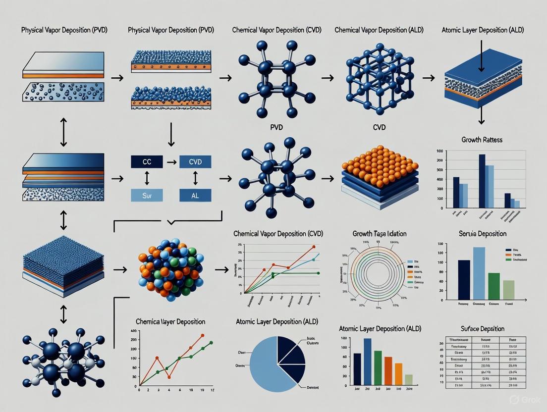
A Guide to Thin Film Growth Techniques
The properties of a thin film are intrinsically linked to its deposition method. The choice of technique determines the film's microstructure, density, purity, adhesion, and ultimately, its performance in a biomedical device.
Physical Vapor Deposition (PVD)
PVD is a vacuum-based process where the coating material is vaporized from a solid source and transported to the substrate, where it condenses to form a thin film.
- Sputter Deposition: This is the dominant PVD technique, accounting for 47.6% of the PVD market [5]. It involves bombarding a target material with energetic ions (e.g., Ar⁺), causing atoms to be ejected and deposited onto the substrate. It is renowned for excellent adhesion, high density, and the ability to coat complex shapes with uniform layers [6] [5].
- Thermal Evaporation: The source material is heated in a high vacuum until it evaporates, and the vapor condenses on the cooler substrate. It is suitable for high-purity, low-melting-point metals but can have poorer adhesion compared to sputtering [6].
Experimental Protocol Overview: Magnetron Sputtering of a TiN Coating
- Substrate Preparation: Substrates (e.g., Ti-6Al-4V alloy) are polished to a mirror finish, ultrasonically cleaned in acetone and ethanol, and dried.
- Chamber Evacuation: The deposition chamber is evacuated to a high base vacuum (e.g., < 1 × 10⁻⁶ mbar) to minimize contamination.
- Pre-sputter Etching: The substrate surface is often cleaned in-situ via Ar⁺ ion bombardment (e.g., RF bias) to remove native oxides and enhance adhesion.
- Deposition Parameters:
- Target: High-purity Titanium (Ti).
- Sputtering Gas: Argon (Ar).
- Reactive Gas: Nitrogen (N₂). The flow rate is precisely controlled to form the stoichiometric TiN phase.
- Pressure: Maintained in the range of 1-10 mTorr.
- Power: DC or RF power applied to the target (e.g., 200-500 W).
- Substrate Temperature: Can be varied from room temperature to several hundred °C to control film structure.
- Post-deposition Characterization: Film thickness (by profilometry), structure (XRD), morphology (SEM), composition (EDS), and mechanical properties (nanoindentation) are analyzed.
Chemical Vapor Deposition (CVD)
CVD involves the dissociation and/or chemical reaction of gaseous precursors on a heated substrate surface, forming a solid, deposited film. Variants like Plasma-Enhanced CVD (PECVD) allow for lower deposition temperatures, which is critical for substrates that cannot withstand high heat [7] [8].
Experimental Protocol Overview: Plasma-Enhanced CVD (PECVD) of a SiC Film
- Substrate Preparation: Silicon wafers are cleaned in diluted HF to remove native oxide and passivate the surface [8].
- Precursor Introduction: A single-source precursor, such as 1,3,5-trisilacyclohexane (TSCH), is vaporized and introduced into the chamber with a carrier gas [8].
- Plasma Ignition & Deposition:
- Chamber Pressure: Set to a low pressure (e.g., 200 mTorr) to promote a surface-reaction-limited growth regime for smoother films [8].
- Substrate Temperature: Typically in the range of 650–800°C [8].
- RF Power: Applied to generate a plasma, which provides the energy to decompose the precursors, enabling deposition at lower temperatures.
- Post-deposition Annealing: The film may be annealed (e.g., at 850-1100°C in a forming gas or Ar atmosphere) to improve its stoichiometry and reduce optical losses [8].
Atomic Layer Deposition (ALD)
ALD is a variant of CVD based on sequential, self-limiting surface reactions. Precursors are pulsed into the reactor one at a time, separated by purge periods. This cycle allows for atomic-scale control over film thickness, unparalleled conformality even on complex 3D structures, and excellent uniformity [9]. This makes ALD ideal for coating high-aspect-ratio nanostructures and creating ultra-thin, pinhole-free barrier layers.
Growth Mechanisms at the Atomic Scale
The initial stages of thin film formation follow one of three primary growth modes, which are critical for determining the final film morphology and are highly dependent on the interaction between the film and substrate material [10]:
- Volmer-Weber (Island Growth): 3D islands form directly on the substrate. This occurs when the atoms of the deposit are more strongly bound to each other than to the substrate (e.g., metals on ionic crystal substrates) [10].
- Frank-van der Merwe (Layer-by-Layer Growth): Atoms form complete two-dimensional layers one after another. This mode requires strong bonding between the deposit and the substrate and is ideal for producing atomically flat films (e.g., homoepitaxy) [10].
- Stranski-Krastanov (Layer-plus-Island Growth): One or a few complete monolayers form first, followed by the growth of 3D islands on top of this wetting layer. This is common in systems with lattice mismatch (e.g., Ge on Si) and can be used for self-assembled quantum dots [10].
Table 3: Comparative Analysis of Major Thin Film Deposition Techniques
| Parameter | Physical Vapor Deposition (PVD) | Chemical Vapor Deposition (CVD) | Atomic Layer Deposition (ALD) |
|---|---|---|---|
| General Principle | Physical ejection of atoms from a target and condensation on substrate. | Chemical reaction of gaseous precursors on heated substrate. | Sequential, self-limiting surface chemical reactions. |
| Typical Film Quality | High density, good adhesion. Can be polycrystalline. | High purity, conformal, dense. Polycrystalline or amorphous. | Excellent conformality, ultra-thin, pinhole-free. Amorphous or crystalline. |
| Deposition Rate | Medium to High (1-10 nm/s) [10] | Medium to High | Low (0.01-0.1 nm/s per cycle) |
| Substrate Temperature | Low to High (room temp. to >1000°C) | High (often >500°C); lower with Plasma (PECVD) | Low to Medium (room temp. to ~400°C) |
| Advantages | Good for metals & alloys; high deposition rates; no hazardous gases needed. | Excellent step coverage & conformality on complex shapes; wide material selection. | Atomic-level thickness control; best conformality; uniform on high-aspect-ratio structures [9]. |
| Disadvantages | Line-of-sight process can lead to shadowing; poor conformality on complex shapes. | High temperatures; toxic/flammable precursors may be used. | Very slow; expensive precursors; complex process control. |
| Primary Biomedical Use Cases | TiN, DLC, Ag coatings on orthopedic implants, tools. | SiC, Diamond, protective ceramic coatings. | Ultra-thin barrier layers, functionalization of nanoporous materials, drug-eluting implants. |
The Scientist's Toolkit: Essential Research Reagents and Materials
Table 4: Key Research Reagent Solutions for Thin Film Development and Analysis
| Reagent / Material | Function / Role | Example in Context |
|---|---|---|
| High-Purity Target (PVD) | Source material for the thin film. | Titanium (Ti) target for sputtering Ti or reactive sputtering of TiN coatings [2]. |
| Gaseous Precursors (CVD/ALD) | Reactant chemicals in vapor phase that decompose or react to form the solid film. | Silane (SiH₄), hydrocarbons (CH₄), ammonia (NH₃), metal-organic compounds like TSCH for SiC [8]. |
| Inert & Reactive Gases | Create plasma (sputtering), act as carrier gas, or participate in reactive deposition. | Argon (Ar - sputtering gas), Nitrogen (N₂ - reactive gas for nitrides), Hydrogen (H₂ - reducing atmosphere) [8]. |
| Substrates | The base material upon which the thin film is deposited. | Silicon wafers, Ti-6Al-4V alloy, 316L stainless steel, glass slides for testing [2] [8]. |
| Etchants & Cleaners | Prepare substrate surface to ensure good film adhesion and purity. | Hydrofluoric Acid (HF) for oxide removal from Si wafers [8]; acetone & ethanol for ultrasonic degreasing. |
| Characterization Tools | Analyze film properties post-deposition. | Spectroscopic Ellipsometry (thickness, refractive index), SEM/TEM (morphology), XRD (crystallinity), XPS (chemistry) [9] [8]. |
Thin films represent a cornerstone of modern biomedical device engineering, providing the critical interface between synthetic implants and the dynamic biological environment. Their significance lies in their ability to be meticulously engineered at the micro- and nanoscale to confer essential properties such as biocompatibility, corrosion and wear resistance, and bioactivity, which are unattainable by the bulk material alone. The selection and optimization of a deposition technique—be it PVD, CVD, or ALD—are paramount, as the growth mechanism and process parameters directly dictate the film's microstructure and ultimate performance. As the field advances, the integration of novel nanomaterials, multifunctional coatings with therapeutic agents, and smarter, more precise deposition technologies will continue to push the boundaries of what is possible in regenerative medicine and implantology, ultimately leading to longer-lasting and more biologically integrated medical devices.
Thin film growth is a fundamental process in the fabrication of modern devices, from microelectronics and photovoltaic cells to optical coatings. The structural quality and properties of these films are directly determined by the mechanism by which atoms assemble on a substrate. Among the various theoretical frameworks, three classic growth modes—Volmer-Weber, Frank-van der Merwe, and Stranski-Krastanov—form the cornerstone of our understanding of epitaxial growth. These models describe distinct pathways of film formation, governed by the interplay of surface, interface, and strain energies. This guide provides a comparative analysis of these fundamental modes, equipping researchers and scientists with the knowledge to select and optimize growth strategies for specific material systems and applications. The principles outlined are particularly relevant for the development of advanced semiconductor devices, quantum structures, and functional coatings in both research and industrial settings.
Fundamental Growth Modes: Definitions and Thermodynamic Criteria
The growth of epitaxial thin films on a crystal surface is primarily governed by the interplay of surface energies and lattice misfit. The three classical modes—Volmer-Weber (VW), Frank-van der Merwe (FM), and Stranski-Krastanov (SK)—are differentiated by a thermodynamic criterion based on the specific surface energy of the substrate (( \sigmas )), the film (( \sigmaf )), and the film-substrate interface (( \sigma_i )) [11].
The key parameter is the change in surface energy, ( \Delta\sigma = \sigmaf + \sigmai - \sigma_s ), which represents a measure of the substrate's wettability by the film material [11]:
- Frank–Van der Merwe (Layer-by-Layer) Growth: This mode occurs when ( \Delta\sigma \leq 0 ) and lattice misfit is negligible. The condition ( \Delta\sigma \leq 0 ) is equivalent to the adhesion energy between film and substrate atoms (( \psi' )) being greater than or equal to the cohesion energy between film atoms (( \psi )) [11]. In this scenario, atoms are more strongly bound to the substrate than to each other, leading to the formation of smooth, two-dimensional (2D) layers and resulting in atomically sharp interfaces [12] [10]. This is often considered the ideal growth mode for smooth films but is typically limited to systems with perfect lattice matching, often in homoepitaxy [12].
- Volmer–Weber (Island) Growth: This mode is characterized by ( \Delta\sigma > 0 ) (or ( \psi' < \psi )), meaning the atoms of the film are more strongly coupled to each other than to the substrate [11] [13]. This leads to incomplete wetting, where three-dimensional (3D) islands nucleate and grow directly on the substrate surface. This mode is common when there is a significant chemical or lattice mismatch between the film and substrate, such as metals like Au or Ag on ionic substrates like NaCl [10].
- Stranski–Krastanov (Layer-plus-Island) Growth: The SK mode is an intermediate process that begins with ( \Delta\sigma < 0 ) (or ( \psi' > \psi )), but with a non-zero lattice misfit [11]. Growth initiates by the formation of a few complete, two-dimensional wetting layers. However, as these layers thicken, strain energy accumulates elastically. At a critical thickness, it becomes energetically favorable for the system to relieve this strain by nucleating 3D islands on top of the wetting layer [14] [10]. Thus, the growth transitions from an initial 2D (FM) mode to a subsequent 3D (VW-like) mode.
Table 1: Thermodynamic and Kinetic Criteria for Thin Film Growth Modes
| Feature | Volmer-Weber (VW) | Frank-van der Merwe (FM) | Stranski-Krastanov (SK) |
|---|---|---|---|
| Energetic Criterion | ( \Delta\sigma > 0 ) (( \psi' < \psi )) [11] | ( \Delta\sigma \leq 0 ) (( \psi' \geq \psi )) [11] | ( \Delta\sigma < 0 ) & non-zero misfit [11] |
| Chemical Potential Derivative | ( d\mu/dN < 0 ) [11] | ( d\mu/dN > 0 ) [11] | ( d\mu/dN ) changes sign at critical thickness [11] |
| Growth Sequence | 3D islands form directly on substrate | Sequential, complete 2D layers | 2D wetting layer followed by 3D islands [14] |
| Driving Force for 3D Islands | Lower surface energy of islands | Not applicable | Strain relief in the wetting layer [14] |
Comparative Analysis of Growth Modes
A direct comparison of the three growth modes reveals their characteristic morphological evolution, material examples, and technological applications, which are summarized in Table 2 below.
Table 2: Characteristic Morphologies, Material Examples, and Applications of Growth Modes
| Aspect | Volmer-Weber (VW) | Frank-van der Merwe (FM) | Stranski-Krastanov (SK) |
|---|---|---|---|
| Schematic Morphology | 3D islands on bare substrate | Atomically smooth, sequential layers | 3D islands on a thin wetting layer [14] |
| Resulting Film Structure | Rough, polycrystalline or discontinuous initially | Very smooth, continuous, epitaxial | Continuous wetting layer with discrete, coherent islands [14] |
| Typical Material Examples | Au, Ag on NaCl or Si substrates [10]; AlN on Si [10] | Ag on Cu; Au on Pd; GaAs on GaAs (homoepitaxy) [10] | Ge on Si; InAs on GaAs; Ag on Si(111) [14] [10] |
| Primary Applications | Where substrate adhesion is less critical | High-quality epitaxial layers, superlattices, heterostructures [10] | Quantum dots [14], nanostructures for optoelectronics [10] |
The following diagram illustrates the morphological evolution of each growth mode over time, from the initial deposition of adatoms to the final film structure.
Experimental Protocols for Growth Mode Identification
Accurately identifying the operative growth mode during thin film deposition requires a combination of in-situ and ex-situ characterization techniques. The following section outlines standard methodologies for monitoring and distinguishing between VW, FM, and SK growth.
In-situ Monitoring Techniques
In-situ techniques are invaluable for observing real-time growth dynamics without breaking vacuum.
- Reflection High-Energy Electron Diffraction (RHEED): This is a primary technique in Molecular Beam Epitaxy (MBE) systems. The intensity of the RHEED specular spot oscillates with a period corresponding to the completion of each atomic monolayer in ideal layer-by-layer (FM) growth. The damping of these oscillations often signals a transition to 3D growth, as in the SK mode. A spotty pattern instead of streaky one indicates 3D island formation (VW or SK post-transition) [14].
- Auger Electron Spectroscopy (AES): The intensity of Auger peaks from the deposited film material is tracked as a function of deposition time or coverage. In FM growth, this intensity increases in a segmented linear fashion. In SK growth, a distinct break point at a critical adsorbate coverage is observed, followed by a linear segment with a reduced slope, indicating the onset of 3D island nucleation. For VW growth, a single line of low slope is typical from the beginning [14].
- Low-Energy Electron Microscopy/Diffraction (LEEM/LEED): LEEM allows direct real-time imaging of surface morphology, enabling visualization of 2D layer progression or 3D island formation. LEED can provide complementary diffraction information on surface structure and periodicity [14] [11].
Ex-situ Characterization Techniques
After growth, ex-situ techniques provide detailed structural and morphological information.
- Atomic Force/Scanning Tunneling Microscopy (AFM/STM): These scanning probe techniques provide direct, high-resolution (down to the atomic scale) visualization of the surface morphology. They are indispensable for confirming the presence and shape of 3D islands in VW and SK growth, measuring island densities, sizes, and size distributions [14] [11].
- Scanning/Transmission Electron Microscopy (SEM/TEM): SEM offers high-magnification imaging of surface islands. Cross-sectional TEM provides crucial information about the film's cross-section, including the structure of the wetting layer and islands in SK growth, and can reveal defects like dislocations [14].
The workflow for a typical experiment integrating these techniques is shown below.
The Scientist's Toolkit: Essential Reagents and Materials
Successful thin film growth and analysis requires specific instrumentation, reagents, and materials. The following table details key components of a research toolkit for studying classic growth modes.
Table 3: Essential Research Tools for Thin Film Growth Studies
| Tool Category | Specific Examples | Function & Relevance |
|---|---|---|
| Deposition Instruments | Molecular Beam Epitaxy (MBE) [10], Physical Vapor Deposition (PVD) [10], Chemical Vapor Deposition (CVD) [10] | Provide an ultra-high vacuum environment and atomic-scale control for depositing high-purity, epitaxial thin films. MBE is the gold standard for studying fundamental growth modes. |
| In-situ Characterization Tools | RHEED system [14], AES spectrometer [14], LEED/LEEM [14] | Enable real-time monitoring of growth dynamics, surface structure, and chemical composition without exposing the sample to air. |
| High-Purity Materials | Elemental sources (e.g., Ga, As, Ge, Si) for MBE [10], High-purity sputtering targets (e.g., CdTe, AlN) [10], Gaseous precursors for CVD (e.g., silane, methane) [10] | Serve as the source of the film and substrate materials. Purity is critical to minimize defects and contamination during growth. |
| Substrates | Single crystal wafers (e.g., Si, GaAs, Sapphire) [10] | Provide the crystalline template for epitaxial growth. Crystallographic orientation and surface termination are key variables [10]. |
| Computational Resources | Molecular Dynamics (MD) & Kinetic Monte Carlo (kMC) simulation codes [10] [15], High-Performance Computing (HPC) clusters | Used for atomistic modeling of growth processes, providing insights into adatom diffusion, nucleation, and defect formation that are challenging to observe directly [10]. |
Emerging Trends and Machine Learning in Growth Control
The field of thin film growth is being transformed by the integration of machine learning (ML) and advanced computational methods. A significant challenge in traditional growth monitoring is the human expert's inability to detect subtle, real-time changes in data streams, often leading to irreparable film defects before corrective action can be taken [16].
Researchers are now developing ML programs, such as the "RHAAPsody" process, to autonomously analyze in-situ data (e.g., electron diffraction images) and identify critical "change points" during deposition. This approach has demonstrated the ability to flag detrimental changes about a minute faster than human experts, a critical improvement for implementing real-time feedback control [16]. The long-term goal is fully autonomous film growth systems that can predict and adapt growth conditions to correct problems as they emerge [16]. Furthermore, machine learning is aiding in materials characterization, such as the rapid analysis of Raman spectroscopy data for characterizing large-scale bilayer graphene grown in an FM mode [17]. These advancements, coupled with powerful atomistic simulations like Molecular Dynamics (MD) and Kinetic Monte Carlo (kMC), are accelerating the understanding and optimization of complex growth processes for next-generation materials [10] [15].
In the pursuit of advanced thin-film technologies for applications in nanoelectronics, photovoltaics, and catalysis, precise control over film morphology at the atomic scale is paramount. The structural quality and ultimate performance of thin films are governed by fundamental atomic-scale processes—nucleation, coalescence, and adatom diffusion—that occur during the earliest stages of growth [10] [1]. These processes are highly sensitive to deposition conditions and the choice of substrate, leading to varied microstructural outcomes across different material systems and synthesis techniques [18] [10]. This guide provides a comparative analysis of these mechanisms across select material systems, highlighting how specific experimental parameters dictate nucleation kinetics, diffusion barriers, and coalescence behavior, thereby enabling researchers to make informed decisions for tailoring thin-film properties.
Fundamental Growth Mechanisms and Atomic-Scale Processes
Thin film growth initiates when vapor-phase atoms condense on a substrate, setting in motion a series of complex atomic-scale events. The classic growth modes—Volmer-Weber (island formation), Frank-van der Merwe (layer-by-layer), and Stranski-Krastanov (layer-plus-island)—provide a macroscopic framework for understanding film morphology, but their realization is dictated by atomic-scale kinetics and thermodynamics [10].
Adatom Diffusion: Upon landing on the substrate, atoms, known as adatoms, migrate across the surface. This migration is a thermally activated process described by the diffusivity ( D = D0 \exp(-ED/kB TS) ), where ( ED ) is the surface diffusion activation barrier, ( TS ) is the substrate temperature, and ( D_0 ) is the diffusivity prefactor [18]. The rate of this process relative to the vapor arrival rate (F) critically determines the subsequent nucleation and growth behavior.
Nucleation: Migrating adatoms collide and form stable clusters. The density of these nuclei (( N{sat} )) scales with the ratio of the arrival rate to diffusivity, often following ( N{sat} \sim (F/D)^{2/7} ) for a critical cluster size of one atom (i* = 1) [18]. Higher diffusivity (achieved at higher temperatures or on lower-energy substrates) favors the growth of existing islands over the nucleation of new ones, leading to a lower density of larger islands.
Coalescence: As islands grow, they eventually impinge on one another. The process of coalescence, where two islands merge into one to reduce their total surface energy, is critical for forming a continuous film. The time required for coalescence of a pair of islands scales as ( \tau_{coal} \sim R^4 / B ), where R is the island radius and B is a coalescence-rate parameter that scales with the adatom self-diffusivity [18]. If coalescence is slow relative to the rate of new island impingement, an elongated, porous network forms.
The dynamic competition between these processes—dictated by F, TS, and the intrinsic energy landscape of the substrate-film system—controls key transition points in film morphology, such as the nominal thickness at percolation (( \Theta{perc} )) and continuous film formation (( \Theta_{cont} )) [18].
Comparative Analysis of Atomic-Scale Processes Across Material Systems
The following tables synthesize quantitative data and qualitative observations from experimental and simulation studies, highlighting how adatom diffusion, nucleation, and coalescence behaviors vary across different material systems and growth conditions.
Table 1: Comparative Adatom Diffusion and Nucleation Kinetics
| Material System | Diffusion Barrier, E_D (eV) | Attempt Frequency, ν₀ (s⁻¹) | Nucleation Density Scaling | Growth Technique |
|---|---|---|---|---|
| Ag on amorphous Carbon (a-C) [18] | ~0.4 (cluster diffusion) | ~1×10⁹ | ( \Theta_{elong} \sim (D/F)^{1/7} ) | Magnetron Sputtering |
| Cu on amorphous Carbon (a-C) [18] | ~0.6 (cluster diffusion) | ~1×10¹¹ | ( \Theta_{elong} \sim (D/F)^{1/7} ) | Magnetron Sputtering |
| GaN on Graphene/sapphire [19] | Not directly measured | Not directly measured | Nucleation site density scales with ( f_p ) (perforated-area fraction) | Hydride Vapor Phase Epitaxy (HVPE) |
| Pt on Graphene [20] | Governed by NP coalescence | Governed by NP coalescence | Broad, right-skewed PSD from aggregative growth | Atomic Layer Deposition (ALD) |
Table 2: Coalescence Dynamics and Resulting Film Morphology
| Material System | Coalescence Mechanism | Governing Parameters | Resulting Morphology | Key Experimental Evidence |
|---|---|---|---|---|
| Ag/Cu on a-C [18] | Coalescence of immobile clusters | Temperature (T_S), Arrival Rate (F) | Percolated network → Continuous film | In situ sheet resistance & wafer curvature |
| GaN on perforated Graphene [19] | Lateral growth and "front-front" contact of domains | Perforated-area fraction (( f_p )) | Isolated domains → Coalesced film | Time-resolved areal coverage & domain counts |
| Pt on Graphene [20] | Smoluchowski aggregation (NP diffusion & coalescence) | Number of ALD cycles, Temperature | Narrow PSD (T < 100°C) → Broad PSD (T > 100°C) | TEM image analysis of Particle Size Distributions (PSD) |
Experimental Protocols for Probing Atomic-Scale Dynamics
Methodology for Quantifying Diffusion Barriers on Weakly-Interacting Substrates
This protocol, used to study Ag and Cu on amorphous carbon, determines surface diffusivity from characteristic morphological transitions [18].
- Core Principle: The nominal film thickness at the elongation (( \Theta{elong} )) or percolation (( \Theta{perc} )) transition scales with the surface diffusivity (D) and vapor arrival rate (F). By measuring this scaling relationship across temperatures, one can extract the effective diffusion barrier ( ED ) and attempt frequency ( \nu0 ) [18].
- Experimental Procedure:
- Deposition: Deposit metal films (e.g., Ag, Cu) via magnetron sputtering onto weakly-interacting substrates (e.g., a-C) across a temperature range (e.g., 298–413 K) and arrival rates, F (e.g., 0.08–5.38 monolayers/s).
- In Situ Monitoring: Use in situ, real-time sheet resistance and wafer curvature measurements to accurately determine the nominal thickness at percolation (( \Theta{perc} )) and continuous film formation (( \Theta{cont} )).
- Data Analysis:
- Establish the functional relationship between ( \Theta{perc} ) (or ( \Theta{cont} )) and F at a constant T_S.
- Plot ln(D) vs. ( 1/TS ), where D is derived from the scaling analysis. The slope yields the diffusion barrier ( -ED/kB ), and the intercept gives ln(( D0 )).
- Key Insight: The extracted ( ED ) and ( \nu0 ) often correspond to the diffusion of multiatomic clusters, not single adatoms, which is the rate-limiting process for morphological evolution in these systems [18].
Kinetic Monte Carlo (kMC) Modeling of Nucleation on Nanoperforated Masks
This computational protocol quantitatively links engineered surface parameters to nucleation statistics, as demonstrated for GaN growth on O₂-plasma-perforated graphene [19].
- Core Principle: A kMC model coarse-grains atomistic events (adatom arrival, diffusion, attachment at exposed substrate sites, and domain coalescence) to simulate the temporal and spatial evolution of nucleation.
- Simulation Procedure [19]:
- Setup: Initialize a discrete lattice representing the substrate. A fraction of sites, ( f_p ) (the effective perforated-area fraction), are designated as potential nucleation sites.
- Nucleation & Growth Loop: For each time step (routine):
- Nucleation Trials: Select ( No ) random lattice sites. If a site is empty and exposed (within a perforation), it becomes a nucleus with a constant per-step probability ( P0 ).
- Growth: Existing domains grow laterally at a defined rate.
- Coalescence Event: When two growing domains make "front–front" contact, coalescence is recorded.
- Validation: Fit the kMC outputs (nucleation delay time, areal coverage) to experimental data by tuning parameters like ( fp^{eff} ) and ( P0 ).
- Key Insight: The model confirmed that nucleation-site density scales linearly with ( fp ), while nucleation-delay time decreases approximately as ( 1/fp ), identifying ( f_p ) as a single, surface-engineerable parameter governing nucleation [19].
<100 chars: kMC Simulation Workflow>
The Scientist's Toolkit: Essential Research Reagents and Materials
Successful investigation and control of atomic-scale processes require carefully selected substrates, precursors, and analytical tools.
Table 3: Key Research Reagent Solutions for Thin-Film Growth Studies
| Reagent/Material | Function in Experiment | Exemplary Use Case |
|---|---|---|
| Weakly-Interacting Substrates (a-C, Graphene) [18] [20] | Provides a low-energy surface to study intrinsic metal adatom/cluster diffusion without strong chemical bonding. | Quantifying Ag/Cu cluster diffusivity [18]; Studying aggregative growth of Pt NPs [20]. |
| O₂ Plasma [19] | Engineered tool for creating nanoscale perforations (thru-holes) in 2D material masks (e.g., graphene). | Controlling nucleation site density (f_p) for GaN THE (Thru-Hole Epitaxy) [19]. |
| Metalorganic Precursors (e.g., Pt(MeCp)Me₃) [20] | Volatile precursor for self-limiting surface reactions in Atomic Layer Deposition (ALD). | Studying temperature-dependent nucleation & coalescence of Pt nanoparticles [20]. |
| In Situ Diagnostics (Sheet Resistance, Wafer Curvature) [18] | Real-time, non-destructive monitoring of morphological transitions (percolation, stress). | Determining Θperc and Θcont for diffusion coefficient calculation [18]. |
Visualization of Atomic-Scale Processes and Growth Outcomes
The following diagram illustrates the key atomic-scale processes and their connection to final film morphology, integrating the roles of critical parameters like substrate temperature and vapor arrival rate.
<100 chars: From Parameters to Morphology>
The comparative analysis presented herein underscores that the atomic-scale processes of nucleation, coalescence, and adatom diffusion are not universal constants but are highly dependent on the specific film-substrate system and growth conditions. Key differentiators include the dominance of single-atom versus cluster diffusion [18], the mechanism of coalescence (classical sintering vs. Smoluchowski aggregation) [20], and the ability to engineer nucleation sites via substrates like perforated graphene [19]. Mastery of these processes, achieved through integrated experimental and simulation approaches like in situ kinetics and kMC modeling, provides the foundational knowledge required to push the boundaries of thin-film technology in semiconductors, energy storage, and catalysis.
In the study of complex material processes, no single computational method can efficiently capture the full spectrum of atomic interactions and long-timescale evolution. Molecular Dynamics (MD) and Kinetic Monte Carlo (kMC) simulations have emerged as complementary techniques that, when integrated, bridge the gap between atomistic and mesoscopic scales [21]. MD simulations provide insights into atomic-scale dynamics by solving Newton's equations of motion, making them ideal for studying detailed atomistic pathways, defect formation, and surface reconstruction in real-time [10]. In contrast, kMC simulations excel at modeling rare events and thermally activated processes that occur over microsecond to second timescales, which are computationally inaccessible to MD [10]. This powerful combination enables researchers to understand phenomena ranging from thin film growth and catalyst design to ion transport in energy materials, providing a comprehensive framework for predicting and optimizing material properties and functions.
Fundamental Principles: A Comparative Analysis
Table 1: Fundamental Comparison Between MD and kMC Simulations
| Feature | Molecular Dynamics (MD) | Kinetic Monte Carlo (kMC) |
|---|---|---|
| Time Scale | Femtoseconds to nanoseconds [21] [10] | Microseconds to seconds, even years [22] [10] |
| Length Scale | Nanometers (typically up to ~0.01 μm for large systems) [22] | Up to micrometers (∼1 μm) [22] |
| Underlying Principle | Numerical solution of Newton's equations of motion [10] | Stochastic simulation of state-to-state transitions based on rate constants [21] |
| Nature of Simulation | Deterministic (with known atomic forces) | Stochastic (probabilistic) |
| Key Outputs | Atomic trajectories, structural evolution, vibrational frequencies [21] | Microstructural evolution, surface coverage, growth rates [21] |
| Energy Landscape | Samples continuous pathways between states | Jumps between pre-defined (meta)stable states [21] |
| Acceleration Factor | Not applicable; simulates every vibration | Can be >10²² over MD for suitable problems [22] |
| Computational Cost | High for large systems/long times | Generally lower for equivalent simulated time |
Molecular Dynamics: Capturing Atomistic Trajectories
MD simulations track the real-time motion of every atom in a system, making them powerful for observing fundamental atomic-scale processes such as adatom diffusion, defect formation, and surface reconstruction during thin film growth [10]. The accuracy of these simulations hinges on the quality of the interatomic potentials (e.g., Lennard-Jones, Embedded Atom Method), which describe the forces between atoms [10]. A significant limitation of MD is its confinement to short timescales, typically nanoseconds, due to the need to use femtosecond time steps to resolve atomic vibrations [21] [10]. This makes it impossible for MD to directly simulate processes governed by infrequent, thermally activated events, which are common in materials growth and evolution.
Kinetic Monte Carlo: Enabling Mesoscopic Time Scales
kMC simulations overcome the timescale limitation of MD by employing a coarse-grained approach. The system is mapped onto a lattice, and its evolution is modeled as a series of discrete jumps between pre-defined states, such as diffusion hops or reaction events [21]. The core of a kMC model is a catalog of all possible processes and their corresponding rate constants, often derived from MD or Density Functional Theory (DFT) calculations [23] [21]. The algorithm proceeds by randomly selecting and executing one of these processes, with a probability weighted by its rate constant. The system clock is then advanced by an time increment calculated from the total rate of all possible events [21]. This bypasses the simulation of every atomic vibration, allowing kMC to access timescales that are orders of magnitude longer than MD, from microseconds to years [22].
Applications in Thin Film Growth: Insights and Outcomes
Thin film growth is a quintessential multiscale process, and the integration of MD and kMC has been pivotal in unraveling its complex mechanisms. MD provides the foundational understanding of atomistic processes, while kMC simulates the resulting morphological evolution over experimentally relevant timescales.
Table 2: MD and kMC Applications in Thin Film Growth Research
| Research Focus | MD Contribution | kMC Contribution | Key Findings |
|---|---|---|---|
| Cu Thin Film on TiN [24] | Simulated initial island nucleation and atomic impingement during deposition. | Modeled long-time thermal relaxation, island coalescence, and final film structure. | Revealed atomistic mechanisms for a new crystallographic orientation relationship (ORB) at a slightly higher growth temperature. |
| II-VI Semiconductors (e.g., CdTe) [10] | Studied adatom mobility, defect formation, and the effect of substrate termination on atomic bonding. | Modeled nucleation dynamics, growth kinetics, and microstructural evolution over long times. | Identified temperature, substrate crystallography, and termination as critical factors controlling defect densities and film quality. |
| Oxide Thin Film Growth [25] | Used to study ion deposition and short-time surface reorganization. | Combined with MD in an adaptive scheme to simulate growth over ~0.5 ms, identifying diffusion mechanisms. | Methodology successfully modeled growth for different lattice mismatch scenarios, agreeing with experimental results. |
| Helium Bubble Formation in Metals [22] | Revealed fundamental physics: He cluster diffusion and vacancy displacement (on ~100 ns scale). | Accelerated kMC simulated bubble microstructure evolution over years, reaching μm-length scales. | Demonstrated that bubble nucleation is feasible via a defect-trapping mechanism, not just classical self-trapping. |
| Graphene Growth on Cu(111) [26] | Used for structural optimization and calculation of reaction energies for methane dehydrogenation. | Simulated the long-timescale dynamics of carbon atom migration and graphene nucleation. | Provided a multiscale understanding of how CVD parameters (T, P) affect the graphene deposition rate and quality. |
The synergy between MD and kMC is exemplified in the study of copper thin film growth on TiN substrates [24]. A hybrid MD/time-stamped force-bias Monte Carlo (tfMC) approach was used, where MD handled the fast deposition events, and tfMC, a type of kMC, enabled the necessary thermal relaxation. This combined methodology was crucial for understanding why a slight increase in growth temperature (~30°C) led to a completely different crystallographic orientation of the copper film, a phenomenon difficult to explain by experiment alone [24].
Methodological Protocols: From Theory to Implementation
Workflow for a Hybrid MD/kMC Study
The following diagram illustrates a generalized, iterative workflow for a multiscale study integrating MD and kMC to investigate a process like thin film growth.
Detailed Methodologies
Molecular Dynamics Protocol for Deposition
An MD simulation of thin film growth mimics the experimental deposition process [27]. The protocol generally involves:
- System Setup: A substrate crystal (e.g., TiN, Cu) is created and equilibrated. A vacuum layer is added above it [24] [26].
- Deposition: Atoms or molecules are introduced into the simulation box at random positions and orientations above the substrate, mimicking a vapor flux [27]. Their initial velocities are set according to the simulation temperature.
- Dynamics: Newton's equations of motion are integrated using a finite-difference algorithm (e.g., Verlet) with a time step of ~1 femtosecond [24]. The forces on all atoms are computed at each step using an interatomic potential.
- Data Collection: Atomic trajectories are recorded, allowing for post-analysis of phenomena like adsorption, surface diffusion, island nucleation, and defect formation [10].
Kinetic Monte Carlo Protocol for Growth
A lattice kMC simulation for thin film growth involves [21]:
- Lattice Definition: The simulation space is mapped onto a lattice, where each site represents a possible atomic position (e.g., an adsorption site on a crystal surface).
- Process Catalog and Rates: All possible elementary processes are defined (e.g., adsorption, desorption, diffusion hops to neighboring sites). Each process is assigned a rate constant
k_i, typically of the formk_i = ν * exp(-E_a / kT), whereE_ais the activation energy barrier, often obtained from MD or DFT [23] [21]. - Simulation Loop:
- Catalog Update: Identify all possible processes
iin the current system configuration and compute their ratesk_i. - Total Rate: Calculate the cumulative rate
k_tot = Σ k_i. - Event Selection: Generate a random number to select which event occurs, with a probability proportional to
k_i / k_tot. - Time Advancement: Advance the simulation clock by
Δt = -ln(r)/k_tot, whereris a random number between 0 and 1. - Execution: Update the system configuration by executing the selected event.
- Catalog Update: Identify all possible processes
- Output Analysis: The simulation tracks the evolution of surface morphology, island densities, and other mesoscopic properties over time.
Advanced and Accelerated kMC Schemes
For systems with vast disparities in process rates, standard kMC can become inefficient. Accelerated kMC schemes have been developed to overcome this. One method for simulating random diffusion, as in helium bubble formation, is protected random-walk-accelerated kMC [22]. This approach avoids simulating every single diffusion jump. Instead, clusters are moved in large steps, ensuring they don't encounter other clusters during that time (protected diffusion). When clusters come within a critical distance, the simulation switches to a classical, jump-based kMC to handle their encounters and mergers accurately [22]. This hybrid acceleration scheme can achieve speedups of over 10²² relative to MD, enabling simulations spanning years of real time [22].
The Scientist's Toolkit: Essential Research Reagents and Computational Solutions
Table 3: Key Computational Tools and Inputs for MD and kMC Simulations
| Tool/Reagent | Type | Function in Research |
|---|---|---|
| Interatomic Potentials (e.g., MEAM, LJ) [24] [10] | Computational Model | Defines the forces between atoms in MD simulations; accuracy is critical for reliable results. |
| Density Functional Theory (DFT) [26] | Computational Method | Provides high-accuracy data for adsorption energies, reaction pathways, and energy barriers, used to parameterize MD/kMC [21]. |
| Rate Constants Catalog [23] [21] | Model Input | A complete list of all possible elementary processes and their rates is the fundamental input for any kMC simulation. |
| Process Activation Energy (E_a) [21] | Model Parameter | The energy barrier for an elementary process; the most important quantity determining a rate constant in kMC. |
| Time-stamped force-bias MC (tfMC) [24] | Algorithm | A type of Monte Carlo that allows for time association, used in hybrid schemes with MD for efficient simulation of deposition and relaxation. |
| Ab Initio Software (e.g., VASP) [26] | Software Package | Performs quantum-mechanical DFT calculations to obtain parameters for classical simulations. |
MD and kMC simulations are not competing techniques but rather synergistic pillars of modern computational materials science. MD provides the atomistic-resolution "microscope" to observe fundamental processes, while kMC acts as the "time machine" that projects these findings to experimentally relevant scales. As the case studies in thin film growth demonstrate, their combined use offers an unparalleled ability to decode complex experimental observations, predict materials behavior, and rationally design synthesis protocols. The continued development of hybrid MD/kMC methodologies, accelerated algorithms, and more accurate interatomic potentials promises to further solidify their role as indispensable tools for researchers and engineers across disciplines.
In thin film technology, the precise control of growth parameters is a fundamental prerequisite for tailoring the structural, morphological, and functional properties of the final material. These parameters directly govern the atomic-scale processes during deposition, thereby determining the performance of thin films in applications ranging from microelectronics and photovoltaics to sensing and catalysis. Within the broad spectrum of influential factors, growth temperature, substrate interaction, and crystallographic orientation emerge as three particularly critical and interconnected variables. This guide provides a comparative analysis of these parameters, synthesizing experimental data and theoretical insights to offer a structured overview of their individual and synergistic impacts on thin film growth. The objective is to furnish researchers and scientists with a consolidated reference that bridges foundational concepts with recent experimental findings, thereby supporting the optimization of deposition processes for targeted material properties.
The Role of Growth Temperature
Growth temperature is a paramount parameter that directly influences atomic surface mobility, diffusion rates, and nucleation dynamics during thin film deposition. It exerts a profound effect on the crystallinity, defect density, and ultimate functional properties of the grown film.
Experimental Evidence and Comparative Data
The following table summarizes the impact of growth temperature on the properties of different thin film materials, as observed in recent studies.
Table 1: Comparative Impact of Growth Temperature on Thin Film Properties
| Material | Deposition Method | Temperature Range | Key Findings | Reference |
|---|---|---|---|---|
| AlN | Metal-Organic Chemical Vapor Deposition (MOCVD) | 900°C - 1300°C | Crystal quality greatly improved with increasing temperature; Optimal FWHM of 0.033° at 1300°C. | [28] |
| Ni | Radio Frequency (RF) Sputtering | 100°C - 350°C | Saturation magnetization (MS) peaked and coercivity (HC) and Gilbert damping were minimized at 250°C, indicating low defect density. | [29] |
| Mg & Al | Magnetron Sputtering | Room Temperature (RT) to 300°C | Lowest oxygen impurity concentration was achieved at a homologous temperature of 0.4. | [30] |
| General II-VI Semiconductors | Various (Theoretical Review) | - | Higher temperatures promote adatom migration, leading to smoother films and fewer defects; Lower temperatures result in rough, defective films. | [10] |
Detailed Experimental Protocol: Temperature Optimization in AlN MOCVD
The investigation into AlN thin films provides a clear protocol for temperature optimization [28].
- Methodology: The AlN films were grown on 2-inch c-plane (0001) sapphire substrates using a CRIUS 31×2″ CCS MOCVD system. Trimethylaluminum (TMA) and ammonia (NH₃) were used as the precursors for aluminum and nitrogen, respectively.
- Variable Control: The growth temperature was systematically varied from 900°C to 1300°C while other parameters, such as reactor pressure and precursor flow rates, were controlled.
- Characterization & Analysis: The crystal quality of the resulting films was quantitatively assessed using X-ray diffraction (XRD) to measure the Full Width at Half Maximum (FWHM) of the rocking curve. A lower FWHM value indicates superior crystal quality. The study conclusively demonstrated that the FWHM value decreased significantly as the growth temperature increased, with the optimal value of 0.033° achieved at 1300°C. This improvement is attributed to enhanced adatom surface mobility at higher temperatures, allowing atoms to find lower-energy lattice sites and thereby reduce crystal defects.
The Influence of Substrate Interaction
The nature of the interaction between the depositing material and the substrate—encompassing chemical bonding, lattice mismatch, and elastic forces—dictates the initial nucleation stage and subsequent growth mode, ultimately defining the film's morphology and stability.
Modes of Growth and Interaction Strength
The strength of adsorbate-substrate interactions is a primary factor in determining the growth mode [10] [31]:
- Volmer-Weber (Island Growth): Occurs when adsorbate-adsorbate interactions are stronger than adsorbate-substrate interactions, leading to the formation of three-dimensional islands. This is common for metals on ionic substrates or dissimilar materials like AlN on Si [10].
- Frank-van der Merwe (Layer-by-Layer Growth): Takes place when adsorbate-substrate interactions are strong and the materials have similar lattice structures, resulting in the completion of full monolayers before subsequent layers begin. Examples include Ag on Cu and homoepitaxial semiconductor growth [10].
- Stranski-Krastanov (Layer-plus-Island Growth): A hybrid mode where initial layer-by-layer growth is followed by the formation of three-dimensional islands after a critical strain energy is reached, often due to lattice mismatch [10].
Experimental Insights and Data
Table 2: Impact of Substrate Interaction on Thin Film Growth
| Interaction Type / Variable | Impact on Growth Dynamics & Film Properties | Reference |
|---|---|---|
| Strong Elastic Adsorbate-Substrate Interaction | Induces first-order transitions and pattern formation; leads to fewer, larger adsorbate islands and higher surface coverage. | [31] |
| Lattice Mismatch | Can introduce dislocations to relax misfit strain; allowable mismatch is material-dependent (~7% for semiconductors). | [10] |
| Substrate Composition (Multi-component) | Leads to a stationary surface morphology with a higher number of smaller adsorbate islands compared to one-component substrates. | [31] |
| Capping/Passivation | Al-capping layers effectively prevent post-deposition oxygen incorporation in reactive films like Ca; Mg and Al self-passivate. | [30] |
Experimental Protocol: Investigating Adsorbate-Substrate Bonding
Computational studies provide deep insight into how adsorbate-substrate bonding is investigated [31].
- Methodology: A reaction-diffusion model is used to simulate the spatio-temporal evolution of a monoatomic layer during low-pressure condensation. The model incorporates terms for adsorption, desorption, and inter-layer transference, with a free energy functional that includes both adsorbate-adsorbate and elastic adsorbate-substrate interactions.
- Variable Control: The key variable is the strength of the elastic adsorbate-substrate interaction term (Uel) in the model's free energy equation. The system's response to varying this strength is simulated.
- Characterization & Analysis: The output of the simulation is analyzed for pattern formation and morphological transformation. The study found that increasing the interaction strength stabilizes surface structures, increases coverage, and leads to the formation of fewer, larger islands. At high adsorption rates, strong interactions can even result in percolating adsorbate structures.
The Effect of Crystallographic Orientation
The crystallographic orientation of both the substrate and the growing film influences the surface energy, diffusion anisotropy, and the arrangement of defects, thereby affecting the film's microstructure and physical properties.
Key Mechanisms and Experimental Observations
- Pseudomorphism and Strain: A classic phenomenon observed in thin films is pseudomorphism, where a sufficiently thin film can adopt the crystal structure of the substrate, leading to an abnormal, strained structure. For instance, aluminium deposited on platinum can acquire a face-centred tetragonal structure instead of its normal cubic structure [32].
- Defect Propagation: The crystallographic orientation determines how defects such as dislocations and stacking faults evolve. Simulation studies on II-VI semiconductors highlight that orientation affects the arrangement and density of these defects, which in turn impacts electronic properties crucial for devices like photovoltaic cells [10].
- Anisotropic Growth: Different surface terminations possess varying atomic arrangements and reactive sites. For example, during the growth of CdTe, the behavior of adatoms and the resulting defect distribution are significantly different on Cd-terminated surfaces compared to Te-terminated surfaces [10].
Synergistic Interplay of Parameters
The parameters of temperature, substrate interaction, and orientation do not act in isolation. Their synergistic interplay is critical for defining the final film microstructure. For instance, the optimal growth temperature for a material is not a fixed value but is contingent upon the specific substrate and its orientation. A higher growth temperature might be necessary to overcome a large lattice mismatch by providing sufficient energy for adatoms to find lower-defect configurations. Conversely, on a closely matched substrate, excessive temperature could lead to undesirable effects like interdiffusion or roughening. Furthermore, the efficiency of a protective capping layer in preventing impurity incorporation is strongly dependent on the underlying film morphology, which is itself a product of the growth temperature and substrate interaction [30].
The following diagram illustrates the logical relationship between these key growth parameters and their collective impact on the final thin film properties.
The Scientist's Toolkit: Essential Research Reagents and Materials
Table 3: Key Research Reagent Solutions for Thin Film Growth Studies
| Item / Solution | Function & Application | Example from Research |
|---|---|---|
| MOCVD System (e.g., AIXTRON CRIUS) | High-precision deposition of single-crystalline semiconductor thin films from vapor-phase precursors. | Used for growing high-quality, crack-free AlN films on sapphire substrates [28]. |
| High-Purity Sputtering Targets (e.g., Mg, Al, Ca >99.5%) | Source material for physical vapor deposition; purity is critical for minimizing unintentional doping and defects. | Essential for studying impurity incorporation and morphological evolution in sputtered metallic films [30]. |
| c-plane (0001) Sapphire Substrate | A common, stable, and insulating substrate for heteroepitaxial growth of semiconductors like GaN and AlN. | Provided the template for epitaxial growth of AlN thin films in MOCVD studies [28]. |
| Trimethylaluminum (TMA) & Ammonia (NH₃) | Metal-organic and hydride precursors, respectively, for the MOCVD growth of group III-nitride films (AlN, GaN). | Reacted in the vapor phase to form AlN thin films [28]. |
| Aluminum Capping Layer | A thin, protective layer deposited in-situ to prevent post-growth oxidation and impurity uptake of reactive films. | Used to protect Ca films from atmosphere exposure, enabling study of growth-related vs. post-growth impurities [30]. |
| X-Ray Diffractometer (XRD) | Primary tool for characterizing crystal structure, phase, orientation, and quality (via FWHM) of thin films. | Used to measure the crystal quality (FWHM) of AlN and the structure of Ni films [28] [29]. |
The controlled synthesis of thin films with tailored properties hinges on a deep and nuanced understanding of key growth parameters. As this guide has detailed, growth temperature is a master variable controlling kinetic processes and defect formation. The substrate interaction fundamentally sets the stage for the growth mode and initial film morphology. Finally, the crystallographic orientation of the involved materials guides anisotropic growth and defect evolution. The most successful growth strategies emerge from optimizing these parameters not in isolation, but in concert, acknowledging their powerful synergy. Future progress in the field will be accelerated by the integration of advanced simulation techniques like molecular dynamics and kinetic Monte Carlo [10], coupled with emerging autonomous experimentation platforms that use machine learning to rapidly identify optimal growth windows [16]. This combined approach promises to unlock new frontiers in the precision fabrication of thin films for next-generation technologies.
A Toolkit for Innovation: Deposition Methods and Their Biomedical Applications
Physical Vapor Deposition (PVD) represents a cornerstone suite of vacuum deposition techniques used to create high-purity, high-performance thin films and coatings. The fundamental principle of all PVD processes involves the physical transference of material from a solid or liquid source to a vapor phase, followed by condensation and film formation on a substrate surface [33] [34]. These techniques are distinguished from chemical vapor deposition (CVD) by their reliance on physical mechanisms for vapor generation rather than chemical reactions [35]. Operating typically under low or high vacuum conditions, PVD enables precise control over film properties including thickness, adhesion, density, stress, and electrical/optical characteristics, making these methods indispensable across numerous high-technology sectors including microelectronics, photovoltaics, medical devices, and aerospace [33] [34].
The global PVD market, valued at approximately $22.8-$24.67 billion in 2024, demonstrates strong growth prospects with a projected compound annual growth rate (CAGR) of 7.7%-8.3%, potentially reaching $33.1-$33.89 billion by 2029 [36] [37] [38]. This expansion is propelled by several key factors: regulatory limitations on hazardous alternatives like hexavalent chromium electroplating, increasing demand for decorative coatings, digitalization and automation in PVD processes, and growth in end-use industries such as semiconductors, medical devices, and green energy technologies [36] [33]. As the semiconductor industry advances toward more complex architectures and materials, PVD techniques have become increasingly critical for applications ranging from metallization and interconnects to advanced barrier layers [35].
This guide provides a comprehensive technical comparison of three principal PVD techniques: Sputtering, Pulsed Laser Deposition (PLD), and Thermal Evaporation. Within the broader context of thin film growth technique research, we objectively evaluate these methods based on their fundamental mechanisms, experimental parameters, resultant film properties, and suitability for specific applications, with particular relevance to researchers, scientists, and technology development professionals working at the forefront of materials science and device fabrication.
Fundamental Principles and Mechanisms
Cross-Technique Comparative Mechanisms
The following diagram illustrates the fundamental operational principles and workflow relationships between the three PVD techniques compared in this guide.
Individual Technique Mechanisms
Sputtering Deposition
Sputtering deposition operates on the principle of momentum transfer through gaseous plasma ionization [33] [34]. In this process, a target material (cathode) is bombarded with energetic ions of an inert gas, typically argon, created in a glow discharge plasma. This ion bombardment causes atoms from the target to be ejected due to momentum transfer and deposited onto a substrate [34] [39]. Variants include DC magnetron sputtering (for conductive materials), RF sputtering (for dielectric materials), and advanced techniques like High-Power Impulse Magnetron Sputtering (HiPIMS) that generate highly ionized plasma for denser films with minimal defects [33] [34]. Reactive sputtering, performed in the presence of reactive gases like oxygen or nitrogen, enables the formation of compound films such as oxides or nitrides [33].
Pulsed Laser Deposition (PLD)
PLD utilizes high-energy laser ablation for vapor generation [39] [37]. An intense pulsed laser beam is focused onto a target material, causing rapid localized heating, vaporization, and the creation of a plasma plume containing atoms, ions, and molecules from the target [39]. This ablation plume expands rapidly toward and condenses on the substrate, forming a thin film. The process offers exceptional stoichiometric transfer for multi-component materials, as all elements are vaporized nearly simultaneously regardless of their vapor pressures [39]. The high energy of ablated species often results in high-quality crystalline films, with the pulsed nature enabling precise thickness control through the number of laser pulses [39].
Thermal Evaporation
Thermal evaporation relies on resistive or electron-beam heating to vaporize source materials [33] [34]. In resistive thermal evaporation, the source material is heated in a refractory metal boat or basket until it reaches its evaporation temperature, producing a vapor stream that travels line-of-sight to condense on the substrate [34]. Electron-beam (e-beam) evaporation uses a focused electron beam to locally heat and vaporize material from a specific spot on a source ingot, enabling deposition of high-melting-point materials [34] [39]. Both approaches are characterized by relatively high deposition rates and are preferred for applications sensitive to plasma damage, though they typically offer poorer step coverage compared to sputtering techniques [33].
Experimental Protocols and Methodologies
Standardized Experimental Setup
The table below outlines the core experimental protocols and system configurations for implementing each PVD technique, providing researchers with foundational methodologies for laboratory application.
Table 1: Standard Experimental Protocols for PVD Techniques
| Experimental Parameter | Sputtering Deposition | Pulsed Laser Deposition (PLD) | Thermal Evaporation |
|---|---|---|---|
| Vacuum Requirements | Base pressure: 10⁻⁶ to 10⁻⁸ Torr; Sputtering pressure: 1-100 mTorr with Ar gas [34] | High vacuum: 10⁻⁶ to 10⁻⁸ Torr base pressure; Possible background gas for reactive deposition [39] | High vacuum: 10⁻⁶ to 10⁻⁸ Torr to minimize contamination and gas scattering [34] |
| Substrate Preparation | Standard cleaning (solvents, plasma); Possible heating (RT-500°C); Possible bias application [33] | Standard cleaning; Often heated (RT-900°C) for crystalline growth; Precise positioning [39] | Standard cleaning; Moderate heating (RT-300°C) to improve adhesion; No bias typically [34] |
| Source Configuration | Planar or rotary target (metal, ceramic); RF or DC power supply; Magnetron for enhanced efficiency [33] [34] | Dense, sintered target of desired composition; Rotating for uniform erosion; Laser-transparent window [39] | Crucible (W, Mo, Ta, C) or e-gun hearth; Wire, basket, or boat for resistively heated materials [34] |
| Process Control | Power (100W-5kW); Pressure; Gas flow; Substrate bias; Rotation for uniformity [33] | Laser energy density (1-10 J/cm²); Repetition rate (1-100 Hz); Substrate temperature; Target-substrate distance [39] | Source temperature (monitored); Deposition rate (0.1-50 nm/s via quartz crystal); Substrate rotation [34] |
Advanced Process Variations
Beyond standard configurations, several advanced process variations have been developed to address specific material and application requirements:
Sputtering: Reactive sputtering processes introduce controlled flows of reactive gases (e.g., O₂, N₂) alongside argon to deposit compound films such as oxides, nitrides, or oxynitrides. This requires precise gas flow control and plasma monitoring to maintain stoichiometry and avoid target poisoning [33]. High-Power Impulse Magnetron Sputtering (HiPIMS) utilizes short, high-power pulses to generate extremely dense, highly ionized plasmas, resulting in films with superior density, smoothness, and adhesion compared to conventional DC magnetron sputtering [34].
Pulsed Laser Deposition: For complex oxide materials, background oxygen pressure is frequently employed during deposition to maintain oxygen stoichiometry in the growing film. The use of dual-laser systems or hybrid approaches combining PLD with other techniques like radical source assistance can further enhance film quality and control over crystallinity [39].
Thermal Evaporation: Multi-source evaporation employs separate crucibles for different materials, allowing for sequential or co-deposition of layered structures or alloys. For e-beam evaporation, sweeping beam patterns and multiple hearth crucibles enable sequential deposition of different materials without breaking vacuum, which is particularly valuable for complex device structures [34].
Performance Comparison and Experimental Data
Quantitative Technique Comparison
The table below provides a comprehensive comparison of the three PVD techniques across critical performance parameters, synthesizing data from experimental studies and industrial applications.
Table 2: Quantitative Performance Comparison of PVD Techniques
| Performance Parameter | Sputtering Deposition | Pulsed Laser Deposition (PLD) | Thermal Evaporation |
|---|---|---|---|
| Typical Deposition Rate | 0.01-2 nm/s (varies with power, pressure, material) [33] | ~0.01-0.1 nm/pulse; Overall rate lower due to pulsed nature [39] | 0.1-50 nm/s; Generally the highest rates among PVD [34] |
| Film Uniformity | Excellent (1-2% over 150-200mm); Enhanced with substrate rotation [33] | Good on small areas; Decreases with increasing substrate size [39] | Good for point sources; Requires planetary fixtures for large areas [34] |
| Step Coverage & Conformality | Moderate; Improved with bias, pressure control, and source positioning [33] | Line-of-sight; Limited conformality similar to evaporation [39] | Poor (line-of-sight); Non-uniform on complex topography [34] |
| Film Density & Pinholes | Moderate to high density; HiPIMS produces densest films with minimal defects [34] | Typically very high density; Low pinhole density [39] | Lower density than sputtering; Columnar microstructure common [34] |
| Adhesion Strength | Excellent (high energy of deposited atoms ~1-10 eV) [33] | Good to excellent (high energy species in plume) [39] | Moderate; Often requires adhesion layers or substrate heating [34] |
| Stoichiometry Transfer | Good for alloys; Excellent for reactive deposition of compounds [33] | Exceptional for multi-component materials (congruent vaporization) [39] | Poor for alloys with component vapor pressure differences [34] |
Material-Specific Performance Data
Experimental studies provide quantitative insights into the performance of each technique with specific materials:
Sputtering Performance: Research on tungsten thin films demonstrates that HiPIMS with a 100 μs pulse length produces significantly denser films with minimal defects compared to conventional DC magnetron sputtering (dc-MS), leading to stable sensor performance and remarkable TCR (temperature coefficient of resistance) values, particularly for annealed samples [34]. HiPIMS films maintained superior initial characteristics even without thermal treatment, offering both time and cost efficiency [34].
Thermal Evaporation Applications: Studies on electron beam evaporation for depositing Al₂O₃ insulation layers emphasize the pivotal role of thermally grown alumina in enhancing electrical insulation performance under high-temperature conditions, with insulation resistance exceeding 2 MΩ attained at 800°C [34]. This demonstrates the capability of certain thermal evaporation processes to produce thermally resilient coatings for demanding applications.
PLD for Complex Materials: The unique advantage of PLD in preserving complex stoichiometries makes it particularly suitable for depositing advanced materials such as high-temperature superconductors, multiferroic oxides, and complex semiconductor heterostructures that require precise cation ratios for optimal performance [39].
Research Reagent Solutions and Essential Materials
Successful implementation of PVD techniques requires specific materials and reagents tailored to each deposition method. The following table catalogues essential research solutions with their specific functions in the PVD process.
Table 3: Essential Research Reagent Solutions for PVD Techniques
| Material/Reagent | Primary Function | Application Notes |
|---|---|---|
| High-Purity Targets (Sputtering, PLD) | Source material for thin film deposition | 2-8 inch diameters, 99.95%-99.999% purity; Metallic, ceramic, or composite for sputtering; Dense, sintered for PLD [33] [39] |
| Evaporation Sources (Thermal Evaporation) | Source material in wire, pellet, or powder form | 99.99%-99.999% purity; Formed to fit specific crucibles/boats; Alloys often challenging due to preferential evaporation [34] |
| Ultra-High Purity Process Gases | Plasma generation (Ar) and reactive deposition (O₂, N₂) | Argon: 99.9995%-99.9999%; Reactive gases: 99.995%-99.999%; Gas purification filters often employed [33] [34] |
| High-Vacuum Components | Creation and maintenance of deposition environment | Diffusion pumps, turbomolecular pumps, cryopumps; Vacuum gauges; High-temperature substrates heaters [34] |
| Specialized Substrates | Support for thin film growth | Si, SiO₂/Si, sapphire, glass, specialized crystals (MgO, LAO); Often with specific orientations and surface finishes [35] |
| Surface Characterization Standards | Quality verification and process calibration | Certified thickness standards, roughness specimens, composition standards for SEM, AFM, XPS, XRD analysis [34] |
Application-Specific Recommendations
Technique Selection Guidance
The choice between sputtering, PLD, and thermal evaporation depends significantly on the specific application requirements, material systems, and available resources. The following diagram illustrates the decision-making workflow for selecting the most appropriate PVD technique based on key project parameters.
Industry-Specific Applications
Each PVD technique has found particular prominence in specific industrial sectors based on its unique advantages:
Sputtering Dominance: The sputtering segment leads the PVD market and is widely employed in semiconductor manufacturing for barrier layers (e.g., Tantalum Nitride - TaN, Titanium Nitride - TiN), seed layers (Copper - Cu, Cobalt - Co), and interconnect metals [36] [33]. It's also extensively used for transparent conductive oxides (Indium Tin Oxide - ITO) in displays and photovoltaics, hard coatings (TiN, TiAlN) on cutting tools, and decorative coatings on consumer goods [36] [33].
Thermal Evaporation Applications: Thermal evaporation remains important for optical coatings, specific metal layers in electronics where plasma damage is a concern, some compound semiconductor applications, and decorative coatings [33]. Its simplicity, relatively low cost, and high deposition rates make it suitable for applications where extreme conformity and precise stoichiometric control are not primary requirements [33] [34].
PLD Research Applications: PLD is predominantly utilized in research and development environments for depositing complex multi-component materials such as high-temperature superconductors, multiferroics, complex oxides, and other materials where stoichiometric preservation is critical [39]. Its ability to work with multiple targets in sequence makes it valuable for creating novel multilayer structures and combinatorial materials research [39].
This comparative analysis demonstrates that sputtering, pulsed laser deposition, and thermal evaporation each occupy distinct and valuable positions within the PVD technology ecosystem. Sputtering emerges as the most versatile and widely adopted technique, particularly in industrial settings where large-area uniformity, excellent adhesion, and moderate-to-high deposition rates are paramount. Pulsed laser deposition offers unparalleled capability for stoichiometric transfer of complex multi-component materials, making it indispensable for advanced materials research despite limitations in scalability. Thermal evaporation provides a straightforward, cost-effective approach with the highest deposition rates for applications where line-of-sight limitations and potential stoichiometry deviations are acceptable trade-offs.
The continuing evolution of these technologies—through developments such as HiPIMS for sputtering, hybrid PVD processes, and advanced process control with AI/ML—ensures they will remain fundamental to semiconductor, energy, medical, and aerospace applications [36] [33]. For researchers and technology developers, the selection among these techniques should be guided by specific material requirements, performance specifications, and economic considerations, with the understanding that hybrid approaches and process innovations continue to expand the capabilities of physical vapor deposition for increasingly demanding applications in advanced manufacturing and materials science.
Chemical Vapor Deposition (CVD) and Atomic Layer Deposition (ALD) for Ultra-Thin, Conformal Coatings
The relentless drive for device miniaturization and the emergence of complex three-dimensional architectures in fields ranging from semiconductors to quantum computing have fundamentally shifted the requirements for thin-film deposition technologies. Conventional techniques often struggle to uniformly coat high-aspect-ratio structures or control thickness at the atomic scale. In this context, Chemical Vapor Deposition (CVD) and Atomic Layer Deposition (ALD) have emerged as two cornerstone technologies for producing ultra-thin, conformal coatings. ALD, a variant of CVD, has gained prominence where atomic-level precision is paramount. This guide provides an objective comparison of these two powerful techniques, framing them within the broader research landscape of thin-film growth and providing the experimental data and methodologies essential for researchers and development professionals.
Table 1: Core Characteristics of CVD and ALD
| Feature | Chemical Vapor Deposition (CVD) | Atomic Layer Deposition (ALD) |
|---|---|---|
| Fundamental Mechanism | Continuous chemical reactions of precursors mixed in gas phase at substrate surface [40] [41] | Sequential, self-limiting surface reactions from separated precursor pulses [42] [43] |
| Growth Nature | Continuous and linear [41] | Cyclical and saturative (sub-monolayer per cycle) [43] |
| Typical Deposition Rate | High ( nm/min to µm/min ) | Slow ( 0.1-0.2 nm/cycle, typically 1-2 Å/cycle ) [44] [41] |
| Thickness Control | Good, but less precise than ALD | Excellent, digital control via cycle count [42] [43] |
| Conformality on 3D Structures | Good, but can vary with aspect ratio | Exceptional, uniform on complex geometries and high-aspect-ratio structures [42] [43] |
| Primary Advantage | High growth rate, material versatility | Atomic-scale precision, unparalleled conformality [40] [43] |
| Typical Process Temperature | 600-1000°C (Thermal CVD); Lower for PECVD [41] | 150-350°C (Thermal ALD); Can be lower with plasma enhancement [44] [41] |
Fundamental Mechanisms and Experimental Protocols
Understanding the distinct reaction mechanisms of CVD and ALD is crucial for selecting the appropriate technique. The experimental workflows differ significantly, which directly impacts the properties of the resulting film.
CVD Reaction Mechanism and Workflow
In a standard CVD process, precursor gases are simultaneously introduced into a reaction chamber where they mix and react either in the gas phase or on a heated substrate surface to form a solid film. The process relies on continuous chemical reactions, and growth rate is influenced by factors such as precursor concentration, temperature, and flow dynamics [40] [41]. This can lead to challenges in achieving uniform thickness on non-planar surfaces, as the reactant concentration can deplete within deep trenches or around complex features.
ALD Reaction Mechanism and Workflow
ALD is defined by its sequential, self-limiting surface reactions. A typical thermal ALD cycle for a metal oxide, such as Al₂O₃ using trimethylaluminum (TMA) and water (H₂O), consists of four distinct steps [44] [40]:
- Precursor A Exposure: Pulse of TMA vapor into the chamber. The TMA molecules chemisorb onto the substrate's surface functional groups (e.g., -OH), forming a saturated monolayer and releasing reaction by-products.
- Purge I: An inert gas (e.g., N₂) purges the chamber, removing all excess TMA and reaction by-products.
- Precursor B Exposure: Pulse of H₂O vapor into the chamber. The H₂O reacts with the adsorbed TMA monolayer, converting it to Al₂O₃ and regenerating the surface functional groups.
- Purge II: A second inert gas purge removes excess H₂O and any remaining by-products.
This cycle is repeated n times to achieve a desired film thickness, with each cycle adding a sub-monolayer of material (the Growth Per Cycle, or GPC) [43]. The self-limiting nature of each half-reaction is the key to ALD's exceptional conformality and thickness control.
Diagram 1: The sequential, self-limiting reaction cycle of Atomic Layer Deposition (ALD).
Performance Comparison and Experimental Data
The fundamental differences in mechanism translate directly to measurable differences in film performance, particularly regarding conformality, thickness control, and material properties. The following table summarizes quantitative and qualitative comparisons based on experimental and industrial data.
Table 2: Comparative Performance Data for CVD and ALD
| Performance Metric | CVD Coating | ALD Coating | Experimental Context & Notes |
|---|---|---|---|
| Thickness Uniformity | Moderate to Good | Excellent (typically ±1-2% across wafer) [43] | ALD's self-limiting growth ensures uniformity independent of precursor flux once saturated. |
| Conformality on HAR Structures | Good, but can be line-of-sight limited | Exceptional (Step coverage >95% on structures with >1000:1 aspect ratio) [43] | Conformality is quantified as the ratio of film thickness at the bottom of a feature to that at the top. |
| Growth Rate | High (nm/min to µm/min range) | Slow (0.1-0.2 nm/cycle, Å/cycle range) [41] | ALD's low growth rate is a trade-off for its precision. Throughput can be improved with Spatial ALD [45]. |
| Film Density | High | High, often pinhole-free [42] | Both techniques can produce high-quality, dense films, though ALD is noted for producing pinhole-free films at ultra-thin dimensions. |
| Crystallinity | Can be crystalline (epitaxial) | Often amorphous, can be crystalline at higher temperatures | CVD is widely used for epitaxial growth (e.g., MOCVD). ALD films are often amorphous but can be crystallized. |
| Typical Film Thickness Range | Dozens of nm to several µm | <1 nm to ~100 nm (thicker films are time-consuming) [44] | ALD is the preferred technology for nanometre-size layer thickness or pinhole-free layers [40]. |
| Industrial Throughput | High for moderate thicknesses | Lower for traditional temporal ALD; improved with Spatial ALD [45] | Spatial ALD separates precursors in space rather than time, enabling deposition rates up to two orders of magnitude faster [45]. |
Advanced Experimental Protocols
Protocol: Thermal ALD of Al₂O₃ using TMA and H₂O
This is a standard process for depositing high-quality aluminum oxide dielectric films [44] [40].
- Objective: To deposit a conformal, pinhole-free Al₂O₃ film of precisely controlled thickness.
- Substrate Preparation: Clean substrate (e.g., Si wafer) to ensure a hydrophilic surface with ample -OH termination.
- Reactor Conditions: Set reactor temperature within the "ALD window" (e.g., 150-300°C). The process can be run at 150°C using a plasma-enhanced approach [44].
- Precursors: Trimethylaluminum (TMA, Al(CH₃)₃) as the metal source and deionized water (H₂O) as the oxidant.
- Cycle Parameters:
- TMA Pulse: 0.1 seconds.
- First N₂ Purge: 5 seconds.
- H₂O Pulse: 0.1 seconds.
- Second N₂ Purge: 5 seconds.
- Growth Calibration: The Growth Per Cycle (GPC) for this chemistry is typically ~1.0 Å/cycle [44]. To deposit a 10 nm film, approximately 100 cycles are required.
- Characterization: Film thickness is measured by spectroscopic ellipsometry. Conformality can be verified by cross-sectional SEM or TEM on patterned wafers with high-aspect-ratio trenches.
Protocol: CVD of TiN from TiCl₄ and NH₃
This process is commonly used for depositing conductive titanium nitride diffusion barriers.
- Objective: To deposit a conformal TiN film as a diffusion barrier.
- Reactor Conditions: Typical reactor temperature is 400-700°C.
- Precursors: Titanium Tetrachloride (TiCl₄) and Ammonia (NH₃).
- Process Parameters: Both precursors are introduced simultaneously into the chamber. Flow rates, chamber pressure, and temperature are critically controlled to dictate film properties, growth rate, and uniformity.
- Challenges: The use of chloride precursors can lead to corrosive by-products and chlorine incorporation in the film. Achieving uniform coverage on very high-aspect-ratio structures is more challenging than with ALD.
The Researcher's Toolkit: Essential Reagent Solutions
The success of CVD and ALD processes is heavily dependent on the selection of appropriate precursor chemistries.
Table 3: Key Research Reagents and Their Functions
| Reagent Solution | Function in Deposition | Common Examples & Notes |
|---|---|---|
| Metal-Organic Precursors | Provide the metal cation for the thin film. | Trimethylaluminum (TMA for Al₂O₃), Tetrakis(dimethylamido)titanium (TDMAT for TiN). Must have sufficient volatility and thermal stability [41]. |
| Metal Halide Precursors | Alternative source for metal cations. | Titanium Tetrachloride (TiCl₄ for TiN or TiO₂), Tungsten Hexafluoride (WF₆ for W). Can be highly reactive but may produce corrosive by-products [41]. |
| Oxidants | React with metal precursors to form metal oxide films. | H₂O, O₃, O₂ plasma. Ozone and plasma allow for lower process temperatures and improved film quality [44]. |
| Nitrogen Sources | React with metal precursors to form metal nitride films. | Ammonia (NH₃), N₂ plasma, hydrazine (N₂H₄). Plasma enhancement is often used to crack strong N≡N bonds at lower temperatures. |
| Reducing Agents | Used for the deposition of pure metal films. | Molecular hydrogen (H₂), formalin (H₂CO), plasma species. React with metal precursors, removing the ligand shell and leaving behind the metal. |
| Inert Purge Gas | Separates precursor pulses in ALD, removes reaction by-products. | Nitrogen (N₂), Argon (Ar). Must be high purity to prevent unwanted reactions or contamination [44] [43]. |
CVD and ALD are complementary, rather than competing, technologies in the research and development of ultra-thin conformal coatings. The choice between them is dictated by the specific application requirements.
- CVD is the preferred method when high deposition rates and thicker films are needed, and where extreme conformality on the most challenging nanostructures is not the primary driver. It offers a broader established base of processes for a wide range of materials.
- ALD is unmatched for applications demanding atomic-scale thickness control, perfect conformality on complex 3D architectures, and the synthesis of pinhole-free films at a nanometre-scale thickness [42] [43]. Its primary trade-off is a lower deposition rate, though advancements like Spatial ALD are actively addressing this bottleneck [45].
The ongoing evolution of both techniques, particularly in precursor chemistry and reactor design, continues to expand their capabilities. For researchers, the decision framework rests on prioritizing the triad of precision, conformality, and throughput, with ALD excelling in the first two and CVD in the third, guiding their selection in the sophisticated toolkit of thin-film growth techniques.
Thin-film technology is a cornerstone of modern science and industry, enabling advancements in sectors ranging from semiconductors and photovoltaics to biomedical devices and protective coatings [1]. The properties of these films—typically ranging from nanometers to several micrometers in thickness—are profoundly influenced by their deposition methods [1]. Among the various fabrication techniques, solution-based processing offers a unique combination of cost-effectiveness, versatility, and scalability [46] [47]. This guide provides an objective comparison of three predominant solution-based methods: spin coating, dip coating, and spray coating. Framed within broader research on thin-film growth techniques, this analysis is designed to assist researchers, scientists, and development professionals in selecting the optimal deposition strategy based on specific application requirements, with a particular emphasis on scalability and performance.
Core Principles and Methodologies
Spin Coating Process
Spin coating is a rapid and widely used technique for depositing uniform films on flat substrates [48] [46]. The process begins by depositing a small volume of coating solution onto the center of a substrate. The substrate is then rotated at high speed, generating centrifugal force that spreads the solution into a uniform liquid film. Excess solution is ejected from the edge of the substrate, and simultaneous solvent evaporation leads to the formation of a solid thin film [46]. The final thickness is primarily determined by the spin speed, with higher speeds producing thinner films, and is also influenced by solution viscosity, concentration, and solvent evaporation rate [46]. The process can be broken down into four key stages: deposition, spin-up, spin-off, and evaporation [46].
Dip Coating Process
Dip coating involves a series of controlled immersion and withdrawal steps [46] [47]. The substrate is first immersed into a reservoir of the coating solution. After a brief dwelling period to ensure complete wetting, the substrate is withdrawn from the solution at a constant, controlled speed. A thin layer of liquid adheres to the substrate surface, and as the solvent evaporates, it leaves behind a solid film [46]. The withdrawal stage is critical, as the balance between viscous drag (entraining the liquid upward) and gravitational force (draining the liquid back into the bath) determines the wet film thickness [46]. This is often described by the Landau-Levich equation, where thickness is proportional to the withdrawal speed and solution viscosity [46]. The final drying and possible post-heat treatment stages complete the process.
Spray Coating Process
Spray coating is a non-contact, direct deposition method where a coating solution is atomized into fine droplets using a gas stream (pneumatic) or ultrasonic vibrations [49] [46]. These droplets are then directed toward the substrate surface, where they coalesce, wet the surface, and dry to form a solid film [46]. This technique is highly versatile and can be adapted for both laboratory-scale research and industrial roll-to-roll (R2R) production [48] [47]. Key parameters influencing film quality include nozzle speed, spray angle, distance to the substrate, and the atomization pressure or frequency [49]. Advanced variants like ultrasonic spray coating (USSC) offer exceptional control over droplet size and distribution, enabling the fabrication of ultra-thin coatings below 15 nm [50].
Comparative Performance Analysis
The following tables summarize the key characteristics and performance metrics of the three coating techniques, synthesizing data from experimental studies and reviews.
Table 1: Qualitative comparison of coating technique attributes.
| Feature | Spin Coating | Dip Coating | Spray Coating |
|---|---|---|---|
| Principle | Centrifugal spreading [46] | Immersion & withdrawal [46] | Atomization & deposition [46] |
| Scalability | Poor (Batch processing) [48] | Moderate [47] | Excellent (R2R compatible) [48] [47] |
| Uniformity | Excellent on flat, small substrates [48] | Good on simple geometries [47] | Good to Moderate, requires parameter optimization [49] |
| Material Utilization | Low (High waste) [48] | Low (High solution volume required) [48] | High (Up to 50% less than dip-coating) [49] |
| Substrate Compatibility | Rigid, flat substrates [48] | Flat, tubular, or complex shapes [48] [47] | Any shape, including flexible and 3D [46] |
| Complexity & Cost | Low (Simple setup) [48] | Low (Simple setup) [48] | Moderate to High (Parameter-sensitive) [48] |
Table 2: Quantitative performance data from experimental studies.
| Parameter | Spin Coating | Dip Coating | Spray Coating |
|---|---|---|---|
| Typical Film Thickness Range | Nanometers to a few microns [48] [46] | Nanometers to microns [46] | Nanometers to micrometers; Ultrathin (<15 nm) possible [46] [50] |
| Key Controlling Parameters | Spin speed, acceleration, time [46] | Withdrawal speed, viscosity [46] | Nozzle speed, distance, atomization pressure [49] |
| Reported Performance Example (Application) | ITO films: >80% transmittance, low sheet resistance [51] | Anti-reflective coatings on glass [47] | Membrane fabrication: 50% less precursor use vs. dip-coating [49] |
| Throughput | Low (Single substrate) [46] | Moderate | High (Large-area, continuous) [46] |
| Industrial Maturity | High for R&D and microelectronics [52] | High for specific applications (e.g., optical glass) [47] | High in sectors like automotive; growing in energy devices [49] |
Detailed Experimental Protocols
Protocol: Optimizing ITO Thin Films via Spin Coating and Spray Coating
This protocol is adapted from a study that systematically compared spin coating (for lab-scale optimization) and spray coating (for scale-up) of Indium Tin Oxide (ITO) transparent conductive films [51].
- Objective: To fabricate ITO thin films with high optical transmittance (>80%) and low electrical sheet resistance.
- Materials:
- Precursors: Indium nitrate hydrate and tin(II) acetate.
- Solvent: Anhydrous ethanol.
- Substrates: Glass substrates (e.g., 30 × 30 cm for spray coating).
- Methodology – Spin Coating (Lab-Scale Optimization):
- Solution Preparation: Dissolve precursors in ethanol with a molar ratio of 80% Indium to 20% Tin. Stir for 2 hours at room temperature and age for 48 hours. Filter before use.
- Deposition: Dynamically dispense the precursor solution onto a stationary or slowly rotating substrate. Spin at a pre-determined speed (e.g., 3000 rpm) for 30 seconds.
- Post-treatment: Anneal the wet film immediately after deposition. Systematically vary annealing temperature (e.g., 450–600 °C) and time (e.g., 15–60 minutes) to optimize crystallinity and remove organic residues.
- Multi-layer Deposition: Repeat the coating and annealing steps to build thickness and improve conductivity.
- Methodology – Spray Coating (Scale-Up):
- Solution Preparation: Use the same optimized precursor solution.
- Deposition: Use a spray coating system with a nozzle. Maintain a constant distance (e.g., 20–30 cm) from the substrate. Pass the nozzle over the substrate at a constant speed with a 50% overlap between passes. The substrate can be heated (e.g., 400 °C) during deposition to facilitate solvent evaporation and film densification.
- Post-treatment: Anneal the deposited film using the optimized temperature and time profile determined from spin-coating experiments.
- Characterization: Measure sheet resistance with a four-point probe and optical transmittance with a UV-Vis spectrophotometer.
Protocol: Fabricating Anti-Fouling Membranes via Spray Coating
This protocol is based on research using spray coating to fabricate and modify membranes for water treatment, demonstrating enhanced performance and reduced material consumption [49].
- Objective: To create a thin, uniform, and defect-free selective layer on a porous substrate to improve permeance and fouling resistance.
- Materials:
- Coating Solution: Polymer solution (e.g., Polyvinylidene fluoride (PVDF)) or a reactive precursor for interfacial polymerization.
- Substrate: Porous polymeric or ceramic support membrane.
- Methodology:
- Substrate Preparation: Clean the substrate surface to ensure good adhesion.
- Spray-Assisted Fabrication:
- For polymer solutions, directly spray the coating material onto the substrate. Precisely control the spray angle, nozzle speed, and nozzle-to-substrate distance to ensure uniformity.
- For interfacial polymerization, spray an aqueous amine solution onto a substrate saturated with an organic acid chloride solution (or vice versa). The reaction at the interface forms an ultra-thin polyamide layer.
- Advanced Patterning (s-NIPS): To create patterned membranes for enhanced permeance, pass the substrate through a patterned casting knife and immediately spray with a non-solvent (spray-assisted non-solvent induced phase separation). This technique creates patterns without reducing pore size or porosity, reported to improve permeance by up to 50% [49].
- Post-treatment: Rinse and dry the membrane as required.
- Characterization: Evaluate membrane performance via flux and rejection tests. Analyze surface morphology and thickness using Scanning Electron Microscopy (SEM).
Decision Workflow for Technique Selection
The following diagram illustrates a logical pathway for selecting the most appropriate coating technique based on project requirements.
The Scientist's Toolkit: Essential Research Reagents and Materials
Successful thin-film deposition relies on a careful selection of materials. The following table details key components used in solution-based processing.
Table 3: Key research reagents and materials for solution-based thin-film deposition.
| Item | Function | Example Materials & Notes |
|---|---|---|
| Precursor Salts | Source of functional film material. | Indium nitrate hydrate & tin(II) acetate for ITO [51]; metal halides (e.g., PbI₂) for perovskites [53]. Purity critically affects film properties. |
| Solvents | Dissolve precursors and control solution rheology. | Anhydrous ethanol [51], Gamma-butyrolactone (GBL), N,N-Dimethylformamide (DMF), Dimethyl sulfoxide (DMSO) [53]. Choice affects drying kinetics and film morphology. |
| Polymeric Materials | Form the matrix of the functional layer. | Polyvinylidene fluoride (PVDF), Polytetrafluoroethylene (PTFE), Polyethersulfone (PES) for membranes [49]; PEDOT:PSS for conductive layers [50]. |
| Dopants / Additives | Modify electrical, optical, or morphological properties. | Tin acetate as a dopant in ITO [51]; large macromolecular polymers to enhance membrane antifouling characteristics [49]. |
| Substrates | Support for the thin film. | Glass, silicon wafers, flexible polymers, metallic foils. Surface energy and roughness must be compatible with the coating solution. |
Spin coating, dip coating, and spray coating are all powerful solution-based techniques for thin-film fabrication, each with distinct advantages and ideal application domains. Spin coating remains the uncontested method for achieving the highest uniformity on small, flat substrates in a research and development context, despite its limitations in scalability and material efficiency [48] [46]. Dip coating offers simplicity and unique capabilities for coating complex geometries and is well-established for specific industrial applications like optical coatings [46] [47]. Spray coating emerges as the most versatile and scalable technique, compatible with a wide range of substrates and geometries, and is particularly suited for industrial-scale manufacturing and emerging applications like patterned membranes and ultra-thin electronics [49] [50].
The choice of technique is not a matter of superiority but of alignment with project goals. Researchers must weigh the critical factors of substrate geometry, required film quality, material efficiency, and, most importantly, the pathway to scalable fabrication. The ongoing evolution of these methods, including the development of hybrid approaches and advanced process control, will continue to expand the possibilities for next-generation thin-film devices across energy, environmental, and biomedical fields.
Metal-organic framework (MOF) thin films have emerged as critical components for advanced applications in separation technologies, catalysis, sensing, and flexible electronics. The fabrication of high-quality, continuous MOF thin films with controlled thickness and crystallinity remains a significant challenge in the field. Among various deposition techniques, meniscus-guided coating (MGC) has gained substantial attention as a scalable, rapid, and controllable method for producing large-area MOF thin films. This technique leverages the fundamental principles of evaporative crystallization and fluid dynamics to achieve film formation within timescales of seconds to minutes, significantly faster than traditional solvothermal methods that require hours or even days [54] [55].
MGC techniques, including solution shearing, blade coating, and dip coating, utilize an evaporation front where solute concentration increases as the solvent evaporates, leading to supersaturation and subsequent crystallization. The intrinsic directionality of these processes enables control over film morphology, crystal orientation, and thickness by adjusting various parameters such as blade speed, temperature, gap height, and solution concentration [56]. This review provides a comprehensive comparison of meniscus-guided coating techniques against alternative MOF film fabrication methods, with detailed experimental protocols and performance data to guide researchers in selecting appropriate methodologies for their specific applications.
Comparative Analysis of MOF Thin Film Fabrication Techniques
Table 1: Comparison of Major MOF Thin Film Fabrication Techniques
| Technique | Processing Speed | Thickness Control | Film Quality & Morphology | Scalability | Equipment Requirements | Key Applications |
|---|---|---|---|---|---|---|
| Meniscus-Guided Coating (Solution Shearing) | Seconds to minutes [55] | Excellent (nanometer to micrometer) [54] | Continuous large-area films; tunable crystallinity [57] | High - compatible with R2R processing [56] | Moderate (precision blade, temperature stage) | Sensors, separations, flexible electronics [57] |
| Layer-by-Layer (LbL) Growth | Hours to days [54] | Excellent (angstrom-level precision) [54] | Highly ordered, uniform films | Moderate | Specialized precursors and sometimes vacuum systems [54] | Electronics, precise separation layers |
| Solvothermal Synthesis | Hours to days [58] | Poor | Often discontinuous crystals; poor film morphology [54] | Low | High-pressure, high-temperature reactors | Fundamental studies, powder synthesis |
| Vapor-Phase Deposition (CVD/ALD) | Minutes to hours [59] | Excellent (angstrom-level precision) [59] | Conformal, pinhole-free films [59] | High for CVD; Moderate for ALD | High (vacuum systems, specialized precursors) [54] [59] | Microelectronics, gas separation membranes [59] |
| Spin Coating | Minutes | Moderate | Limited by solution viscosity; often discontinuous | Low to moderate | Low to moderate (spin coater) | Research-scale devices |
Meniscus-guided coating, particularly the solution shearing approach, offers a compelling balance between processing speed, thickness control, and scalability. Unlike vapor-phase deposition methods that require specialized precursors and vacuum systems [54] [59], solution shearing operates under ambient conditions with standard precursor solutions. Compared to layer-by-layer growth which is time-consuming and labor-intensive [54], solution shearing can produce continuous films within minutes [55]. The technique also outperforms conventional solvothermal methods in controlling film morphology and avoiding discontinuous crystal formation [54].
The primary limitations of meniscus-guided coating include its sensitivity to solvent evaporation dynamics and potential mass transfer limitations [54]. Additionally, achieving perfect monolayer control comparable to atomic layer deposition may be challenging. However, for most applications requiring large-area, continuous polycrystalline films with moderate thickness control, solution shearing presents an optimal combination of performance and practicality.
Fundamental Principles and Mechanisms
Physics of Meniscus-Guided Coating
Meniscus-guided coating operates on principles of fluid mechanics and evaporative crystallization. In a typical solution shearing setup, a precursor solution is confined between a moving blade and a heated substrate, forming a meniscus that serves as the evaporation front [56]. As the blade translates across the substrate, solvent evaporates through the meniscus, increasing solute concentration until supersaturation is achieved, triggering nucleation and crystal growth [54] [55].
The fluid dynamics in the meniscus region are governed by a combination of Capillary flow, Marangoni flow, and evaporation-induced supersaturation. The coffee-ring effect, where solute is transported to the contact line by capillary flow, can be counteracted by Marangoni flow arising from surface tension gradients [56]. Understanding these competing flows is essential for achieving uniform film deposition. The shear field established under the coating blade, which resembles classical Couette flow, can induce molecular alignment in the deposited films, influencing their final morphology and properties [56].
Microkinetic Analysis of MOF Crystallization
Recent advances have integrated in situ characterization techniques with microkinetic models to elucidate the complex mechanisms of MOF film formation during evaporative crystallization. Studies combining solution shearing with grazing incidence wide-angle X-ray scattering (GIWAXS) have revealed the profound effects of evaporation and linker mass transfer limitations on MOF growth dynamics [54].
Microkinetic modeling of UiO-66 film formation has demonstrated that the process involves both bulk phase reactions in the solution (forming secondary building units - SBUs) and surface reactions on the substrate. The model incorporates a series of reaction mechanisms, including SBU formation, nucleation, growth, and deprotonation, providing quantitative insights into how reaction conditions influence nucleation and growth mechanisms [54]. This combined experimental-computational approach enables rational design of MOF thin films with tailored structures and properties.
Experimental Protocols and Parameters
Standard Solution Shearing Procedure for UiO-66 Films
Table 2: Key Parameters for Solution Shearing of UiO-66 Thin Films [55]
| Parameter | Typical Range | Impact on Film Properties |
|---|---|---|
| Blade Speed | 0.1-10 mm/s | Controls film thickness; higher speed typically produces thinner films |
| Substrate Temperature | 35-100°C | Affects evaporation rate and crystal nucleation |
| Blade-Substrate Gap | 50-500 μm | Determines solution volume and meniscus shape |
| Precursor Concentration | 0.1-1.0 M | Influences film coverage and crystal size |
| Solvent Composition | DMF, water, methanol mixtures | Affects solubility, evaporation rate, and crystal morphology |
| Reaction Time | Seconds to minutes | Determines extent of crystallization |
The fundamental workflow for solution shearing involves several critical steps. First, substrates (typically glass, ITO-coated glass, or silicon wafers) are thoroughly cleaned through sonication in methanol or other solvents and may undergo surface treatment to enhance hydrophilicity or promote MOF adhesion [57] [58]. For UiO-66 synthesis, the precursor solution consists of zirconium propoxide (metal source) and terephthalic acid (linker) dissolved in dimethylformamide (DMF), often with acetic acid as a modulator [57].
The solution shearing process itself involves placing the precursor solution between the shearing blade and substrate, then translating the blade across the substrate at a controlled speed. The gap height, blade speed, and substrate temperature are precisely regulated to control the meniscus dynamics and evaporation rate [55]. Following deposition, films are typically rinsed with fresh solvent to remove unreacted precursors and dried under controlled conditions.
Composite Film Fabrication: Polymer-MOF Integration
Solution shearing has been successfully extended to fabricate polymer-MOF composite films, offering enhanced mechanical properties and additional functionality. In a demonstrated approach, composite thin films of UiO-66 and piezoelectric polymer P(VDF-TrFE) were fabricated by incorporating the polymer into the MOF precursor solution [57]. The polymer concentration during MOF synthesis significantly influences composite properties, with optimal results achieved at specific weight ratios (e.g., 91 wt% P(VDF-TrFE)) [57].
This method enables enhanced areal coverage (increasing from 70% to 100%) and improved piezoelectric performance while maintaining the porosity and crystallinity of the MOF component. Solid-state NMR spectroscopy confirmed polymer-MOF interactions, suggesting that polymer strands reside in close proximity to UiO-66 pores, supporting a mixed surface coating and pore infiltration model [57].
Diagram 1: Workflow for meniscus-guided coating of MOF thin films, highlighting critical parameters that control film properties.
Performance Comparison and Experimental Data
Structural and Morphological Properties
Meniscus-guided coating produces MOF films with distinct structural characteristics compared to other deposition methods. For UiO-66 films fabricated via solution shearing, scanning electron microscopy reveals continuous, polycrystalline films with complete substrate coverage when optimized parameters are employed [55]. The crystalline volume fraction and grain size can be precisely controlled through adjustment of temperature and initial reactant concentrations, as quantified through in situ X-ray scattering studies [54].
The integration of polymers into MOF matrices through solution shearing significantly enhances film mechanical properties. Composite P(VDF-TrFE)-UiO-66 films demonstrate improved flexibility and areal coverage compared to pure MOF films, making them suitable for flexible electronics and sensing applications [57]. Solid-state NMR analysis provides evidence of polymer-MOF interactions, with polymer strands infiltrating MOF pores rather than simply forming a separate phase [57].
Functional Performance in Applications
The functional performance of meniscus-guided MOF films has been evaluated across various applications. For sensing applications, composite P(VDF-TrFE)-UiO-66 films exhibit enhanced piezoelectric response, with the optimal composition (91 wt% polymer) generating output voltages up to 9.1 V with a sensitivity of 0.26 V/N under applied pressure [57]. This represents a significant improvement over pure polymer or MOF films alone.
In separation applications, the continuity and thickness control afforded by solution shearing enables the fabrication of membranes with enhanced selectivity. While specific separation factors for solution-sheared MOF films were not provided in the search results, the ability to grow full coverage UiO-66 films on porous supports such as anodic aluminum oxide (AAO) demonstrates promise for membrane applications [55].
The microkinetic models developed for evaporative crystallization provide quantitative insights into the relationship between processing parameters and final film properties. These models reveal that linker mass transfer limitations significantly impact growth rates and final crystalline volume fractions, enabling predictive optimization of film characteristics for specific applications [54].
The Scientist's Toolkit: Essential Research Reagents and Materials
Table 3: Essential Research Reagents for Meniscus-Guided MOF Film Fabrication
| Material/Reagent | Function/Application | Examples/Specifications |
|---|---|---|
| Zirconium Propoxide | Metal precursor for Zr-based MOFs | 70% solution in 1-propanol for UiO-66 synthesis [57] |
| Terephthalic Acid (H₂BDC) | Organic linker for UiO-66 | 98% purity, benzene-1,4-dicarboxylic acid [57] |
| Dimethylformamide (DMF) | Solvent for precursor solutions | Anhydrous, 99.8% purity [57] [58] |
| Acetic Acid | Modulator for crystal growth control | Glacial, ≥99% purity [57] |
| Poly(vinylidene fluoride-trifluoroethylene) | Piezoelectric polymer for composites | P(VDF-TrFE) for enhanced flexibility and piezoelectricity [57] |
| Trichloro(octadecyl)silane (OTS) | Surface treatment for shearing blade | ≥90% purity for hydrophobic coating [57] |
| Anodic Aluminum Oxide (AAO) | Porous substrate for separation membranes | Specific pore sizes depending on application [55] |
| ITO-coated Glass Slides | Conductive substrates for electronic applications | 1.1 mm thick, 100 Ω/sq surface resistance [57] |
Successful implementation of meniscus-guided coating requires careful selection and preparation of substrates. Glass, silicon wafers, and ITO-coated glass are commonly used, with specific cleaning protocols involving sequential sonication in solvents, piranha solution treatment for organic residue removal, and oxygen plasma or UV-ozone treatment to enhance hydrophilicity [57] [58]. For the shearing blade itself, typically fabricated from silicon wafers, surface treatment with OTS creates a hydrophobic surface that improves meniscus control and prevents undesirable adhesion [57].
Diagram 2: Mechanism of evaporative crystallization during meniscus-guided coating, showing key stages and controlling parameters.
Meniscus-guided coating represents a versatile and scalable platform for MOF thin film fabrication, offering significant advantages in processing speed, thickness control, and large-area uniformity compared to many conventional methods. The integration of in situ characterization techniques with microkinetic modeling provides unprecedented insights into the fundamental mechanisms of MOF crystallization during evaporative deposition, enabling rational design of film properties [54].
Future developments in meniscus-guided coating for MOF films will likely focus on expanding the range of processable MOF compositions, optimizing multi-component and composite systems, and enhancing integration with device manufacturing processes. The combination of computational screening with experimental parameter optimization presents a promising approach for accelerating the development of tailored MOF films for specific applications [58]. As the understanding of fluid dynamics and crystallization mechanisms in meniscus-guided systems deepens, this technique is poised to become a standard method for manufacturing MOF-based devices across energy, environmental, and electronic applications.
The advancement of modern biomedicine is increasingly fueled by progress in materials science and engineering, particularly in the domain of thin-film technologies. Thin films, defined as material layers ranging from nanometers to several micrometers in thickness deposited on substrates, possess unique properties—including high surface-area-to-volume ratios, tunable porosity, and enhanced reactivity—that make them indispensable for a new generation of biomedical devices [1]. These technologies form the foundational layer for active implantable drug delivery systems, highly sensitive biosensors, biocompatible implantable coatings, and multifunctional lab-on-a-chip (LoC) devices [60] [61] [62]. The growth and quality of these thin films directly dictate the performance, reliability, and longevity of the biomedical devices they enable.
This guide provides a comparative analysis of key thin-film growth techniques, framed within the context of their specific biomedical applications. For researchers and drug development professionals, selecting an appropriate deposition method involves careful consideration of multiple parameters, including film conformity, thickness control, scalability, and biocompatibility. We synthesize current research to objectively compare techniques through structured data presentation, detailed experimental protocols, and visualizations of core concepts, aiming to bridge the gap between fundamental thin-film research and its translational application in healthcare.
Comparative Analysis of Thin-Film Growth Techniques
The performance of a thin film in a biomedical application is profoundly influenced by the deposition technique used. Each method offers distinct advantages and limitations in terms of structural control, material compatibility, and operational feasibility [1] [10].
Table 1: Comparison of Major Thin-Film Growth Techniques for Biomedical Applications
| Technique | General Principles | Thickness Control | Key Advantages | Major Limitations | Exemplary Biomedical Use Cases |
|---|---|---|---|---|---|
| Chemical Vapor Deposition (CVD) | Gaseous precursors react on a heated substrate to form a solid film [1]. | Moderate to High | Conformal coatings on complex 3D structures; high purity and density [1] [10]. | High temperatures often required; precursor toxicity and cost [54]. | Protective coatings for implants; MOF thin films for sensing [54]. |
| Atomic Layer Deposition (ALD) | Sequential, self-limiting surface reactions from vapor-phase precursors [10]. | Excellent (Atomic-level) | Unmatched conformity and thickness uniformity; low defect density [10]. | Very slow deposition rate; limited material selection and high cost [10]. | Ultrathin, pinhole-free barriers on implantable electronics [10]. |
| Physical Vapor Deposition (PVD) | Vaporization of a solid source (e.g., via sputtering or evaporation) followed by condensation on a substrate [1]. | Moderate | High-quality, dense films; relatively high deposition rates [1]. | Line-of-sight process leading to poor step coverage; potential for pinholes [1]. | Electrodes for electrochemical biosensors; metallic coatings [1]. |
| Solution-Based Processing (e.g., Spin Coating, Dip Coating) | Deposition from a liquid precursor solution, followed by solvent removal [1] [10]. | Low to Moderate | Low-cost, scalable; compatible with temperature-sensitive substrates [10]. | Poor thickness uniformity on uneven surfaces; potential for structural polydispersity [10]. | Polymer coatings for drug elution; hydrogel films for biosensing [1]. |
| Evaporative Crystallization | Film formation driven by solvent evaporation and solute concentration gradients [54]. | Moderate | Rapid synthesis; scalable for large-area applications like meniscus-guided coating [54]. | Difficult to control nucleation uniformly; film morphology sensitive to environmental conditions [54]. | Rapid fabrication of metal-Organic Framework (MOF) thin films for sensing [54]. |
Material Considerations and the Scientist's Toolkit
The selection of substrate and thin-film materials is equally critical to the deposition process. Material choices must satisfy stringent requirements for biocompatibility, mechanical flexibility, and chemical stability [61] [62].
Table 2: Essential Research Reagent Solutions and Materials for Thin-Film Biomedical Devices
| Material Category | Specific Examples | Key Properties | Primary Functions | Application Examples |
|---|---|---|---|---|
| Elastomers | Polydimethylsiloxane (PDMS), Ecoflex [62] | Highly flexible, stretchable, gas permeable, biocompatible [62] | Microfluidic channel fabrication; flexible substrates for wearables [62] | Wearable sweat sensors; organ-on-a-chip devices [61] [62] |
| Hydrogels | Poly(ethylene glycol) (PEG), Alginate, Hyaluronic Acid [62] | Soft, hydrated, stimuli-responsive, drug-loading capability [63] [62] | Drug release matrix; biosensing interface; tissue-mimicking component [63] | Glucose-responsive insulin delivery systems [63] |
| Thin-Film Polymers | Parylene C, Polyimide [62] | High chemical stability, excellent insulation, MEMS-compatible [62] | Flexible substrates; protective encapsulation layers [62] | Implantable microelectrodes; insulation for neural probes [62] |
| Inorganic Films | Metal-Organic Frameworks (MOFs), Silicon Nitride [54] | High porosity, large surface area, tunable functionality [54] | Selective sensing; filtration membranes; catalytic surfaces [54] | MOF thin films for chemical sensing and separation [54] |
| Conductive Materials | Gold, Platinum, Conductive Hydrogels [61] | High electrical conductivity, biocompatibility | Electrodes for electrochemical sensing; electrical stimulation | Electrochemical biosensors in LoC devices [61] [63] |
Experimental Protocols for Key Applications
Protocol: Fabrication of a Microfluidic LoC Biosensor Using Soft Lithography
This protocol details the creation of a common PDMS-based microfluidic device for biosensing, a cornerstone technique in developing wearable and implantable diagnostic tools [61] [62].
- Master Mold Fabrication: A silicon wafer is spin-coated with a negative photoresist (e.g., SU-8) to a desired thickness. The wafer is then exposed to UV light through a photomask defining the microchannel pattern. After development, the exposed cross-linked photoresist remains, forming a positive relief of the channel network that serves as the master mold [62].
- PDMS Molding and Curing: A mixture of PDMS oligomer and cross-linking agent (typically 10:1 ratio) is poured onto the master mold and degassed in a vacuum desiccator to remove bubbles. The PDMS is then cured in an oven at ~65-80°C for 1-2 hours [62].
- Bonding and Assembly: The cured PDMS layer is peeled from the mold, and inlet/outlet ports are punched. The patterned PDMS surface and a flat substrate (glass or another PDMS layer) are treated with oxygen plasma. This treatment activates the surfaces, allowing them to form an irreversible, sealed bond when brought into contact [62].
- Sensor Functionalization: For electrochemical detection, electrodes (e.g., gold or carbon) are integrated into the microchannels. The electrode surface is then modified with a bio-recognition element, such as glucose oxidase for glucose sensing. This can be achieved via drop-casting or covalent immobilization using linkers like 1-ethyl-3-(3-dimethylaminopropyl)carbodiimide (EDC) and N-hydroxysuccinimide (NHS) [61] [63].
Protocol: Glucose-Responsive Insulin Delivery via a Smart Hydrogel Film
This protocol describes a "closed-loop" drug delivery system, where the film itself acts as both sensor and actuator, eliminating the need for external electronics [63].
- Hydrogel Synthesis: A monomer solution is prepared containing a biocompatible polymer like Poly(acrylic acid) (PAA) or a hybrid network.
- Enzyme Incorporation: The enzyme Glucose Oxidase (GOx) is dissolved and mixed thoroughly into the monomer solution. The catalytic action of GOx is the core sensing mechanism [63].
- Drug Loading: Insulin is added to the solution, becoming entrapped within the polymer matrix.
- Film Formation and Cross-linking: The mixture is cast into a thin film, and cross-linking is initiated, either chemically (using a cross-linker like N,N'-methylenebisacrylamide) or via UV irradiation, to form a solid hydrogel matrix [63].
- Mechanism of Action (Experimental Validation):
- Stimulus: When blood glucose levels rise, glucose diffuses into the hydrogel film.
- Sensing Reaction: GOx catalyzes the oxidation of glucose to gluconic acid, consuming oxygen and releasing protons (H⁺) in the process [63].
- Actuation: The local pH decrease (increase in H⁺) causes the pH-responsive polymer network to protonate, leading to a swelling of the hydrogel due to increased electrostatic repulsion and osmotic pressure.
- Drug Release: The swelling of the hydrogel matrix increases the mesh size, allowing the entrapped insulin to diffuse out at a rate proportional to the glucose concentration [63].
Protocol: Investigating MOF Thin-Film Growth via In Situ GIWAXS
This advanced protocol combines synthesis with real-time characterization to elucidate growth mechanisms, crucial for optimizing film properties [54].
- Precursor Solution Preparation: A solution containing the metal salt (e.g., Zirconium chloride for UiO-66) and organic linker (e.g., Terephthalic acid) in a suitable solvent is prepared [54].
- Solution Shearing Setup: The substrate is placed on a temperature-controlled stage. The precursor solution is confined between a shearing blade and the substrate.
- In Situ GIWAXS Measurement: As the blade moves, creating a thin, evaporating liquid film, a synchrotron X-ray beam is directed at the liquid-substrate interface at a grazing incidence angle. A 2D detector collects diffraction patterns at sub-second intervals [54].
- Data and Microkinetic Analysis: The evolving GIWAXS patterns are analyzed to determine the onset of crystallization, crystal orientation, and phase evolution. This experimental data is then fit to a microkinetic model that incorporates bulk phase reactions (e.g., secondary building unit formation) and surface reactions to quantify the rates and contributions of different growth mechanisms under varying temperatures and precursor concentrations [54].
Visualizing Core Concepts and Workflows
The following diagrams illustrate key logical relationships and experimental workflows in the field of thin-film based biomedical devices.
Closed-Loop Implantable Drug Delivery System
Thin-Film Growth Modes
Experimental Workflow for MOF Thin-Film Analysis
Precision Engineering: Strategies for Optimizing Film Quality and Performance
In thin-film technology, the microstructure of a deposited layer is a paramount determinant of its functional properties. Whether a film is amorphous, polycrystalline, or epitaxial influences critical characteristics such as electrical conductivity, optical response, and mechanical stability [1]. This control is achieved through the deliberate selection and optimization of deposition conditions and parameters.
This guide provides a comparative analysis of the strategies employed to achieve these three distinct microstructures. It outlines the fundamental growth mechanisms, summarizes key experimental data, and details the protocols that guide the formation of each structure, providing a framework for researchers to tailor thin-film properties for specific applications in semiconductors, photovoltaics, and beyond.
Fundamental Growth Mechanisms and Microstructure Comparison
The final microstructure of a thin film is a direct result of the kinetic and thermodynamic processes that occur during deposition. The interplay between adatom mobility and the interaction with the substrate defines three classical growth modes, which are foundational to understanding microstructural development.
Figure 1: Relationship between deposition parameters, growth modes, and final microstructure. Low adatom mobility, often from low-temperature deposition, leads to amorphous films. Volmer-Weber and Stranski-Krastanov modes typically yield polycrystalline films, while Frank-van der Merwe growth is essential for epitaxy.
The three primary growth modes are characterized as follows:
- Volmer-Weber (Island Growth): Three-dimensional islands form directly on the substrate surface due to strong cohesive bonds between film atoms compared to adhesive bonds with the substrate [10]. This mode is common for metals like gold and silver on ionic substrates and often results in polycrystalline films [10].
- Frank-van der Merwe (Layer-by-Layer Growth): Film atoms are deposited in complete, two-dimensional layers one after another [10]. This occurs when the film wets the substrate effectively, requiring similar crystal structures and small lattice misfits (e.g., Ag on Cu, or homoepitaxy) [10]. This is the ideal mode for epitaxial film growth.
- Stranski-Krastanov (Layer-plus-Island Growth): An initial two-dimensional monolayer forms, followed by the development of three-dimensional islands on top [10]. This is an intermediate mode, observed in systems like Ag on Si(111), and can lead to polycrystalline or sometimes epitaxial structures depending on subsequent layers [10].
Table 1: Comparison of Key Characteristics for Amorphous, Polycrystalline, and Epitaxial Thin Films.
| Characteristic | Amorphous | Polycrystalline | Epitaxial |
|---|---|---|---|
| Atomic Structure | Short-range order only, disordered | Long-range order within grains, random grain orientation | Single-crystal, continuous long-range order aligned with substrate |
| Typical Deposition Methods | PVD (low-T), CVD, Sputtering [64] [65] | PVD, Sputtering, CVD [65] [1] | MBE, PLD, HT-CVD [66] [10] |
| Key Controlling Parameters | Low substrate temperature, high deposition rate [64] | Intermediate temperature, deposition rate [65] | High temperature, low deposition rate, lattice matching [66] [10] |
| Lattice Misfit Tolerance | Not applicable (no long-range order) | High (grains relax independently) | Very low (typically < 2% for perfect epitaxy) [10] |
| Grain Boundaries | None | High density | None |
| Common Materials | Organic small molecules (e.g., NPB, CBP) [64], SiO₂ | CdTe, CdS [10] [65], metals | LSMO, GaAs, TiN [66] [10] |
| Primary Applications | OLED layers [64], protective coatings | Photovoltaics (CdTe cells) [10], sensors | High-speed electronics, quantum devices, optoelectronics |
Experimental Protocols for Microstructural Control
Achieving Amorphous Growth in Organic Thin Films
Amorphous films, particularly of small organic molecules, are critical for OLED technology. A key protocol involves Physical Vapor Deposition (PVD) under specific conditions to prevent crystallization [64].
Detailed Experimental Protocol:
- Preparation: Use pre-synthesized organic materials (e.g., NPB, CBP, BPhen). Load into a thermal evaporation boat and place a cleaned substrate (e.g., ITO-glass) in the deposition chamber [64].
- System Evacuation: Pump down the deposition chamber to a high vacuum (base pressure typically < 10⁻⁶ Torr) to minimize contamination and gas-phase scattering [64].
- Substrate Temperature Control: Activate substrate cooling or heating. For amorphous growth, maintain a low substrate temperature (often near room temperature). This reduces adatom surface mobility, preventing them from finding ordered, crystalline lattice sites [64] [10].
- Deposition: Heat the source material to its sublimation point using a resistive heater. Precisely control the deposition rate (e.g., 0.1 - 1.0 Å/s) using a quartz crystal microbalance. A moderately high rate can further suppress ordering [64].
- Film Formation: The deposited molecules land on the substrate with limited mobility, forming a disordered, amorphous structure. The film thickness is typically controlled to be between 10-100 nm for OLED applications [64].
Validation: Confirmation of amorphous structure is achieved through characterization techniques such as X-ray diffraction (showing a broad halo pattern) and spectroscopic ellipsometry (to determine isotropic refractive index) [64].
Controlling Polycrystalline Growth for Photovoltaics
Polycrystalline thin films, like CdTe for solar cells, require a balance between adatom mobility for crystal growth within grains and conditions that promote grain nucleation.
Detailed Experimental Protocol (Sputter Deposition of CdS/CdTe):
- Preparation: Place a CdTe target in a magnetron sputter gun. Use a glass substrate, clean it thoroughly, and load it into the chamber [65].
- System Evacuation and Gas Introduction: Pump down to a base vacuum, then introduce an inert process gas, typically Argon, at a controlled pressure (e.g., a few mTorr) [65].
- Substrate Heating: Heat the substrate to an intermediate temperature. For CdS, a temperature of ~250°C is used to promote grain growth and densification while maintaining a polycrystalline structure [65].
- Plasma Ignition and Deposition: Apply a high voltage to create a plasma. Ar⁺ ions bombard the target, ejecting CdTe atoms which travel to the substrate. The deposition rate and sputtering pressure are controlled to define the kinetic energy of arriving species, which influences grain size and density [65].
- Growth and Coalescence: Initially, discrete 3D islands nucleate (Volmer-Weber growth). As deposition continues, these islands grow and eventually coalesce to form a continuous, polycrystalline film with a columnar grain structure [65].
Validation: Atomic Force Microscopy (AFM) is used to track the evolution of surface morphology and grain size at different thicknesses, confirming the polycrystalline nature and columnar growth [65].
Achieving Epitaxial Growth via Pulsed Laser Deposition
Epitaxial growth demands the highest level of control, requiring a near-perfect atomic arrangement on a crystalline substrate.
Detailed Experimental Protocol (Pulsed Laser Deposition of Complex Oxides):
- Substrate Selection and Preparation: Choose a single-crystal substrate (e.g., SrTiO₃, LaAlO₃) with a crystal structure and lattice parameter closely matching the film. Pre-treat the substrate with annealing and chemical etching to achieve an atomically flat, terminated surface [66].
- Target and Chamber Preparation: Place a high-purity, sintered ceramic target of the film material (e.g., Lanthanum Strontium Manganese Oxide, LSMO) in the chamber. Pump the chamber to an ultra-high vacuum (UHV) base pressure (<10⁻⁹ Torr) [66].
- In-situ Substrate Heating: Heat the substrate to a high temperature (e.g., 600-800°C for oxides). This provides ample thermal energy for adatoms to diffuse across the surface and find the lowest-energy, crystallographically aligned lattice sites [66].
- Laser Ablation and Deposition: Focus a high-energy pulsed laser (e.g., KrF excimer) onto the rotating target. The laser pulses ablate the material, creating a forward-directed plume of atoms and ions that travels to the substrate. The low deposition rate afforded by pulsed delivery allows for orderly layer-by-layer (Frank-van der Merwe) growth [66].
- Oxygen Pressure Control: For oxide films, introduce a precise background pressure of oxygen (e.g., 10⁻⁴ - 10⁻² Torr) during deposition to maintain the correct film stoichiometry [66].
- Post-Deposition Annealing and Cooling: After deposition, anneal the film in an oxygen atmosphere to anneal any point defects, then cool slowly to minimize thermal stress from differences in thermal expansion coefficients [66].
Validation: In-situ Reflection High-Energy Electron Diffraction (RHEED) is used to monitor the growth in real-time, with intensity oscillations confirming layer-by-layer growth. Post-deposition X-ray diffraction (XRD) θ-2θ scans and φ-scans confirm epitaxial alignment and crystallinity [66].
Comparative Analysis of Key Parameters and Properties
The properties of a thin film are directly governed by its microstructure. The following table synthesizes experimental data from the literature, providing a direct comparison of how different microstructures impact material performance.
Table 2: Comparison of Experimental Data and Resulting Film Properties.
| Property / Material | Microstructure | Experimental Data & Conditions | Performance Implication |
|---|---|---|---|
| Organic Films (e.g., NPB, CBP) | Amorphous [64] | PVD at low T; Density: ~1.1-1.3 g/cm³ (slightly lower than crystalline); Isotropic refractive index [64]. | Ideal for uniform OLED light emission; carrier mobility is limited by disorder. |
| CdTe / CdS Solar Cells | Polycrystalline [10] [65] | Sputtered on glass at 250°C; Grain size increases with thickness; Efficiency below theoretical limit [10]. | Grain boundaries act as recombination centers, limiting photovoltaic efficiency [10]. |
| Titanium Dioxide (TiO₂) | Epitaxial / Polycrystalline [16] | Grown by PVD; Structure varies with conditions; In-situ RHEED and ML used for monitoring [16]. | Crystal structure (anatase, rutile) determines catalytic and electronic properties. |
| Titanium Nitride (TiN) on MgO | Epitaxial [66] | Grown by PLD; Large lattice misfit (~22.5%) handled by Domain Matching Epitaxy (DME) [66]. | Enables high electrical conductivity and stability in a heteroepitaxial structure. |
| LSMO on LaAlO₃ | Epitaxial [10] | Grown by PLD/HPCVD; Follows Stranski-Krastanov mode; Specific oxygen partial pressure required [10]. | Yields superior magnetic and electronic properties for device applications. |
The Scientist's Toolkit: Essential Research Reagents and Materials
The experimental protocols rely on a set of key materials and tools, each serving a specific function in the growth process.
Table 3: Key Research Reagent Solutions and Essential Materials.
| Item | Function in Thin-Film Growth | Relevance to Microstructure |
|---|---|---|
| Single-Crystal Substrates (SrTiO₃, LaAlO₃, Si) | Provides a crystallographic template for film growth. | Essential for epitaxial growth; the lattice parameter and symmetry dictate the strain and orientation of the film [66] [10]. |
| Organic Small Molecules (NPB, CBP, BPhen) | Functional materials used as sources in PVD. | Their molecular shape and deposition conditions favor the formation of stable amorphous films for OLEDs [64]. |
| II-VI Compound Targets (CdTe, CdS) | Sputtering or PLD targets for photovoltaic absorber/window layers. | Inherently form polycrystalline films on amorphous substrates like glass; grain size is a critical performance parameter [10] [65]. |
| High-Purity Metal Targets (Ti, Al) | Sources for depositing metallic thin films or nitride/oxide formation. | Can form any microstructure (amorphous, polycrystalline, or epitaxial) depending on the substrate and deposition energy [66]. |
| In-situ Diagnostics (RHEED, QCM) | RHEED: Monitors surface crystallography during growth. QCM: Measures deposition rate in real-time. | Critical for epitaxy to confirm layer-by-layer growth and for all processes to ensure precise, reproducible deposition rates [16]. |
The strategic control of thin-film microstructure is a cornerstone of modern materials science and device engineering. As this guide has detailed, achieving amorphous, polycrystalline, or epitaxial films requires a deliberate selection of deposition techniques and a precise tuning of parameters such as temperature, rate, and substrate choice.
The future of this field lies in the refinement of these strategies, particularly through the integration of advanced simulation techniques like molecular dynamics and kinetic Monte Carlo [10], and the emergence of autonomous experimentation driven by machine learning [16]. These approaches promise to accelerate the optimization of deposition processes, enabling the discovery of new materials and the fabrication of ever-more sophisticated thin-film devices for the technologies of tomorrow.
Managing Intrinsic Stress, Dislocation Density, and Defects to Enhance Film Adhesion and Stability
In thin-film technology, the management of intrinsic stress, dislocation density, and defects is a fundamental determinant of performance across semiconductor, photovoltaic, and protective coating applications. These structural factors directly govern critical properties including film adhesion, operational stability, and electronic performance. Intrinsic stress develops during deposition from lattice mismatches and thermal expansion coefficient differences between film and substrate, influencing dislocation formation and defect propagation [67]. Dislocations and other extended defects can create charge trapping sites, reduce breakdown voltages in electronic devices, and initiate mechanical failure through crack propagation, ultimately compromising device longevity and reliability [10] [68].
This guide provides a comparative analysis of advanced strategies for controlling these critical parameters across different material systems and deposition techniques. By examining experimental data from recent studies, we objectively evaluate the performance of various approaches—including composition engineering, growth parameter optimization, and novel defect engineering techniques—against conventional methods. The insights presented herein aim to equip researchers with methodologies to enhance thin-film adhesion and stability for next-generation devices.
Comparative Analysis of Defect Management Techniques
Composition Engineering and Doping
Table 1: Performance Comparison of Composition Engineering Approaches
| Material System | Technique | Key Modification | Impact on Defects/Stress | Experimental Outcome | Limitations/Drawbacks |
|---|---|---|---|---|---|
| Multicomponent Perovskites (e.g., Cs₀.₅(FA₀.₈₃MA₀.₁₇)₀.₅Pb(I₀.₈₃Br₀.₁₇)₃) [69] | Solution Processing | Multiple A-site cation & mixed halide incorporation | Increased ion migration activation energy; Stabilized 3D perovskite α-phase; Suppressed phase segregation | Enhanced operational stability; PCE > 25% | Lead content presents toxicity concerns; Complex stoichiometry control |
| Fluorine-doped ZrO₂ [70] | Solution-based Spin Coating | Anionic F⁻ doping (10% optimal) | Reduced oxygen vacancies (Vo) from 16.4% to 15.8%; Reduced hydroxyl groups from 13.65% to 12.12% | Low leakage current density (~10⁻⁸ A/cm²); Stable capacitance (20 Hz–1 MHz); Reduced TFT hysteresis | Increased surface roughness at higher doping levels (15% F: RMS = 0.753 nm) |
| Conventional Single-Cation Perovskites (e.g., MAPbI₃) [69] | Solution Processing | Single A-site cation | High intrinsic ion migration; Phase instability under stressors | Rapid degradation under heat, light, humidity | Poor thermal & environmental stability |
| Undoped ZrO₂ [70] | Solution-based Spin Coating | None | High Vo and -OH concentration; Significant polarization | Strong capacitance-frequency dependence; High dielectric losses; Large I-V hysteresis | Unsuitable for low-voltage, stable TFT applications |
Experimental Protocol: Fluorine-Doped ZrO₂ Dielectric Fabrication and Assessment [70]
- Objective: To investigate the effect of fluorine doping on the defect density and electrical stability of solution-processed ZrO₂ dielectrics.
- Materials: Zirconyl nitrate hydrate (ZrO(NO₃)₂·xH₂O), Hydrofluoric Acid (HF, 49%), 2-methoxyethanol, Indium Zinc Oxide (IZO) precursors (In(NO₃)₃·xH₂O, Zn(NO₃)₂·xH₂O).
- Methodology:
- Precursor Preparation: A 0.3 M ZrO(NO₃)₂ solution in 2-methoxyethanol is mixed with HF at varying molar ratios (0%, 5%, 10%, 15%) to create the F:ZrO₂ precursor. The IZO channel precursor (0.05 M) is prepared separately.
- Device Fabrication: The F:ZrO₂ solution is spin-coated onto an n++ Si wafer at 3000 rpm for 30s, then annealed at 200°C for 20 minutes. This process is repeated three times to build thickness, followed by a final anneal at 500°C for 1 hour. For TFTs, the IZO channel is subsequently spin-coated and annealed at 300°C.
- Characterization:
- X-ray Photoelectron Spectroscopy (XPS): Used to quantify atomic concentrations and chemical states, specifically the ratio of lattice oxygen (M-O), oxygen vacancies (V₀), and hydroxyl groups (-OH).
- Atomic Force Microscopy (AFM): Measures surface morphology and roughness.
- Electrical Analysis: A semiconductor parameter analyzer measures leakage current density and transistor transfer characteristics. Capacitance-frequency measurements are taken with an LCR meter.
Growth Parameter Optimization and Advanced Deposition
Table 2: Comparison of Deposition Techniques and Growth Parameters
| Technique / Material | Critical Growth Parameters | Impact on Stress & Defects | Resulting Film Properties |
|---|---|---|---|
| Molecular Beam Epitaxy (MBE) [10] [68] | Ultra-high vacuum; Precise temperature control; Low deposition rates | Minimizes point defects and impurities; Enables layer-by-layer growth for low dislocation densities | Highest quality single-crystalline films; Superior interface sharpness; Precisely controlled stoichiometry |
| Chemical Vapor Deposition (CVD) & Field-Enhanced CVD [7] | Precursor chemistry; Growth temperature/pressure; External fields (plasma, electric, magnetic) | Fields can enhance adatom mobility, reducing defect density; Can induce anisotropic strain for property tuning | Versatile; High-purity films; Field enhancement allows lower temperature processing & kinetic stabilization |
| Solution-Based Methods (Spin-coating, Chemical Bath Deposition) [70] [71] | Annealing temperature/time; Precursor concentration; Solution chemistry (e.g., pH) | Higher risk of pinholes and voids; Organic residues can create charge traps; Thermal stress during annealing | Low-cost, scalable; Suitable for non-extreme environments; Moderate electrical performance (e.g., ZnO mobility: 50-100 cm²/V·s [71]) |
| Hybrid MBE of BaSnO₃ on SrTiO₃ [68] | High substrate temperature (~900°C); Precise stoichiometric control | Lattice mismatch induces strain, leading to threading dislocations and Ruddlesden-Popper (RP) faults | High structural quality; Defect type and density can be engineered for specific functions (e.g., conductive dislocations) |
Experimental Protocol: Defect Engineering via Nanoscale Substrate Patterning [68]
- Objective: To nucleate and control the location of extended defects (dislocations, RP faults) in perovskite oxide thin films.
- Materials: (001) SrTiO₃ substrate, Precursors for BaSnO₃ or SrSnO₃, Gallium source for Focused Ion Beam (FIB).
- Methodology:
- Substrate Patterning: A Ga⁺ Focused Ion Beam (FIB) is used to write patterns (e.g., parallel lines) onto the SrTiO₃ substrate surface. The ion dose (DI) controls the modification, creating either amorphous ridges (low dose) or trenched channels (high dose, DI ≥ 3×10¹⁷ ions/cm²).
- Recrystallization: The patterned substrate is annealed either in-situ within the MBE chamber or ex-situ at high temperature (600–950°C). This recrystallizes the amorphous SrTiO₃, but leaves behind a high density of imperfections that serve as nucleation sites for defects.
- Film Growth: BaSnO₃ or SrSnO₃ films are grown epitaxially on the patterned substrate using hybrid MBE. The defects nucleate preferentially at the patterned lines and propagate through the film thickness.
- Characterization:
- Scanning Transmission Electron Microscopy (STEM): Used with high-angle annular dark-field (HAADF) imaging and energy-dispersive X-ray (EDX) spectroscopy to directly visualize and identify the atomic structure of threading dislocations and RP faults.
- X-ray Diffraction (XRD): Confirms the epitaxial relationship and overall film crystallinity.
Defect and Interface Engineering
Table 3: Defect Engineering Strategies for Property Enhancement
| Strategy | Mechanism | Targeted Material/System | Key Outcome |
|---|---|---|---|
| Nanoscale Substrate Patterning [68] | Pre-defined surface perturbations act as controlled nucleation sites for extended defects. | Perovskite oxides (BaSnO₃, SrSnO₃) on SrTiO₃ | Nanometer-level location specificity for 1D dislocations and 2D RP faults; Enables defect-driven property modulation. |
| Multiple Cation/Halide Incorporation [69] | Increases activation energy for ion migration; Optimizes Goldschmidt tolerance factor for phase stability. | Halide Perovskites (e.g., FA/MA/Cs, Br/I) | Suppresses halide segregation and lead leakage; Markedly improves intrinsic stability against heat and light. |
| Anionic Doping (e.g., F⁻ in ZrO₂) [70] | Fills oxygen vacancies (V₀) due to similar ionic radius to O²⁻; Reduces hydroxyl groups. | Metal Oxide Dielectrics (ZrO₂, Al₂O₃) | Passivates electron traps; Reduces leakage current and capacitance-frequency dependence. |
| Strain Engineering via Substrate Selection [67] | Uses lattice mismatch to impose compressive or tensile biaxial strain. | General epitaxial thin films | Can modify band structure, Curie temperature, and transport properties; limited by critical thickness for relaxation. |
Visualization of Concepts and Workflows
Thin Film Stress and Defect Relationships
Diagram 1: Relationship between stress, defects, and device performance.
Substrate Patterning for Defect Engineering
Diagram 2: Workflow for defect engineering via substrate patterning.
The Scientist's Toolkit: Essential Research Reagents and Materials
Table 4: Key Reagents and Materials for Thin-Film Defect Studies
| Item | Function / Relevance | Example Application |
|---|---|---|
| Fluorine-doped ZrO₂ Precursor [70] | Source for high-k dielectric films; F-dopant passivates oxygen vacancies. | Fabricating stable metal-oxide TFTs with minimal hysteresis. |
| Multication Perovskite Precursors (FAI, MABr, CsI, PbI₂) [69] | Enables formation of stable, multi-component perovskite lattices with suppressed ion migration. | Creating high-efficiency (>25% PCE) and stable perovskite solar cells. |
| SrTiO₃ (001) Single Crystal Substrate [68] | Standard substrate for epitaxial growth of perovskite oxides; suitable for nanoscale patterning. | Defect engineering studies in BaSnO₃ and SrSnO₃ thin films. |
| Gallium Focused Ion Beam (FIB) [68] | Tool for nanoscale patterning of substrate surfaces to nucleate defects with location specificity. | Creating predefined patterns for dislocation and RP fault nucleation. |
| Hybrid MBE Sources [68] | Provides precise, atomic-level control over stoichiometry during film growth. | Growing high-quality, epitaxial perovskite oxide films with engineered defect structures. |
| 2-Methoxyethanol [70] | Solvent for solution-based precursor preparation in spin-coating. | Preparing F:ZrO₂ and IZO precursor solutions for device fabrication. |
In the field of thin-film technology, the precise control of deposition parameters is not merely a procedural necessity but a fundamental determinant of material performance. The properties of thin films—ranging from structural integrity and electrical conductivity to optical characteristics and mechanical hardness—are profoundly influenced by the conditions under which they are formed [1]. Parameters such as temperature, pressure, and deposition rate govern atomic-scale processes including nucleation, diffusion, and grain growth, thereby dictating the final film's microstructure and functionality [10] [72]. This guide provides a systematic comparison of how these critical parameters are optimized across major thin-film deposition techniques, offering researchers a foundational framework for selecting and refining processes for specific applications. The objective analysis presented herein, supported by experimental data, is situated within the broader context of advancing thin-film growth techniques for next-generation technologies in semiconductors, photovoltaics, and protective coatings.
Comparative Analysis of Deposition Techniques and Parameters
Thin-film deposition techniques are broadly categorized into physical vapor deposition (PVD), chemical vapor deposition (CVD), and solution-based methods. Each technique employs distinct mechanisms and operates under unique parameter constraints, making them suitable for different applications.
Table 1: Overview of Major Thin-Film Deposition Techniques
| Deposition Technique | General Process Description | Typical Deposition Temperature | Pressure Regime | Control over Deposition Rate | Key Material Applications |
|---|---|---|---|---|---|
| Spin Coating [48] | Solution spread via centrifugal force | Low (Ambient, often with post-deposition heat treatment) | Ambient | Indirect (via spin speed & solution viscosity); very high initial speed | Polymers, organic photovoltaics, photoresists |
| Dip Coating [48] | Substrate withdrawal from solution | Low (Ambient, often with post-deposition heat treatment) | Ambient | Governed by withdrawal speed | Protein coatings, protective layers, tribological coatings |
| Sputtering (PVD) [72] | Ejection of target material via plasma bombardment | Low to High (Room temp. to >500°C) | Medium to High Vacuum (mTorr range) | Controlled by power, pressure, and substrate bias; moderate | Metals, nitrides, oxides for hard coatings and electronics |
| Thermal Evaporation (PVD) [10] | Thermal heating of source material in vacuum | Variable (Substrate can be heated or cooled) | High Vacuum (<10⁻⁵ Torr) | Controlled by source temperature; moderate | Pure metals, simple compounds for optical and electronic layers |
| Chemical Vapor Deposition (CVD) [10] [1] | Chemical reaction of gaseous precursors | High (Often 300°C to 1000°C) | Low to Atmospheric Pressure | Controlled by precursor flow and temperature; variable | High-purity semiconductors, diamond-like carbon, complex oxides |
| Atomic Layer Deposition (ALD) [10] [73] | Sequential, self-limiting surface reactions | Low to High (50°C to 400°C) | Medium to High Vacuum | Inherently slow, cycle-dependent; precise atomic-level control | Ultra-thin, high-k dielectrics, conformal coatings on complex 3D structures |
The Critical Role of Temperature
Temperature is a pivotal parameter across all deposition techniques, primarily governing adatom mobility and reaction kinetics. At higher substrate temperatures, adsorbed atoms (adatoms) possess sufficient kinetic energy to migrate to stable lattice sites, promoting layer-by-layer growth and reducing surface roughness. Conversely, lower temperatures restrict adatom diffusion, resulting in rougher films with higher defect densities and potentially amorphous structures [10].
The influence of temperature on film morphology is classically described by the Thornton model for PVD processes, which outlines distinct structural zones [72]:
- Zone 1 (Low T/Tₘ): At low homologous temperatures (T/Tₘ < 0.1, where Tₘ is the melting point), limited surface diffusion results in porous, fibrous structures with fine grains.
- Zone T (Transition): At intermediate temperatures (0.2 < T/Tₘ < 0.4), the structure becomes denser with columnar grains.
- Zone 2 (High T/Tₘ): At higher temperatures (T/Tₘ > 0.4), bulk diffusion becomes active, leading to dense, columnar microstructures with smooth surfaces [72].
In chemical methods like CVD and ALD, temperature directly controls the rates of precursor decomposition and surface reactions. For instance, in Al₂O₃ ALD using trimethylaluminum (TMA) and water, the growth per cycle can vary with deposition temperature, often showing an increase to a saturation point before potentially declining at excessively high temperatures due to precursor desorption or decomposition [73].
The Influence of Pressure and Gas Flow
The pressure regime during deposition directly affects the mean free path of particles, film stoichiometry, and the incorporation of impurities. In sputtering (PVD), argon gas pressure influences the energy of particles arriving at the substrate. Lower pressures result in a longer mean free path, allowing sputtered atoms and ions to arrive with higher kinetic energy, which can lead to denser films through increased surface mobility and atomic peening [72]. Higher pressures increase collision frequency, scattering the flux and potentially reducing density and creating porous, columnar structures.
In reactive sputtering and CVD processes, the partial pressures and flow rates of reactive gases (e.g., N₂, O₂) are critical for achieving correct film stoichiometry. For example, in Cr-N coating deposition, varying the nitrogen flow rate can change the phase composition (Cr vs. Cr₂N vs. CrN) and directly influence mechanical properties like hardness [72]. Similarly, in a study on SiOx thin films deposited via magnetron sputtering, the oxygen partial pressure was a key parameter determining the film's composition and, consequently, its optical properties [74].
Optimization of Deposition Rate
The deposition rate is a key productivity metric, but its optimization must be balanced against film quality. Excessively high rates can lead to defective, porous films, while very low rates are often impractical for manufacturing.
In solution-based techniques like spin coating, the rate is intrinsically high but unsuitable for large-scale or patterned coating [48]. Methods like slot die coating offer a better compromise, providing relatively high, controllable speeds with minimal waste, making them promising for roll-to-roll manufacturing of devices like organic photovoltaics [48].
In vapor phase techniques, the rate is carefully tuned. For example, in Al₂O₃ ALD, a slow, cycle-dependent growth rate (e.g., ~1.1 Å/cycle) is inherent to the self-limiting mechanism. A full factorial Design of Experiments (DOE) study identified that while deposition temperature was the most significant factor affecting the growth rate, its interaction with purging time was also critical [73]. This highlights that parameters are not independent; optimal deposition rate is found by understanding factor interactions, not just individual effects.
Table 2: Experimental Data on Parameter Effects from Literature
| Deposition Technique | Parameter Studied | Experimental Variation | Observed Effect on Film Properties | Source |
|---|---|---|---|---|
| Al₂O₃ ALD [73] | Deposition Temperature | 100°C to 300°C | Growth per cycle increased from ~0.68 Å/cycle to a peak of ~0.79 Å/cycle before saturing/declining; significant interaction with purge time. | [73] |
| Magnetron Sputtering (CrN) [72] | Bias Voltage (UB) | 0 V to -200 V | Lattice parameter increased by 2.6%; structure changed from open dendritic to dense; hardness first increased then decreased. | [72] |
| Magnetron Sputtering (SiOx) [74] | Oxygen Partial Pressure | Varied | High power and low oxygen pressure favored formation of hypoxic, sub-stoichiometric SiOx films, altering optical constants. | [74] |
| Arc Evaporation (TiAlN) [72] | Substrate Temperature | Varied | Thermal stresses calculated; intrinsic stresses shifted towards tensile mode with increasing temperature, affecting adhesion. | [72] |
Experimental Protocols for Parameter Optimization
A systematic approach to parameter optimization is crucial for reproducible and high-quality thin films. The following protocols outline established methodologies.
Design of Experiments (DOE) for Atomic Layer Deposition
A recent study on Al₂O₃ ALD provides a robust protocol for a multi-parameter optimization study [73].
- Objective: To systematically investigate the main and interaction effects of four process parameters on the growth rate of Al₂O₃ ALD thin films.
- Parameters and Levels: A two-level full factorial DOE was employed for four factors:
- Deposition Temperature (e.g., 150°C vs. 250°C)
- Inert Gas Flow Rate (e.g., 50 sccm vs. 100 sccm)
- Precursor Pulsing Time (e.g., 0.1 s vs. 0.3 s)
- Purging Time (e.g., 5 s vs. 15 s)
- Experimental Execution: With two replicates for each of the 16 experimental conditions, 32 thin-film samples were deposited using a thermal ALD reactor with TMA and water as precursors.
- Characterization and Analysis: The growth rate (Å/cycle) was measured for each sample. Statistical analysis (e.g., Analysis of Variance - ANOVA) was then performed to identify which factors and factor interactions were statistically significant.
- Key Finding: The study concluded that deposition temperature was the only statistically significant main factor, but significant interactions were found between temperature and purging time, and between pulsing time and purging time [73]. This demonstrates the power of DOE in revealing complex parameter relationships that a one-factor-at-a-time approach would miss.
In-Situ Characterization for Sputtering Processes
The integration of real-time, in-situ characterization techniques allows for direct monitoring of film growth and property evolution.
- Technique: Spectroscopic Ellipsometry [74]
- Objective: To non-destructively characterize the composition, thickness, and optical properties of non-stoichiometric silicon oxide (SiOx) films during or immediately after deposition.
- Experimental Workflow:
- Film Deposition: SiOx thin films are deposited via mid-frequency magnetron sputtering, where parameters like sputtering power and oxygen partial pressure are varied.
- Optical Modeling: A sophisticated modeling framework combining the Tauc-Lorentz and Bruggeman effective medium approximation (BEMA) models is applied to the ellipsometry data.
- Parameter Extraction: The models enable the extraction of key parameters such as refractive index (n), extinction coefficient (k), film thickness, and compositional information by treating the SiOx film as a mixture of amorphous silicon (a-Si) and silicon dioxide (SiO₂).
- Validation: The results are cross-validated using independent techniques like profilometry, X-ray photoelectron spectroscopy (XPS), and FT-IR spectroscopy [74].
- Outcome: This protocol provides insights into how deposition parameters influence film properties, enabling real-time optimization. For instance, it was shown that high sputtering power and low oxygen partial pressure favor hypoxic, sub-stoichiometric SiOx films [74].
The following diagram illustrates the logical workflow for optimizing thin film deposition parameters, integrating both DOE and in-situ characterization methods.
The Scientist's Toolkit: Essential Research Reagents and Materials
Table 3: Key Research Reagent Solutions and Materials for Thin-Film Deposition
| Item Name | Function/Application | Specific Example |
|---|---|---|
| Trimethylaluminum (TMA) | A highly reactive, volatile precursor serving as the aluminum source in the ALD of alumina (Al₂O₃) films. | Al₂O₃ ALD for high-k dielectrics, encapsulation layers [73]. |
| Magnetron Sputtering Targets | High-purity (e.g., 99.99% or higher) solid sources that are eroded by plasma to generate the vapor flux for PVD. | Metals (Cr, Ti), ceramics (SiO₂, TiN), compound semiconductors [72] [74]. |
| Spin-Coating Solutions | Dissolved or dispersed functional materials in a solvent, formulated for specific viscosity and concentration. | Polymer blends (e.g., for OLEDs), photoresists, perovskite precursors for solar cells [48]. |
| Spectroscopic Ellipsometer | A non-destructive metrology tool for precisely measuring film thickness, optical constants (n, k), and composition. | In-situ monitoring of film growth; quality control of deposited thin films [74]. |
| Reactive Gases (N₂, O₂, Ar) | Used to create specific plasma environments (Ar) or to react with metal vapors to form compound films (N₂ for nitrides, O₂ for oxides). | Reactive sputtering of TiN or SiO₂; as a purge gas in ALD [72] [73]. |
The optimization of temperature, pressure, and deposition rate is a complex, interdependent process that lies at the heart of thin-film science and technology. As demonstrated, the "optimal" set of parameters is not universal but is intrinsically tied to the specific deposition technique and the desired film properties. The emergence of sophisticated experimental design methods like DOE and advanced in-situ characterization tools like spectroscopic ellipsometry is empowering researchers to move beyond traditional trial-and-error approaches. These methods enable a deeper, more predictive understanding of parameter-property relationships, paving the way for the rational design of thin films with bespoke characteristics for advanced applications in photovoltaics, microelectronics, and beyond. Future progress will likely rely on the increased integration of computational modeling, machine learning, and high-throughput experimentation to navigate the vast multi-parameter space efficiently.
The advancement of thin film technologies and additive manufacturing (AM) processes is critically dependent on precise quality control during fabrication. Traditional methods for defect detection often rely on human supervision and post-process characterization, which are inefficient and prone to human error. The integration of machine learning (ML) and deep learning (DL) approaches has ushered in a new paradigm for real-time process monitoring and defect detection, enabling immediate intervention and significant reductions in material waste [16] [75] [76]. This transformation is particularly impactful in fields requiring high precision, such as the development of next-generation electronics and quantum computing devices, where thin films are essential components [16] [77].
This guide provides a comparative analysis of emerging ML-driven methodologies across two key domains: thin film growth and additive manufacturing. We objectively compare the performance of various algorithms and architectures, supported by experimental data and detailed protocols, to serve as a reference for researchers and scientists selecting appropriate tools for their specific applications.
Comparative Analysis of ML Approaches for Defect Detection
ML-based defect detection systems can be broadly categorized by their algorithmic approach, computational requirements, and real-time performance. The following table summarizes the performance metrics of several prominent models and techniques identified in recent literature.
Table 1: Performance Comparison of Selected ML Models for Defect Detection
| Model/Technique | Application Domain | Key Performance Metrics | Computational Efficiency/Notes |
|---|---|---|---|
| RHAAPsody (PNNL) [16] [77] | Thin Film Growth (TiO₂) | Identifies "change points" ~60 seconds faster than human experts. | Enables real-time analysis of electron diffraction patterns. |
| Visual Foundation Model (VFM) with Fuzzy Control [75] | Material Extrusion AM | Enables real-time closed-loop control for defect mitigation. | Addresses time delay and variable response in control systems. |
| Attention-LightNet (VGG-based) [78] | Laser Sintering AM | Accuracy: 98.40%, Precision: 97.45%, Recall: 99.40%, F1-score: 98.42% | 0.797 GMACs; lightweight, suitable for low-end devices. |
| Enhanced YOLOv8n [79] | Extrusion 3D Printing | mAP@0.5: 91.7%, Speed: 71.9 FPS | Optimized for real-time performance on edge devices. |
| EfficientDet-Lite in Digital Twin [80] | Additive Manufacturing | Average Precision (AP): 92% (91% defected, 94% non-defected) | Integrated with Unity, OctoPrint, and Raspberry Pi. |
| Fine-tuned ResNet (from MicroNet) [81] | WSe₂ Thin Film Analysis | R² Value: 0.99 for predicting crystal coverage. | Demonstrates efficacy of transfer learning for materials characterization. |
The choice of model is a trade-off between accuracy, speed, and computational resources. Lightweight models like Attention-LightNet and YOLOv8n are engineered for real-time deployment, potentially on the manufacturing equipment itself [78] [79]. In contrast, more complex frameworks like the Visual Foundation Model focus on creating robust, closed-loop control systems that not only detect but also automatically correct defects [75].
Experimental Protocols and Methodologies
A critical factor in the success of any ML application is the rigor of its experimental design. Below, we detail the methodologies from several key studies to provide a blueprint for researchers.
Protocol: ML-Driven Thin Film Growth Monitoring (PNNL)
- Objective: To use ML for real-time identification of structural change points during thin film deposition, a crucial step toward autonomous experimentation [16] [77].
- Material System: Titanium dioxide (TiO₂) thin films, chosen for their structural variability under different growth conditions [16].
- Data Acquisition: Films were deposited atom by atom via molecular beam epitaxy. Reflection High-Energy Electron Diffraction (RHEED) patterns were captured once per second to visualize the evolving crystalline structure and surface topography of the growing film [16] [77].
- ML Analysis (RHAAPsody): The ML pipeline converted the instrument measurements into a format suitable for rapid analysis. The algorithm then performed a second-by-second comparison of the diffraction data to identify subtle "change points" where the film's growth behavior began to deviate. These points were flagged for potential intervention [16].
- Validation: The output of the RHAAPsody algorithm was compared against the analysis of a human film growth expert reviewing the same dataset. The ML system identified the same change points but did so approximately one minute faster, a critical advantage for real-time feedback [77].
Protocol: Real-Time Defect Detection in Additive Manufacturing
- Objective: To achieve high-precision, real-time detection of multiple defect categories in extrusion-based 3D printing [79].
- Hardware Setup: A custom extrusion 3D printer was integrated with a high-performance industrial camera (Hikvision MV-CA-10GM/GC, 6-megapixel) for image acquisition [79].
- Dataset Curation:
- Defect Categories: Four primary defects were targeted: scratches, holes, over-extrusion, and impurities.
- Image Collection & Processing: A total of 3,550 valid defect images were collected. Each image was cropped to remove irrelevant backgrounds, denoised with a Gaussian filter (7x7 kernel), and annotated using LabelImg software to record the location and category of each defect [79].
- Data Splitting: The dataset was randomly split into 80% for training (2,840 images), 10% for validation (355 images), and 10% for testing (355 images) [79].
- Model Training & Improvement:
- Base Model: The YOLOv8n (nano) model was selected for its small size and speed.
- Enhancements: The researchers improved the model by replacing the localization loss function and integrating an attention mechanism into the backbone network to boost detection capability [79].
- Quantification: A defect quantification method was added to provide information on the number and actual size of defects based on image pixels [79].
The Scientist's Toolkit: Essential Research Reagents and Materials
Successful implementation of ML for process monitoring requires both computational tools and specialized materials. The following table lists key solutions and their functions based on the cited research.
Table 2: Key Research Reagent Solutions and Materials for ML-Driven Process Monitoring
| Item Name | Function/Application | Example/Note |
|---|---|---|
| Titanium Dioxide (TiO₂) | Model material system for thin film growth studies. | Used for its balance of simplicity and complex structural variability [16]. |
| Polylactic Acid (PLA) | Standard filament material for Fused Filament Fabrication (FFF). | Common material in studies of defect detection for extrusion-based AM [75] [76]. |
| Tungsten Trioxide (WO₃) & V₂O₅ | Materials for flexible electrochromic devices (ECDs). | Sputtered thin films used to validate ML predictions of electrochromic properties [82]. |
| Hikvision MV-CA-10GM/GC Camera | High-quality image acquisition for visual defect detection. | 6-megapixel industrial camera used for dataset creation [79]. |
| RHEED System | In-situ characterization of crystalline structure during thin film growth. | Provides real-time, second-by-second diffraction images for ML analysis [16] [77]. |
| Raspberry Pi | Edge computing device for deploying real-time detection models. | Enables on-printer inference for closed-loop control in digital twin systems [80]. |
| MicroNet & ImageNet | Pre-trained models for transfer learning. | Used to fine-tune models for specific tasks like crystal coverage analysis in WSe₂ films [81]. |
Workflow and System Architecture Diagrams
The following diagrams illustrate the logical workflows and system architectures for ML-driven process monitoring described in the research.
Workflow for ML-Driven Thin Film Monitoring
Closed-Loop Defect Detection and Mitigation System
The integration of machine learning into real-time process monitoring represents a fundamental shift in how researchers and manufacturers approach quality control. As evidenced by the comparative data and methodologies, the field is advancing rapidly on multiple fronts—from the development of ultra-lightweight models for edge deployment to the creation of sophisticated closed-loop control systems capable of autonomous decision-making [78] [75]. The common thread is the move away from reactive, post-hoc inspection toward proactive, in-situ defect mitigation. For researchers in thin film growth and additive manufacturing, the tools and protocols detailed in this guide provide a foundation for implementing these transformative technologies, ultimately accelerating the development of next-generation materials and devices.
The advancement of modern electronics, energy storage, and sensing technologies is intimately linked to the development of advanced functional thin films. Two material classes at the forefront of this research are lead-free ferroelectric materials and metal-organic frameworks (MOFs), both of which face significant synthesis challenges that limit their practical application. This case study provides a comparative analysis of the thin film growth techniques for these materials, focusing on overcoming prevalent challenges in fabrication and performance optimization.
For lead-free ferroelectrics, the primary challenge lies in replicating the exceptional electromechanical properties of lead-based counterparts while avoiding toxicity, requiring innovative approaches to enhance energy storage density and efficiency [83]. For MOFs, the difficulties center on controlling film formation mechanisms during rapid evaporative crystallization processes to achieve uniform, continuous coatings with precise thickness control [54]. This analysis examines how researchers are addressing these challenges through advanced synthesis strategies, in situ characterization techniques, and computational modeling, providing a framework for comparing the progress in these distinct but equally promising material systems.
Performance Comparison of Thin Film Technologies
Table 1: Performance Comparison of Lead-Free Ferroelectric Thin Films
| Material System | Fabrication Method | Energy Density (J/cm³) | Efficiency (%) | Breakdown Strength (kV/cm) | Key Advantages |
|---|---|---|---|---|---|
| BNT-BMN [84] | Chemical Solution Deposition (CSD) | 61 | 70 | High (exact value not specified) | Lead-free, scalable, low-cost processing |
| KNN-SNZ Ceramics [85] | Conventional solid-state sintering | 14 | 89 | 760 | Heterogeneous structure, high efficiency |
| KNN-SNZ Ceramics [85] | Conventional solid-state sintering | 7 | 92 | 500 | Excellent performance at moderate fields |
| Sodium Niobate (NaNbO₃) [83] | Strain-tuning on substrate | N/A | N/A | N/A | Three-phase coexistence, lead-free |
Table 2: Performance and Growth Challenges of MOF Thin Films
| MOF Material | Fabrication Method | Growth Challenge | Solution Strategy | Key Application Potential |
|---|---|---|---|---|
| UiO-66 [54] | Solution Shearing with Evaporative Crystallization | Rapid crystallization (seconds-minutes) limiting mechanistic understanding | In situ GIWAXS with microkinetic modeling | Separation technologies, catalysis, sensing |
| Various MOFs (HKUST-1, UiO-66, MOF-525, NU-901) [54] | Meniscus-guided Coating | Controlling film morphology and thickness during fast synthesis | Controlled supersaturation and evaporation tuning | Large-area, conductive, highly crystalline films |
| General MOFs [86] | Electrochemical Methods | Achieving uniform thickness, low roughness, high homogeneity | Voltage-controlled phase/morphology tuning | Sensor devices, micro-pattern devices |
Experimental Protocols and Methodologies
Lead-Free Ferroelectric Thin Films
Chemical Solution Deposition (CSD) of BNT-BMN Thin Films [84]
The 0.15Bi(Mg₂/₃Nb₁/₃)₀.₉₉Mn₀.₀₁O₃-0.85Bi₀.₅Na₀.₅Ti₀.₉₉Mn₀.₀₁O₃ (BNT-BMN) thin films were fabricated using chemical solution deposition on Pt/TiO₂/SiO₂/Si substrates. The process involved careful optimization of thermal parameters:
- Precursor Solution Preparation: Stoichiometric amounts of precursors were dissolved in appropriate solvents to form a homogeneous solution.
- Deposition: The precursor solution was deposited onto the substrate via spin-coating.
- Thermal Processing: Films were subjected to systematic variation in crystallization temperatures (650°C, 680°C, 700°C) and heating rates (1°C/s, 10°C/s, 30°C/s) to optimize microstructure.
- Characterization: Grazing Incidence X-ray Diffractometry (GIXD) confirmed phase purity, while Scanning Electron Microscopy analyzed surface morphology and grain size. Energy Dispersive Spectroscopy verified homogeneous element distribution.
- Electrical Measurement: Polarization-electric field (P-E) hysteresis loops were measured at 1 kHz using a standardized ferroelectric test system.
Strain-Tuning Approach for Sodium Niobate [83]
Researchers developed an alternative to chemical tuning for enhancing lead-free ferroelectric properties:
- Substrate Selection: Sodium niobate (NaNbO₃) thin films were grown on carefully selected substrates with mismatched lattice parameters.
- Strain Induction: The structural mismatch between the film and substrate created controlled strain as the sodium niobate atoms attempted to match the substrate structure.
- Phase Boundary Manipulation: This mechanical strain, rather than chemical composition changes, induced the formation of three coexisting crystalline phases, optimizing ferroelectric properties at room temperature.
Metal-Organic Framework (MOF) Thin Films
Solution Shearing with In Situ Characterization [54]
The evaporative crystallization of UiO-66 films was studied using an advanced experimental approach:
- Solution Shearing Setup: A blade-shearing technique created a one-dimensional evaporation front, enabling precise control over the crystallization region.
- In Situ GIWAXS: Grazing Incidence Wide-Angle X-ray Scattering measurements at a synchrotron beamline captured crystal formation dynamics at sub-second timescales.
- Microkinetic Modeling: A comprehensive reaction-based kinetic model incorporating bulk phase reactions, surface reactions, and evaporative dynamics was developed and validated against experimental data.
- Parameter Optimization: The effects of temperature, initial reactant concentrations, and evaporation rates on growth mechanisms were systematically investigated.
Thin Film Growth Mechanisms and Workflows
Fundamental Growth Modes for Thin Films
Diagram 1: Fundamental thin film growth modes observed in material deposition [10].
MOF Thin Film Growth via Solution Shearing
Diagram 2: Integrated experimental-computational workflow for MOF thin film growth [54].
The Scientist's Toolkit: Essential Research Reagents and Materials
Table 3: Key Research Reagent Solutions for Thin Film Development
| Material/Reagent | Function | Application Examples |
|---|---|---|
| Bi₀.₅Na₀.₅TiO₃ (BNT) | Base ferroelectric material with high polarization | Lead-free ferroelectric thin films for energy storage [84] |
| K₀.₅Na₀.₅NbO₃ (KNN) | Lead-free piezoelectric base material | KNN-based ceramics for high-energy-density capacitors [85] |
| Sodium Niobate (NaNbO₃) | Strain-tunable lead-free ferroelectric | Phase boundary engineering via substrate-induced strain [83] |
| Zr-based MOF Precursors | Metal clusters for stable MOF formation | UiO-66 synthesis with high porosity and structural stability [54] |
| Organic Linkers (e.g., terephthalate) | Molecular bridges connecting metal clusters | Formation of crystalline MOF structures with tailored functionality [54] |
| Manganese Dopants (Mn) | Leakage current reducer in ferroelectric films | Enhases efficiency in BNT-BMN systems by controlling charge transport [84] |
Discussion: Comparative Analysis of Challenges and Solutions
The development of both lead-free ferroelectric and MOF thin films shares common challenges in controlling crystallization processes and structural properties, though the specific manifestations and solutions differ significantly.
Temperature and Processing Control emerges as a critical factor across both material systems. For lead-free ferroelectrics, crystallization temperature dramatically influences grain size, porosity, and ultimately breakdown strength [84]. Similarly, MOF film growth is highly dependent on temperature-induced evaporation dynamics and solute concentration gradients [54]. The optimal thermal processing conditions must balance crystallization kinetics with defect formation, requiring precise control impossible through traditional methods alone.
Advanced Characterization Integration represents another common theme in addressing these challenges. The solution-shearing approach for MOFs combined with in situ GIWAXS enables real-time monitoring of crystallization events occurring at sub-second timescales [54]. This mirrors the extensive structural characterization employed in ferroelectric optimization, where techniques like Grazing Incidence X-ray Diffractometry and Raman Spectroscopy verify phase purity and domain structure [84].
Computational Guidance has become indispensable for both material systems. The microkinetic model developed for MOF growth incorporates surface reactions, evaporative dynamics, and mass transfer limitations to predict crystallization behavior [54]. Similarly, atomistic simulations including molecular dynamics (MD) and kinetic Monte Carlo (kMC) methods provide insights into thin film growth mechanisms for semiconductor applications [10], approaches equally applicable to ferroelectric systems.
Novel Synthesis Strategies differentiate the solutions for these material classes. Ferroelectric research has pioneered strain-engineering as an alternative to chemical tuning, successfully creating three-phase coexistence in sodium niobate films [83]. MOF development has advanced meniscus-guided coating techniques that control supersaturation and evaporation to fabricate large-area, highly crystalline films [54]. Both approaches represent significant departures from traditional synthesis methods.
This comparative analysis demonstrates that overcoming challenges in lead-free ferroelectric and MOF thin films requires increasingly sophisticated, integrated approaches that combine advanced synthesis techniques, real-time characterization, and computational modeling. The field is moving beyond traditional trial-and-error methods toward fundamentally informed design strategies.
For lead-free ferroelectrics, the development of strain-tuning approaches and heterogeneous structure design has enabled remarkable improvements in energy storage density and efficiency, addressing the critical performance gap with lead-based materials. For MOFs, the integration of solution-shearing with in situ characterization and microkinetic modeling has provided unprecedented insights into rapid crystallization processes, enabling more controlled fabrication of functional thin films.
The convergence of experimental and computational methods across both material systems points toward a future of autonomous experimentation, where machine learning algorithms can rapidly identify optimal growth conditions and even suggest novel synthesis approaches [16]. This integrated methodology represents the most promising path forward for overcoming persistent challenges in thin film technology and unlocking the full potential of these advanced materials for electronic, energy storage, and sensing applications.
Head-to-Head: A Systematic Framework for Comparing Thin Film Techniques
Thin film deposition is a foundational process in the fabrication of modern devices, from semiconductors and photovoltaics to optical coatings and sensors. The performance of these devices is critically dependent on the properties of the thin films, which are in turn governed by the deposition technique employed. For researchers and engineers, selecting an appropriate deposition method requires a careful balance of multiple criteria, including thickness uniformity, scalability, cost, and material compatibility [10] [1]. This guide provides a comparative analysis of major thin-film growth techniques, offering an objective evaluation to inform decision-making for research and development applications. The analysis is framed within a broader thesis on thin film growth techniques, synthesizing insights from experimental studies and industrial practices to serve the needs of researchers, scientists, and development professionals.
Comparative Analysis of Thin Film Deposition Techniques
An objective comparison of deposition technologies is essential for selecting the optimal process for a given application. The following analysis evaluates common techniques against the key criteria of thickness uniformity, scalability, cost, and material compatibility, with quantitative data summarized in Table 1.
Table 1: Comparative Analysis of Thin Film Deposition Techniques
| Deposition Technique | Typical Thickness Uniformity | Scalability (Throughput) | Relative System & Process Cost | Key Material Compatibility & Limitations |
|---|---|---|---|---|
| Thermal Evaporation [87] [88] | Low (Poor without planetary fixturing or masks) | Medium (Batch processing) | Low | Metals, insulators. Limited to materials that can be thermally vaporized without decomposition. |
| E-Beam Evaporation [87] | Low to Medium (Requires enhancements for uniformity) | Medium (Batch processing) | Medium | High-melting-point materials. Similar material limitations as thermal evaporation. |
| Magnetron Sputtering [87] [88] | Medium to High (Good with large cathodes or substrate rotation) | High (Compatible with high automation and in-line systems) | Medium | Excellent versatility for metals, alloys, insulators, semiconductors, and compounds. |
| Ion Beam Sputtering (IBS) [88] | Very High (Excellent, achieved with planetary motion) | Low (Inherently slow process) | High | Produces high-purity films; used for precision optical coatings. |
| Chemical Vapor Deposition (CVD) [1] [87] | High (Conformal coatings on complex shapes) | Medium to High (Single-wafer or batch) | Medium | Semiconductors (Si, Ge), dielectrics (SiO₂, Si₃N₄), and complex oxides. Requires volatile precursors. |
| Plasma-Enhanced CVD (PECVD) [87] | High (Conformal coatings) | Medium to High | Medium | Hydrogenated amorphous silicon (a-Si:H), silicon nitride. Enables lower deposition temperatures than CVD. |
| Atomic Layer Deposition (ALD) [10] [87] | Very High (Atomic-scale control, exceptional conformality) | Low (Sequential, self-limiting reactions are slow) | Medium to High | High-k dielectrics (HfO₂, Al₂O₃), quantum dots. Ideal for ultra-thin, pinhole-free films. |
| Spin Coating [48] | High on flat substrates (Centrifugal force driven) | Low (Batch processing, high material waste) | Low | Polymer blends, photoresists, solution-processable materials. Limited to flat, small substrates. |
| Slot Die Coating [48] | High (Precision control of wet film thickness) | High (Compatible with roll-to-roll (R2R) processing) | Medium (High initial investment) | Low-viscosity solutions for organic photovoltaics (OPVs), LEDs. Low material wastage. |
Key Insights from Comparative Data
Thickness Uniformity: Achieving uniform film thickness is a primary factor in coating yield. Techniques like Ion Beam Sputtering (IBS) and Atomic Layer Deposition (ALD) offer the highest uniformity and precision, making them suitable for demanding applications like precision optics and nanoscale electronics [88]. However, this often comes at the cost of speed and expense. For other methods, uniformity can be enhanced with hardware solutions; for instance, thermal and e-beam evaporation can be equipped with planetary fixturing and masks to improve uniformity, while magnetron sputtering benefits from large cathodes or substrate rotation [88].
Scalability and Throughput: For industrial manufacturing, scalability and high throughput are critical. Magnetron sputtering stands out for its compatibility with high levels of automation and in-line systems, providing a high throughput for semiconductors and optoelectronics [88]. Similarly, solution-based techniques like slot die coating are highly scalable and directly compatible with roll-to-roll (R2R) processing, which is attractive for manufacturing flexible electronics and solar cells at a lower cost [48]. In contrast, methods like IBS and ALD are inherently slower and better suited for R&D or applications where extreme precision outweighs the need for speed.
Cost Considerations: The cost of ownership includes initial system investment, operational expenses, and material utilization. Thermal evaporation and spin coating are generally the most cost-effective for small-scale research due to their simplicity [87] [48]. However, spin coating has high material waste, which becomes prohibitive at a large scale. Physical Vapor Deposition (PVD) methods like sputtering can require a larger initial investment but offer better material utilization and durability, which can lead to a lower cost of ownership for high-volume production [89] [88]. The complexity of techniques like IBS translates to a higher system cost [88].
Material Compatibility: The choice of technique is often constrained by the material to be deposited. Thermal and e-beam evaporation are suitable for a range of metals and some insulators but are limited to materials that can be vaporized without decomposing [87]. Sputtering is one of the most versatile techniques, capable of depositing a wide array of materials, including metals, alloys, semiconductors, and insulating compounds [87]. Chemical methods like CVD and ALD can deposit complex compounds and multi-materials but require specific, often costly, volatile precursors [1] [87]. Solution-based methods are primarily for materials that can be dissolved or dispersed in a solvent [48].
Experimental Protocols for Technique Evaluation
A rigorous, comparative evaluation of deposition techniques requires standardized characterization methods to measure the key parameters discussed. Below are detailed experimental protocols for assessing thickness uniformity, a critical performance metric.
Protocol 1: Thickness Mapping via Spectroscopic Ellipsometry
This method is ideal for non-destructive, high-resolution thickness measurement on thin films (nanometer-scale) on smooth substrates [90].
- Objective: To measure the thickness profile of a thin film sample with high accuracy and create a two-dimensional thickness map to evaluate uniformity.
- Materials and Equipment:
- Deposition system (e.g., sputter coater, evaporator).
- Substrate (e.g., silicon wafer).
- Multichannel Spectroscopic Ellipsometer (SE). A rotating-element design allows for fully parallel data collection as a function of photon energy [90].
- Computer with ellipsometry data modeling software.
- Methodology:
- Substrate Preparation: Clean the substrate (e.g., a silicon wafer) using a standard procedure (e.g., RCA clean) to ensure an atomically smooth, contamination-free surface.
- Film Deposition: Deposit the thin film using the technique and parameters under investigation.
- Ellipsometry Setup: Calibrate the spectroscopic ellipsometer. The instrument typically consists of a light source, a polarizer, a sample stage, a rotating analyzer (or compensator), and a spectrometric detector [90].
- Data Collection:
- Place the coated sample on the stage.
- Perform ellipsometry measurements (acquiring the Psi (Ψ) and Delta (Δ) spectra) at multiple pre-defined points across the substrate in a grid pattern (e.g., a 5x5 or 9-point map). Automated stages are used for this purpose.
- Data Analysis:
- Use an optical model (e.g., a substrate/film/ambient model) in the analysis software to fit the experimental Ψ and Δ spectra at each measurement point.
- The model regression provides the film thickness at every point.
- Calculate the mean thickness (
t_avg), standard deviation (σ), and the thickness uniformity as a percentage variation:Uniformity (%) = ±(σ / t_avg) * 100%.
- Data Interpretation: A lower percentage value indicates superior thickness uniformity. This method is highly accurate but requires a well-characterized optical model for the film material.
Protocol 2: Cross-sectional Analysis via Scanning Electron Microscopy (SEM)
This method provides a direct visualization of the film's cross-section and is suitable for thicker films (>\~100 nm).
- Objective: To directly measure film thickness and observe the film's microstructure and interface quality.
- Materials and Equipment:
- Deposed sample.
- Scanning Electron Microscope (SEM).
- Sample preparation tools for cleaving or focused-ion-beam (FIB) milling.
- Methodology:
- Sample Preparation: Carefully cleave the coated substrate to create a clean cross-section. Alternatively, for precise location-specific analysis, use a Focused Ion Beam (FIB) to mill a trench and expose the cross-section.
- SEM Imaging:
- Mount the sample in the SEM chamber to view the cross-section.
- Acquire high-resolution micrographs at multiple locations along the cross-section.
- Use the SEM's scale bar or calibration to measure the thickness at each location.
- Data Analysis: Measure the thickness from the micrographs at each sampled location. Calculate the average thickness, standard deviation, and uniformity percentage as in Protocol 1.
- Data Interpretation: SEM provides a direct measurement and can reveal additional features like columnar growth, density, and interface roughness [91]. However, it is a destructive technique and may be slower for large-area uniformity assessment.
The following workflow diagram illustrates the logical decision path for selecting a deposition technique based on primary application requirements, integrating the key criteria of uniformity, scalability, cost, and material compatibility.
Decision Workflow for Thin Film Technique Selection
The Scientist's Toolkit: Essential Research Reagent Solutions
Successful thin film research and development relies on more than just deposition equipment. The following table details key materials, reagents, and substrates that constitute the essential toolkit for experimental work in this field.
Table 2: Essential Research Reagents and Materials for Thin Film Research
| Item | Function & Application | Examples & Specifications |
|---|---|---|
| Sputtering Targets [87] [89] | Source material for PVD deposition via sputtering. High purity is critical for film quality. | Metals (Au, Al, Cu), alloys, oxides (ITO, ZnO). Purity: 99.9% (3N) to 99.999% (5N5). |
| Evaporation Sources [87] [89] | Source material for thermal or e-beam evaporation. | Metal pellets, tungsten wire coils, crucibles. Materials: Al, Cr, Ag. |
| CVD/ALD Precursors [1] [92] | Gaseous or volatile liquid chemicals that react to form the desired thin film in chemical deposition. | Metalorganics (e.g., Trimethylaluminum for Al₂O₃), metal halides, hydrides (e.g., SiH₄ for Si). |
| High-Purity Solvents [48] | Dissolve or disperse non-volatile materials for solution-based deposition techniques. | Chloroform, Toluene, Dimethylformamide (DMF). Anhydrous grade (>99.8%) for optimal film morphology. |
| Functional Inks & Solutions [48] | Coating solutions containing the active material for printed electronics or photovoltaics. | Polymer solutions (e.g., P3HT:PCBM for OPVs), nanoparticle dispersions (e.g., perovskite inks). |
| Standard Substrates [90] [48] | The base material onto which the thin film is deposited. Choice affects film adhesion, growth, and stress. | Silicon wafers, glass slides (e.g., ITO-coated for transparent electrodes), flexible PET. |
| Characterization Wafers [88] [90] | Monitored substrates used specifically for post-deposition analysis of film properties. | Prime grade, single-side polished Si wafers for ellipsometry; TEM grids for electron microscopy. |
The landscape of thin film deposition techniques is diverse, with no single method being universally superior. The optimal choice is a strategic decision that depends on the specific priorities of the research or development project. Techniques like Ion Beam Sputtering (IBS) and Atomic Layer Deposition (ALD) deliver exceptional thickness uniformity and precision for cutting-edge applications. In contrast, Magnetron Sputtering and Slot Die Coating offer the scalability and throughput required for successful industrial manufacturing. For initial R&D and prototyping, Spin Coating and Thermal Evaporation provide cost-effective and accessible pathways. Finally, Chemical Vapor Deposition (CVD) techniques remain indispensable for coating complex 3D structures with high conformity. By applying the comparative data, experimental protocols, and selection workflow outlined in this guide, researchers and professionals can make informed, objective decisions to advance their work in thin film technology.
Thin film growth techniques are foundational to advancements in numerous high-technology fields, from semiconductors and photovoltaics to biomedical devices and protective coatings [93]. The properties and performance of these thin films are intrinsically tied to the deposition method employed, each offering a distinct set of advantages and limitations. This guide provides an objective comparison of three dominant thin-film deposition categories: Physical Vapor Deposition (PVD), Chemical Vapor Deposition (CVD), and Solution Processing. Understanding the technical trade-offs—involving factors such as film quality, conformality, material compatibility, process complexity, and cost—is crucial for researchers and development professionals to select the optimal technique for their specific application [94] [95].
Core Principles and Deposition Mechanisms
Physical Vapor Deposition (PVD)
PVD is a vacuum-based deposition process where a solid precursor material is physically vaporized and transported to the substrate, where it condenses to form a thin film [94] [96]. The process is characterized by a ballistic transport of material from the source to the substrate with minimal collisions, requiring low-pressure conditions [96]. The primary PVD techniques are:
- Sputtering: A target material is bombarded with high-energy ions, causing atoms to be ejected and deposited onto the substrate [95] [33]. Variants include DC magnetron sputtering and High-Power Impulse Magnetron Sputtering (HiPIMS) [33].
- Evaporation: The source material is heated to its vaporization point using resistive heating or an electron beam. The vapor then condenses on the substrate [95] [33].
Chemical Vapor Deposition (CVD)
CVD involves introducing gaseous precursors into a reaction chamber where they undergo chemical reactions on or near the heated substrate surface to form a solid thin film [94] [97]. Unlike PVD, CVD relies on chemical reactions rather than physical displacement. Key variants include:
- Low-Pressure CVD (LPCVD): Operates at reduced pressures to enhance film uniformity and quality [98].
- Plasma-Enhanced CVD (PECVD): Utilizes plasma to allow chemical reactions to occur at lower substrate temperatures, beneficial for temperature-sensitive materials [98].
Solution Processing
Solution-based methods represent a different paradigm, relying on the deposition of liquid-phase materials onto a substrate, followed by drying or thermal treatment to form a solid film [47]. These are typically non-vacuum, ambient-pressure processes. Common techniques include:
- Spin-Coating: A small amount of solution is placed on a substrate, which is then rotated at high speed. Centripetal force and surface tension spread the solution into a uniform film, with excess solvent ejected [47].
- Dip-Coating: The substrate is immersed in a coating solution and then withdrawn at a controlled speed, with the film thickness influenced by withdrawal speed, viscosity, and surface tension [47].
- Spray-Coating: The coating solution is atomized into a mist and sprayed onto the substrate, allowing for the coating of larger areas [47].
Comparative Analysis of Technique Performance
The following tables summarize the key characteristics and quantitative data for the three deposition techniques, highlighting their performance trade-offs.
Table 1: Comparative overview of thin film deposition techniques
| Feature | Physical Vapor Deposition (PVD) | Chemical Vapor Deposition (CVD) | Solution Processing |
|---|---|---|---|
| Deposition Mechanism | Physical transfer and condensation [94] [98] | Chemical reaction of gaseous precursors [94] [98] | Evaporation and solidification of liquid precursors [47] |
| Process Environment | High vacuum [96] [98] | Vacuum or atmospheric reactors [97] | Ambient pressure (typically) [47] |
| Typical Deposition Temperature | Relatively low (up to ~450°C) [95] | High (e.g., often >900°C for thermal CVD) [95] | Low (room temperature to ~300°C) [47] |
| Coating Conformality | Directional (line-of-sight), less conformal [98] [97] | Highly conformal, excellent for complex geometries [98] [97] | Varies; generally less conformal on complex 3D structures [47] |
| Primary Material Suitability | Metals, alloys, ceramics, dielectrics [94] [98] | Oxides, nitrides, semiconductors, 2D materials like graphene [98] | Polymers, organic-inorganic hybrids (e.g., perovskites), nanoparticle inks [47] |
| Relative Equipment & Operational Cost | High [33] | High [47] | Low to moderate [47] |
| Scalability | Good for planar substrates, roll-to-roll possible [33] | Excellent for batch processing [97] | Excellent for roll-to-roll and large-area coating [47] |
Table 2: Quantitative performance data for PVD, CVD, and Solution Processing
| Performance Metric | PVD | CVD | Solution Processing |
|---|---|---|---|
| Film Thickness Range | Few nm to hundreds of µm [96] | Few nm to hundreds of µm | Few nm to a few µm [47] |
| Typical Deposition Rate | 1 - 100 nm/s [96] | Varies; can be very high | Varies; highly method-dependent |
| Film Density | High, dense layers [96] | High, dense layers | Can be porous; depends on formulation & processing |
| Film Purity | High [94] | High, but prone to impurity incorporation [94] | Can contain organic residues or solvents |
| Uniformity Control | High on flat surfaces [94] | Excellent and uniform [95] [97] | Good on flat, small areas; can be uneven [47] |
| Adhesion to Substrate | Excellent [94] [98] | Superior adhesion due to chemical bonding [97] | Moderate to good; can be substrate-dependent |
Experimental Protocols for Technique Evaluation
Protocol for Sputter Deposition (PVD)
This protocol outlines the steps for depositing a titanium nitride (TiN) film using reactive DC magnetron sputtering, a common PVD process [33].
- Substrate Preparation: Clean substrates (e.g., silicon wafers) sequentially in acetone, isopropanol, and deionized water using an ultrasonic bath for 10 minutes each. Dry with a stream of inert nitrogen gas.
- Load Lock Evacuation: Transfer substrates into the load lock chamber and pump down to a base pressure of ≤ 1 × 10⁻⁶ Torr to minimize contamination.
- Reactive Sputtering Process:
- Admit high-purity Argon (Ar) and Nitrogen (N₂) gases into the main deposition chamber, maintaining a total pressure of 1-10 mTorr and an N₂/Ar flow ratio optimized for TiN stoichiometry.
- Ignite the plasma by applying a DC power of 500-1000 W to the high-purity Ti target.
- Initiate deposition by opening the shutter to the substrate. Maintain the substrate holder at a controlled temperature (e.g., 300°C) and, if applicable, apply a substrate bias voltage.
- Deposit for a predetermined time (e.g., 30 minutes) to achieve the desired film thickness, monitored in-situ by a quartz crystal microbalance.
- Post-Process Venting: After deposition, close the shutter, turn off the power and gas flows. Allow the chamber to cool before venting with inert gas and retrieving the coated substrates.
Protocol for Plasma-Enhanced CVD (PECVD)
This protocol describes the deposition of a silicon nitride (Si₃N₄) film, a common dielectric, using PECVD to leverage lower process temperatures [98].
- Substrate Loading and Pump Down: Load pre-cleaned substrates and evacuate the chamber to a base pressure of ≤ 1 × 10⁻⁶ Torr.
- Temperature Stabilization: Heat the substrate stage to the target process temperature, typically between 250-400°C.
- Precursor Introduction and Plasma Ignition:
- Introduce precursor gases, typically silane (SiH₄) and ammonia (NH₃), into the chamber, maintaining a precise flow ratio and a working pressure of 100 mTorr to 1 Torr.
- Apply RF power (e.g., 13.56 MHz) to generate a plasma, initiating the chemical reaction that forms Si₃N₄ on the substrate surface.
- Deposition and Cool-Down: Deposit for a calculated time to achieve the target thickness. Terminate the process by turning off the RF power and precursor gases. Allow the substrates to cool under vacuum before retrieval.
Protocol for Spin-Coating (Solution Processing)
This protocol is for depositing a thin film of a metal halide perovskite for photovoltaic research [47].
- Solution Preparation: Prepare a precursor solution by dissolving lead iodide (PbI₂) and methylammonium iodide (MAI) in a polar aprotic solvent like dimethylformamide (DMF) at a typical concentration of 1.0-1.5 M. Filter the solution through a 0.45 µm PTFE syringe filter.
- Static Dispense: Place the substrate (e.g., glass/ITO) on the vacuum chuck of a spin-coater. Dispense a set volume (e.g., 50-100 µL) of the precursor solution onto the stationary substrate, ensuring it covers the central area.
- Spinning Cycle: Initiate a two-step spin program:
- Spread Cycle: Low speed (e.g., 1000 rpm) for 10 seconds to uniformly spread the solution.
- Thinning Cycle: High speed (e.g., 4000-6000 rpm) for 20-30 seconds to thin the film and eject excess solution.
- Post-Treatment: Immediately after spinning, transfer the film to a hotplate for thermal annealing (e.g., 100°C for 10 minutes) to crystallize the perovskite and remove residual solvent.
Workflow and Logical Pathway for Technique Selection
The following diagram illustrates a logical decision-making workflow for selecting an appropriate thin-film deposition technique based on key application requirements.
The Scientist's Toolkit: Essential Research Reagents and Materials
Table 3: Key research reagents and materials for thin-film deposition
| Item Name | Function/Description | Typical Examples |
|---|---|---|
| High-Purity Target (PVD) | Solid source material that is vaporized via sputtering or evaporation. Composition defines film chemistry. | Metals (Ti, Al, Cu), Ceramics (TiO₂, SiO₂), Alloys (TiAl, CoCr) [94] [33] |
| Gaseous Precursors (CVD) | Reactive gases that decompose or react on the substrate to form the solid film. | Silane (SiH₄), Ammonia (NH₃), Metal-organic compounds [94] [97] |
| Solution Precursors (Sol-Gel) | Liquid-phase compounds dissolved in a solvent, forming the film upon drying and annealing. | Metal alkoxides, Salt solutions, Perovskite precursors (PbI₂, MAI) [47] |
| Vacuum System | Creates the necessary low-pressure environment for PVD and many CVD processes to control particle mean free path and purity. | Pumps, chambers, pressure gauges [96] |
| Plasma Source | Generates plasma for sputtering (PVD) or to enable low-temperature reactions in PECVD. | RF or DC power supplies, magnetrons [33] [98] |
| Substrate Heater | Controls the substrate temperature, a critical parameter for film growth, crystallization, and stress. | Resistive heaters, ceramic heaters [95] [10] |
The choice between PVD, CVD, and Solution Processing is a fundamental one in thin-film research and development, with no single technique being universally superior. PVD excels in depositing high-purity, well-adhered metallic and ceramic films with relatively low-temperature processing. CVD is unmatched for coating complex, three-dimensional geometries with highly uniform and conformal layers, albeit often at high temperatures. Solution Processing offers a cost-effective and scalable route for large-area and temperature-sensitive applications, particularly with organic and hybrid materials, though it may compromise on ultimate film purity and density. The optimal path forward often lies in a hybrid approach, combining techniques to leverage their respective strengths, and is guided by a clear understanding of the application's specific requirements for performance, cost, and scalability.
Cadmium Telluride (CdTe) is a leading material in thin-film photovoltaic technology, prized for its ideal bandgap of approximately 1.5 eV and a high absorption coefficient exceeding 10⁴ cm⁻¹ [99] [100]. The choice of deposition technique is critical to unlocking the full potential of the material, directly influencing the structural and electrical properties of the resulting films. Close-Spaced Sublimation (CSS) and Radio Frequency (RF) Magnetron Sputtering are two of the most prevalent vacuum-based methods for depositing high-quality CdTe thin films [99]. This guide provides an objective, data-driven comparison of these two techniques, framing the analysis within the broader research objective of optimizing thin-film growth for high-efficiency, cost-effective solar cells. It is designed to inform researchers and scientists in their selection and refinement of deposition protocols.
The fundamental principles and standard experimental protocols for CSS and RF Sputtering differ significantly, leading to variations in the resulting CdTe films.
Close-Spaced Sublimation (CSS)
CSS is a thermal evaporation technique characterized by high-temperature processing. A typical CSS experimental protocol involves the following parameters [99] [101]:
- Source & Substrate Temperature: The CdTe source and substrate are heated to high temperatures, typically around 600 °C and 550 °C, respectively [99].
- Ambient Pressure: The process occurs in a controlled atmosphere, often 1 to 10 Torr of Argon gas [99] [101].
- Source-Substrate Distance: The separation is very small, typically on the order of 1 mm, enabling efficient transfer of material [99].
- Process Duration: Deposition times are relatively short, often around 8 minutes [99]. The high temperatures facilitate the growth of large, crystalline grains.
RF Magnetron Sputtering
RF Sputtering is a plasma-based deposition technique that can be performed at lower substrate temperatures. A standard protocol is as follows [99] [102]:
- RF Power: The power applied to the CdTe target typically ranges from 70 W to 250 W, with studies indicating 90 W as an optimum for high crystallinity [99] [102].
- Ambient Pressure: The process is conducted under a low Argon pressure, typically between 0.46 Pa (~0.0035 Torr) and several mTorr [102].
- Substrate Temperature: A key advantage is the lower substrate temperature, which can range from room temperature to about 220 °C [99] [102].
- Process Duration: Deposition times are generally longer than CSS, for instance, 30 minutes to 2 hours, to achieve desired film thicknesses [99] [102].
The following workflow diagram illustrates the key procedural steps and parameter decision points for each deposition technique:
Comparative Analysis: Structural and Electrical Outcomes
Direct comparative studies reveal that the distinct deposition mechanisms of CSS and RF Sputtering lead to significant differences in film properties.
Structural and Morphological Properties
The high thermal energy inherent to the CSS process promotes the growth of films with superior crystallinity. In contrast, the kinetic energy-driven RF Sputtering process results in different structural characteristics.
Table 1: Comparative Structural and Morphological Properties of CdTe Films
| Property | Close-Spaced Sublimation (CSS) | RF Magnetron Sputtering |
|---|---|---|
| Crystal Structure | Stable cubic (zinc blende) phase [99] | Mixed cubic and hexagonal (wurtzite) phase [99] |
| Crystallite Size | Large grains, approximately ~3 µm [101] | Smaller grains; crystallite size varies with power (e.g., ~42 nm at 90 W) [102] [103] |
| Preferential Orientation | Strong (111) cubic orientation [101] | (111) cubic orientation, but (220) can also be prominent [99] [104] |
| Micro-Strain & Defects | Lower micro-strain and dislocation density [99] | Higher micro-strain and dislocation density [99] |
| Surface Morphology | Larger, well-defined grains [99] | Smooth, dense surfaces with smaller features; good for ultra-thin films [99] [105] |
| Internal Stress | Compressive stress [99] | Tensile stress [99] |
Electrical and Optical Properties
The structural quality of the film directly impacts its electrical performance, particularly in terms of charge carrier transport and collection.
Table 2: Comparative Electrical and Optical Properties of CdTe Films
| Property | Close-Spaced Sublimation (CSS) | RF Magnetron Sputtering |
|---|---|---|
| Bandgap (Eg) | ~1.49 eV [101] | ~1.49 eV, but can be influenced by crystal phase mixture [104] |
| Carrier Concentration | Higher acceptor concentration (~10¹⁵ cm⁻³) [99] | Lower acceptor concentration (~10¹³ cm⁻³) [99] |
| Mobility & Resistivity | Better carrier mobility, lower series resistance [99] | Lower mobility; resistivity highly dependent on sputtering power [99] [102] |
| Jsc (Short-Circuit Current) | High, >18 mA/cm² (for optimized films) [101] | Generally lower than CSS-grown counterparts [99] |
| Voc (Open-Circuit Voltage) | ~0.64 V (for lab-scale cells) [101] | Typically lower than CSS-grown counterparts [99] |
| Reported Solar Cell Efficiency | 16.4% on flexible glass; >22% on rigid glass [99] | Lower than CSS; but >4% demonstrated for ultra-thin (~480 nm) devices [99] [105] |
The Scientist's Toolkit: Essential Research Reagents and Materials
The fabrication of CdTe solar cells, regardless of the deposition method, requires a suite of high-purity materials and reagents. The table below details key items and their functions within a standard device stack.
Table 3: Key Research Reagent Solutions for CdTe Solar Cell Fabrication
| Material / Reagent | Function | Typical Purity / Specifications |
|---|---|---|
| CdTe Target | Absorber layer material for sputtering; source material for CSS | 99.99% to 99.999% (4N to 5N) [102] [105] |
| CdS Target/Powder | n-type window layer material | 99.99% (4N) for sputtering; 99.995% for thermal evaporation [106] [105] |
| Fluorine-Doped Tin Oxide (FTO) Glass | Transparent conductive oxide (TCO) front contact | Sheet resistance: ~8-15 Ω/sq [105] [107] |
| Ultra-Thin Glass (UTG) | Flexible substrate for superstrate configuration | e.g., Schott D263T eco, 100 µm thick [99] [100] |
| Cadmium Chloride (CdCl₂) | Critical post-deposition treatment agent to passivate defects | 99.99% purity [105] |
| Copper (Cu) / Gold (Au) Targets | Back contact materials for forming ohmic contacts | 99.99% purity [105] |
| Argon (Ar) Gas | Sputtering process gas; ambient gas for CSS | High purity grade [99] [102] |
Advanced Experimental Protocols
To ensure reproducibility and high-quality results, adherence to detailed experimental protocols is essential. Below are refined methodologies for key processes cited in this guide.
Protocol: CSS Deposition of CdTe at Optimized Pressure
This protocol is adapted from studies focusing on the effect of deposition pressure, which is a critical parameter for CSS [101].
- Substrate Preparation: Clean ultra-thin glass (UTG) or rigid glass substrates ultrasonically in acetone, methanol, and deionized water, each for 15 minutes. Dry under a nitrogen flow.
- Pre-Deposition: Pre-deposit a ~160 nm CdS window layer on the substrate using a method like RF sputtering [99].
- CSS System Setup: Load a sintered CdTe source (e.g., 2x2x0.5 cm) into the CSS chamber. Set the source-to-substrate distance to 1 mm.
- Pressure Optimization: Evacuate the chamber and then introduce Argon gas to a working pressure optimized between 1.5 and 2.0 Torr [101].
- Heating and Deposition: Ramp the source and substrate temperatures to 600 °C and 550 °C, respectively. Maintain these temperatures for a deposition time of 8 minutes [99].
- Post-Deposition Cooling: After deposition, allow the system to cool naturally to room temperature under an inert atmosphere.
Protocol: RF Sputtering of CdTe with Power Optimization
This protocol outlines the procedure for depositing CdTe films with controlled properties by tuning the RF power [102].
- Target and Substrate Preparation: Use a 2-inch diameter CdTe target (99.99% purity). Clean p-type Si or optical glass substrates ultrasonically in acetone, isopropyl alcohol, and deionized water for 15 minutes each. Dry with nitrogen.
- Pre-Sputtering: Place the substrate in the sputtering chamber, ensuring a target-to-substrate distance of ~9 cm. Evacuate the chamber and introduce Argon gas to maintain a constant working pressure of 0.46 Pa. Pre-sputter the target for 10-15 minutes to remove surface contaminants.
- RF Power Calibration: Set the substrate heater to 220 °C. For a systematic study, deposit films at various RF powers, with literature suggesting 90 W as an optimal starting point for high-quality absorbers [102].
- Film Deposition: Initiate the plasma and deposit the CdTe film for a duration of 30 minutes to achieve a thickness of approximately 1 µm [102].
- Post-Deposition Annealing: After deposition, subject the films to a thermal treatment in a nitrogen atmosphere. Heat to 450 °C at a rate of 5 °C/min and hold for 30 minutes to improve crystallinity [102].
Protocol: CdCl₂ Post-Deposition Treatment
This treatment is universally applied to CdTe films, regardless of deposition method, to enhance grain growth and passivate defects [106].
- CdCl₂ Coating: Deposit a thin layer (~30-50 nm) of CdCl₂ onto the as-deposited CdTe film via thermal evaporation.
- Thermal Annealing: Anneal the stack in air or a controlled atmosphere (e.g., with 6% O₂ in Ar) at 400 °C for 15-20 minutes.
- Residue Removal: After annealing, allow the sample to cool, then rinse thoroughly in deionized water to remove any excess CdCl₂ residue. Dry with nitrogen.
This comparative analysis demonstrates that CSS and RF Sputtering offer distinct advantages tailored to different research and development goals. CSS is the preferred technique for achieving the highest possible device efficiency due to its superior crystalline quality, phase purity, and excellent electrical properties. It is the benchmark for standard, high-performance CdTe solar cells on rigid and UTG substrates.
Conversely, RF Sputtering excels in applications requiring low-temperature processing, ultra-thin layers (<1 µm), and superior film uniformity on sensitive or flexible substrates. While efficiencies may not yet match those of CSS-optimized devices, its compatibility with temperature-sensitive materials and its scalability make it an invaluable tool for next-generation photovoltaic applications, such as semi-transparent BIPV and flexible electronics.
The choice between CSS and RF Sputtering ultimately depends on the specific research objectives, whether they are focused on pushing the boundaries of conversion efficiency or on enabling novel applications through advanced material engineering.
The performance and reliability of functional thin films are intrinsically linked to their structural and electronic properties. For researchers developing advanced materials for applications in electronics, energy storage, and coatings, selecting appropriate characterization techniques is paramount for validating film quality and understanding structure-property relationships. This guide provides a comparative analysis of four cornerstone techniques—X-ray Diffraction (XRD), Scanning Electron Microscopy (SEM), Grazing-Incidence Wide-Angle X-Ray Scattering (GIWAXS), and Dielectric Property Analysis—focusing on their application in validating thin films grown via various deposition methods. We objectively evaluate the capabilities, limitations, and complementary roles of these methods, supported by experimental data and detailed protocols to inform research and development workflows in academic and industrial settings.
Comparative Analysis of Characterization Techniques
The following table summarizes the core attributes, capabilities, and typical applications of each characterization method, providing a quick reference for technique selection.
Table 1: Core Characteristics of Thin Film Validation Methods
| Characterization Method | Primary Information Obtained | Spatial Resolution | Depth of Penetration/Analysis | Key Applications in Thin Films |
|---|---|---|---|---|
| XRD (X-Ray Diffraction) | Crystalline structure, phase identification, lattice parameters, crystallite size, texture, microstrain [108]. | Macroscopic (bulk-average) | Microns to millimeters (depending on geometry and material) | Phase purity analysis (e.g., MAX phases [109] [110]), monitoring phase transitions, quantifying crystallinity in perovskites and oxides [111] [112]. |
| SEM (Scanning Electron Microscopy) | Surface morphology, topography, film thickness, grain size and distribution, elemental composition (with EDS) [110] [111]. | ~1 nm to a few nanometers | Surface topology (few nm for secondary electrons) | Visualizing nanoparticle distribution [110], comparing film homogeneity from different deposition techniques (spin vs. blade coating) [111], failure analysis. |
| GIWAXS (Grazing-Incidence Wide-Angle X-Ray Scattering) | Nanoscale structure, crystal orientation (texture) relative to substrate, preferred crystal plane alignment, paracrystallinity [113]. | Nanoscale (statistically averaged over beam area) | Tens to hundreds of nanometers (grazing incidence confines beam to near-surface) | Probing molecular packing and orientation in polymers and hybrid materials [113], studying thin film texture in complex semiconductors like perovskites. |
| Dielectric Property Analysis | Dielectric constant (k), dielectric loss (tan δ), breakdown strength, electrical energy storage capacity [113] [114]. | Macroscopic (device-level) | Entire film thickness (dependent on electrode geometry) | Evaluating ultra-low-k dielectrics for interconnects [113], characterizing energy density in capacitor films [114], assessing insulating properties. |
A deeper analysis of the quantitative data these techniques generate further highlights their distinct roles and the specific parameters they measure.
Table 2: Quantitative Data Outputs from Characterisation Techniques
| Technique | Measurable Parameters | Exemplary Data from Literature |
|---|---|---|
| XRD | Crystallite Size (Scherrer formula), Lattice Strain, Dislocation Density, Phase Composition (relative intensity) | Doctor-bladed CuS: 44.9 nm crystallite size; Spin-coated CuS: 37.45 nm crystallite size [111]. |
| SEM | Grain Size, Film Thickness, Surface Roughness (qualitative), Porosity | Ti-Al-C multilayer MAX precursor film: ~12 μm thickness, 60-80 nm spherical nanoparticles [110]. |
| GIWAXS | π-Stacking Distance (in conjugated materials), Crystallite Orientation (via azimuthal integration), Layer Spacing | 2DP-F polymer film: diffuse arc at ~1.6 Å⁻¹ corresponding to a layer distance of 0.393 nm [113]. |
| Dielectric Analysis | Dielectric Constant (k), Dielectric Loss (tan δ), Energy Density (J cm⁻³), Charge-Discharge Efficiency (%) | 2DP-F film: k ≈ 1.82-1.85 [113]; Xylitol-PVDF film: 23.64 J cm⁻³ energy density, 84.2% efficiency [114]. |
Experimental Protocols for Method Validation
X-Ray Diffraction (XRD) for Phase Identification
Objective: To identify crystalline phases, determine crystallite size, and assess microstrain in thin films.
Exemplary Protocol (MAX Phase Thin Films) [110]:
- Instrument Setup: Use a diffractometer (e.g., Rigaku) with Cu Kα radiation (λ = 1.5406 Å).
- Measurement Parameters: Set the 2θ range from 20° to 60° with a step size of 0.02°. Use a Bragg-Brentano geometry for powdered samples or isotropic films, noting that texture in thin films can impact peak intensities [108].
- Data Analysis:
- Phase Identification: Match observed diffraction peaks with reference patterns from databases like the ICDS or COD [108].
- Crystallite Size: Apply the Scherrer formula: τ = Kλ / (β cosθ), where τ is the crystallite size, K is the shape factor (~0.9), λ is the X-ray wavelength, β is the full width at half maximum (FWHM) of the peak in radians, and θ is the Bragg angle [108].
- Microstrain & Dislocation Density: Calculate microstrain from the peak broadening and dislocation density from δ = 1 / D², where D is the crystallite size [111].
Scanning Electron Microscopy (SEM) for Morphology and Composition
Objective: To characterize surface morphology, film cross-section, and elemental composition.
Exemplary Protocol (CuS Hole Transport Layers) [111]:
- Sample Preparation: Coat films with a thin conductive layer (e.g., gold or carbon) to prevent charging, unless using a Field-Emission Gun SEM (FEG-SEM) on conductive samples.
- Imaging: Use an acceleration voltage of 5-15 kV. Obtain secondary electron images for topography and backscattered electron images for compositional contrast.
- Elemental Analysis (EDS): Perform energy-dispersive X-ray spectroscopy at an acceleration voltage of 15-20 kV to determine atomic percentages of constituent elements (e.g., Cu and S) and confirm film stoichiometry [111].
Grazing-Incidence Wide-Angle X-Ray Scattering (GIWAXS) for Nanoscale Structure
Objective: To determine nanoscale structure, crystallite orientation, and paracrystallinity in thin films.
Exemplary Protocol (2D Polymer Films) [113]:
- Instrument Setup: Use a synchrotron beamline or laboratory source equipped with a 2D area detector. The incident X-ray beam strikes the sample at a very shallow angle (typically 0.1° - 0.5°), which is above the critical angle for total external reflection but below the substrate's critical angle. This confines the X-ray beam to the near-surface region of the film [113].
- Data Collection: Collect the scattered intensity on a 2D detector. The resulting pattern contains Debye-Scherrer rings (for polycrystalline materials) or arcs (for textured materials).
- Data Analysis: Analyze the 2D pattern to extract information. A diffuse arc in the qz direction can indicate a layered structure with a specific layer distance, calculated from the position of the arc [113]. Azimuthal integration of specific Bragg peaks reveals preferred orientation (texture) of crystallites relative to the substrate.
Analysis of Dielectric Properties
Objective: To measure the dielectric constant, dielectric loss, and energy storage capacity of a thin film.
Exemplary Protocol (Parallel Plate Capacitor) [113]:
- Device Fabrication: Fabricate a Metal-Insulator-Metal (MIM) structure. This involves depositing a bottom electrode (e.g., Pt or Au) on a substrate, depositing the dielectric thin film, and finally depositing a top electrode (e.g., Au) through a shadow mask to define the capacitor area. A "dry-transfer" method can be used for the top electrode to prevent short-circuits [113].
- Electrical Measurement: Use an impedance analyzer or LCR meter.
- Capacitance-Voltage (C-V) Characterization: Measure capacitance (C) as a function of applied DC bias voltage.
- Dielectric Constant Calculation: The dielectric constant (k) is calculated from the capacitance in the accumulation region using the parallel plate capacitor formula: C = k ε₀ A / d, where ε₀ is the vacuum permittivity, A is the electrode area, and d is the film thickness.
- Energy Density Calculation: For energy storage applications, the energy density (U) can be calculated from the D-E loop (electric displacement vs. electric field) obtained from hysteresis measurements: U = ∫ E dD. As reported for composite films, this can yield values such as 23.64 J cm⁻³ [114].
Workflow and Technique Selection Diagrams
The following diagram illustrates a logical workflow for selecting and applying these characterization techniques based on the specific material property of interest.
The complementary nature of these techniques is best exploited in a structured workflow. For instance, XRD provides bulk-average crystal structure, while GIWAXS reveals how those crystals are oriented at the nanoscale relative to the substrate. This relationship is illustrated below.
Essential Research Reagent Solutions
The following table lists key materials and their functions commonly used in the preparation and characterization of thin films, as derived from the cited experimental protocols.
Table 3: Key Research Reagents and Materials for Thin Film Analysis
| Material/Reagent | Function in Experiments | Exemplary Use Case |
|---|---|---|
| Copper Foil Substrate | Serves as a conductive current collector and substrate for film growth. | Used as a substrate for RF-sputtered Ti₃AlC₂ MAX phase thin films for supercapacitor anodes [110]. |
| High-Purity Sputtering Targets (Ti, Al, C, etc.) | Source materials for physical vapor deposition (PVD) of thin films. | Sequential deposition of Ti, Al, and C layers via RF sputtering to create MAX phase precursor films [110]. |
| Polyvinylidene Fluoride (PVDF) | A polymeric matrix for all-organic dielectric capacitor films. | Used as a base for composite dielectric films with xylitol to achieve high energy storage density [114]. |
| Graphite/Reduced Graphene Oxide (rGO) | Conductive component for electrodes in energy storage devices. | Used as a binder-free cathode material in quasi-solid-state supercapacitor cells [110]. |
| Polyvinylpyrrolidone (PVP) | Serves as a binder or gel electrolyte component. | Used in the preparation of a gel polymer electrolyte for supercapacitor assembly [110]. |
| Triformylphloroglucinol (Tp) & HFDA Monomers | Building block monomers for the synthesis of 2D polymer (2DP) films. | Used in a chemical vapor deposition (CVD) process to grow ultra-low-k dielectric 2DP-F films [113]. |
The advancement of biomedical devices and implants is increasingly dependent on precision surface engineering. Thin film deposition techniques enable the modification of material surfaces at micro and nanoscales, granting them specific functional properties crucial for biomedical applications. These properties include enhanced biocompatibility, antibacterial activity, wear resistance, and specific biological interactions. Selecting the appropriate deposition technique is paramount for achieving desired performance in final medical devices, implants, and diagnostic tools. This guide provides a systematic comparison of major deposition techniques—Atomic Layer Deposition (ALD), Physical Vapor Deposition (PVD), Chemical Vapor Deposition (CVD), and Molecular Beam Epitaxy (MBE)—framed within the context of biomedical application requirements. We evaluate these techniques based on critical parameters including film conformity, processing temperature, material versatility, and scalability, supported by experimental data and methodological protocols to inform researchers and development professionals in the biomedical field.
Comparative Analysis of Deposition Techniques
The following analysis synthesizes the operational characteristics, advantages, and limitations of the four primary deposition techniques, with a specific focus on parameters relevant to biomedical applications.
Table 1: Technical Comparison of Thin Film Deposition Techniques for Biomedical Applications
| Parameter | Atomic Layer Deposition (ALD) | Physical Vapor Deposition (PVD) | Chemical Vapor Deposition (CVD) | Molecular Beam Epitaxy (MBE) |
|---|---|---|---|---|
| Film Conformality | Excellent (High-Aspect-Ratio Structures) [35] | Moderate (Line-of-Sight Limitation) [6] | Good (Complex Geometries) [35] | Good (2D Surfaces) [115] |
| Typical Deposition Rate | Very Low (0.1-1 Å/s) | Medium to High (1-10 Å/s) [6] | Medium to High (1-100 Å/s) [35] | Low (0.1-10 Å/s) [115] |
| Processing Temperature | Low (Room Temp. - 400°C) [116] | Medium to High (150-500°C) [6] | High (300-1000°C) [35] [117] | High (400-800°C) [115] |
| Film Quality/Precision | Atomic-scale control, pinhole-free [116] | Dense, high adhesion [6] | High purity, good adhesion [35] | Ultra-high purity, epitaxial [115] |
| Material Versatility | Wide range (oxides, nitrides, metals) [118] | Metals, alloys, some ceramics [119] | Very wide (metals, ceramics, polymers) [117] | Compound semiconductors (III-V, II-VI) [115] |
| Scalability/Cost | High equipment cost, scalable [118] | Mature, cost-effective for large areas [6] | Mature, scalable, precursor costs [117] | Very high cost, limited scalability [115] |
Table 2: Suitability Mapping for Key Biomedical Application Areas
| Biomedical Application | Critical Coating Requirements | Recommended Technique(s) | Justification and Experimental Evidence |
|---|---|---|---|
| Medical Implants & Stents | Biocompatibility, corrosion resistance, uniform coating on complex shapes. | ALD, PVD (Sputtering) | ALD provides pinhole-free, conformal coatings for corrosion barrier [118]. PVD offers dense, wear-resistant layers like TiN [6]. |
| Drug Delivery Systems | Precise nano-scale thickness control, bioresorbability, functional surfaces. | ALD | ALD's sub-nanometer thickness control enables precise tuning of drug release rates and encapsulation [116]. |
| Biosensors & Diagnostic Chips | High electrical quality, surface functionalization, minimal defects. | ALD, CVD (PECVD) | ALD creates uniform high-k dielectrics for FET-based sensors [118]. PECVD is used for silicon nitride passivation layers [117]. |
| Neural Interfaces & Electrodes | Biostability, conductive or insulating layers, conformality on rough surfaces. | ALD, PVD | ALD's Al₂O₃ or TiO₂ films provide stable, conformal insulation [35]. PVD is ideal for depositing noble metal electrodes like Pt or Ir [119]. |
| Advanced Tissue Engineering | Bioactive surfaces, topographical cues, controlled degradation. | CVD, MBE | CVD can deposit diamond-like carbon (DLC) for wear resistance [117]. MBE grows high-quality crystalline films for advanced cell studies [120]. |
Detailed Technique Profiles and Experimental Protocols
Atomic Layer Deposition (ALD)
Profile: ALD is a gas-phase technique based on sequential, self-limiting surface reactions. Its unique mechanism allows for atomic-scale control over film thickness and exceptional conformity on complex, high-aspect-ratio structures, making it ideal for coating intricate biomedical devices [35]. The global ALD market, valued at USD 2.75 billion in 2024 and projected to reach USD 9.88 billion by 2034, reflects its growing importance in high-tech sectors, including medical devices [116] [118]. A key trend is its use for biocompatible coatings (e.g., Al₂O₃, TiO₂, ZnO) on implants and for encapsulating organic materials in flexible bio-electronics [118].
Experimental Protocol: Al₂O₃ Biocompatible Coating on a Coronary Stent
- Objective: To deposit a uniform, pinhole-free alumina (Al₂O₃) film on a metallic stent to enhance corrosion resistance and biocompatibility.
- Materials: ALD reactor, Trimethylaluminum (TMA) precursor, Deionized water (H₂O) precursor, Nitrogen (N₂) purge gas, Metallic stent substrate.
- Methodology:
- Substrate Preparation: The stent is cleaned ultrasonically in acetone and ethanol, then loaded into the ALD reactor chamber.
- Process Parameters: Set substrate temperature to 150-300°C. The following cycle is repeated to achieve the desired thickness (~50 nm requires ~500 cycles).
- ALD Cycle:
- TMA Dose: Pulse TMA into the chamber for 0.1 seconds. The TMA molecules chemisorb onto the substrate surface in a self-limiting reaction.
- Purge 1: Pulse N₂ for 5-10 seconds to remove unreacted TMA and by-products.
- H₂O Dose: Pulse H₂O vapor for 0.1 seconds. It reacts with the surface-bound TMA ligands.
- Purge 2: Pulse N₂ for 5-10 seconds to remove reaction by-products and excess H₂O.
- Post-processing: The coated stent is characterized by ellipsometry for thickness and electrochemical impedance spectroscopy for corrosion resistance.
Physical Vapor Deposition (PVD) / Sputtering
Profile: PVD describes a variety of vacuum deposition methods where a solid material is vaporized and condensed as a thin film on a substrate. Sputtering, a predominant PVD technique, uses plasma to eject atoms from a target material [6]. The global sputter coating market, valued at USD 7.35 billion in 2024, is driven by its use in electronics and medical devices [119]. In biomedicine, PVD is extensively used to deposit adherent, dense films of metals (e.g., Ti, Pt, Ag) and ceramics (e.g., TiN, CrN) for applications ranging from wear-resistant coatings on joint replacements to conductive layers on electrodes [6].
Experimental Protocol: Titanium Nitride Coating on Orthopedic Implant
- Objective: To deposit a hard, wear-resistant TiN coating on a titanium alloy orthopedic implant.
- Materials: Magnetron sputtering system, High-purity Ti target, Argon (Ar) process gas, Nitrogen (N₂) reactive gas, Ti-6Al-4V implant substrate.
- Methodology:
- System Setup: The Ti target is mounted in the cathode, and the implant is fixed on the substrate holder in the vacuum chamber.
- Evacuation & Glow Discharge: The chamber is pumped down to a base pressure of ~10⁻⁶ Torr. Ar gas is introduced, and a plasma is ignited.
- Reactive Sputtering: Ar⁺ ions bombard the Ti target, ejecting Ti atoms. N₂ gas is introduced, which reacts with the sputtered Ti atoms in the gas phase and on the substrate surface to form TiN.
- Process Control: Maintain substrate temperature at ~400-500°C. Process pressure is held at 1-10 mTorr. The N₂/Ar flow ratio is precisely controlled to achieve stoichiometric TiN.
- Characterization: The coating is analyzed for hardness via nanoindentation, adhesion via scratch test, and wear resistance via pin-on-disk testing.
Chemical Vapor Deposition (CVD)
Profile: CVD involves exposing a substrate to volatile precursors, which react and/or decompose on the substrate surface to produce the desired deposit. It is renowned for producing high-purity, high-performance solid materials and coatings [35]. The CVD market, valued at USD 22,410 million in 2025, serves a broad range of applications, including protective coatings for medical components [117]. Plasma-Enhanced CVD (PECVD) is particularly valuable for depositing films like silicon nitride or diamond-like carbon (DLC) at lower temperatures compatible with many biomedical polymers.
Experimental Protocol: Diamond-Like Carbon (DLC) Coating for Wear Resistance
- Objective: To deposit a DLC coating on a surgical tool to enhance hardness and reduce friction.
- Materials: PECVD reactor, Methane (CH₄) or acetylene (C₂H₂) precursor gas, Argon (Ar) carrier gas.
- Methodology:
- Loading: The surgical tool is cleaned and placed in the PECVD chamber.
- Plasma Ignition: The chamber is evacuated, and a radio-frequency (RF) plasma is ignited in an Ar/CH₄ gas mixture.
- Deposition: The hydrocarbon plasma breaks down, creating a variety of ions and radicals that condense on the cooler substrate, forming a dense, amorphous DLC film.
- Parameters: Substrate temperature is kept below 200°C. The RF power and pressure are optimized to control the film's sp³/sp² carbon bond ratio, which dictates hardness and stress.
- Testing: Coating performance is evaluated by measuring coefficient of friction and hardness.
A Scientist's Toolkit: Essential Research Reagent Solutions
Table 3: Key Materials and Reagents for Deposition Processes in Biomedical Research
| Reagent/Material | Function/Description | Common Biomedical Application Examples |
|---|---|---|
| Trimethylaluminum (TMA) | Aluminum precursor for ALD of Al₂O₃. | Biocompatible barrier layer on implants, encapsulation layer [118]. |
| Titanium Tetrachloride (TiCl₄) | Titanium precursor for ALD or CVD of TiO₂. | Photocatalytic antibacterial surfaces, bio-active coatings. |
| High-Purity Titanium Target | Sputtering target for PVD of Ti films. | Adhesion layer for subsequent coatings, base material for nitridation [6]. |
| Medical Grade Stainless Steel 316L | Common substrate for vascular stents and surgical tools. | Base material for PVD and CVD coatings to enhance surface properties. |
| Silane (SiH₄) | Silicon precursor for PECVD of silicon nitride or oxide. | Passivation layer, diffusion barrier on biosensor chips [117]. |
| III-V Element Sources (e.g., Ga, As) | High-purity elemental sources for MBE systems. | Growth of compound semiconductors for advanced optoelectronic biosensors [115] [120]. |
Decision Framework and Workflow Visualization
Selecting the optimal deposition technique is a multi-parameter decision-making process. The following diagram outlines the key logical steps and criteria to guide researchers toward the most suitable technique for their specific biomedical application.
The selection of a thin film deposition technique is a critical determinant in the performance and success of a biomedical device or implant. As this guide demonstrates, no single technique is universally superior; each offers a distinct set of capabilities and trade-offs. ALD is unmatched for low-temperature, conformal, and pinhole-free coatings on complex geometries, ideal for sensitive drug delivery systems and intricate implants. PVD/Sputtering provides robust, wear-resistant, and biocompatible metal and ceramic coatings, perfectly suited for load-bearing surfaces like orthopedic implants. CVD is a versatile workhorse for high-performance protective coatings, while MBE remains the gold standard for research requiring the ultimate in material purity and crystalline perfection for advanced biosensors.
The optimal choice emerges from a careful analysis of the specific application requirements, including substrate thermal stability, geometrical complexity, required film functionality, and economic constraints. By applying the structured comparison tables, experimental protocols, and the decision framework provided herein, researchers and product developers in the biomedical field can make informed, strategic decisions to match the right deposition technology to their specific challenge, thereby accelerating innovation and enhancing device efficacy and safety.
Conclusion
The strategic selection and optimization of thin film growth techniques are paramount for advancing biomedical applications, from creating biocompatible implants to developing sophisticated biosensors and targeted drug delivery systems. A profound understanding of foundational growth mechanisms enables precise control over film properties, while a diverse methodological toolkit allows for tailoring techniques to specific needs. The ongoing integration of machine learning for real-time optimization and the development of novel, lead-free materials promise to further revolutionize the field. Future directions will likely involve increased automation in deposition processes, the exploration of more sustainable material systems, and the refined engineering of multi-functional thin films that can interact dynamically with biological environments, ultimately paving the way for next-generation diagnostic and therapeutic technologies.
References
- 1. A Review of Thin-Film Growth, Properties, Applications ... [https://www.mdpi.com/2227-9717/13/2/587]
- 2. A comprehensive review on biocompatible thin films for ... [https://www.sciencedirect.com/science/article/abs/pii/S0272884221033952]
- 3. Synthesis and Properties of Ti-Based Nanocomposites [https://pmc.ncbi.nlm.nih.gov/articles/PMC12472300/]
- 4. Multifunctional prospects of physical vapor-deposited silver ... [https://www.sciencedirect.com/science/article/pii/S2468217925000243]
- 5. Physical Vapor Deposition Market Size & Forecast, 2025- ... [https://www.coherentmarketinsights.com/market-insight/physical-vapor-deposition-market-490]
- 6. Physical Vapor Deposition Coating System 2025-2033 ... [https://www.archivemarketresearch.com/reports/physical-vapor-deposition-coating-system-478186]
- 7. new perspectives for thin film growth - Journal of Materials ... [https://pubs.rsc.org/en/content/articlelanding/2025/ta/d5ta01081k]
- 8. Scalable Chemical Vapor Deposition of Silicon Carbide ... [https://www.mdpi.com/2076-3417/15/15/8603]
- 9. Emerging applications and trends in atomic layer ... [https://link.springer.com/article/10.1007/s11998-025-01154-z]
- 10. Elucidating thin film growth mechanisms for high-performance ... [https://pubs.rsc.org/en/content/articlehtml/2025/ce/d5ce00244c]
- 11. Stranski–Krastanov mechanism of growth and the effect ... [https://www.sciencedirect.com/science/article/abs/pii/S0039602817301528]
- 12. Frank–Van der Merwe growth [https://en.wikipedia.org/wiki/Frank%E2%80%93Van_der_Merwe_growth]
- 13. Volmer-Weber growth mode [https://eng.thesaurus.rusnano.com/wiki/article1171]
- 14. Stranski–Krastanov growth [https://en.wikipedia.org/wiki/Stranski%E2%80%93Krastanov_growth]
- 15. [1101.3775] Mechanisms of Stranski-Krastanov Growth [https://arxiv.org/abs/1101.3775]
- 16. Growing Thin Films with Machine Learning | Feature [https://www.pnnl.gov/news-media/moving-autonomous-experimentation-growing-thin-films-machine-learning]
- 17. Article Frank-van der Merwe growth in bilayer graphene [https://www.sciencedirect.com/science/article/pii/S2590238521004112]
- 18. Atomic-scale diffusion rates during growth of thin metal ... [https://www.nature.com/articles/s41598-019-43107-8]
- 19. plasma-perforated graphene masks on c-plane sapphire [https://arxiv.org/html/2509.08275v1]
- 20. Understanding and Controlling the Aggregative Growth of ... [https://pmc.ncbi.nlm.nih.gov/articles/PMC5338001/]
- 21. A Practical Guide to Surface Kinetic Monte Carlo Simulations [https://www.frontiersin.org/journals/chemistry/articles/10.3389/fchem.2019.00202/full]
- 22. Accelerated kinetic Monte Carlo method for simulations of ... [https://www.sciencedirect.com/science/article/abs/pii/S0021999124009148]
- 23. A detailed kinetic Monte Carlo study of growth from solution ... [https://www.sciencedirect.com/science/article/abs/pii/S0022024812003843]
- 24. A combined molecular dynamics/Monte Carlo simulation of ... [https://www.sciencedirect.com/science/article/abs/pii/S0169433221020705]
- 25. Kinetic Monte Carlo modeling of oxide thin film growth [https://pubmed.ncbi.nlm.nih.gov/35676130/]
- 26. Multiscale Simulation of Graphene Growth on Cu(111) [https://www.mdpi.com/2079-6412/15/6/656]
- 27. Predictions of molecular orientation and charge mobility in ... [https://www.nature.com/articles/s43246-025-00841-z]
- 28. Effects of growth temperature and reactor pressure on AlN ... [https://www.sciencedirect.com/science/article/pii/S004060902300367X]
- 29. Effect of Growth Temperature on the Structural, ... [https://link.springer.com/article/10.1007/s11664-024-11512-z]
- 30. Effect of Growth Temperature and Atmosphere Exposure ... [https://www.mdpi.com/1996-1944/16/1/414]
- 31. Impact of adsorbate–substrate interaction on nanostructured ... [https://pmc.ncbi.nlm.nih.gov/articles/PMC11956068/]
- 32. Crystal Structure and Orientation in Thin Films [https://www.nature.com/articles/131877a0]
- 33. PVD Market Trend 2025: Growth, Opportunities And Key ... [https://vaccoat.com/blog/pvd-market-trend-2025/]
- 34. Physical Vapor Deposition - an overview [https://www.sciencedirect.com/topics/chemical-engineering/physical-vapor-deposition]
- 35. Recent advances in precision deposition techniques on ... [https://www.sciencedirect.com/science/article/abs/pii/S2352492825020070]
- 36. Global Physical Vapor Deposition Market [https://www.bccresearch.com/pressroom/mfg/global-physical-vapor-deposition-market?srsltid=AfmBOoqXH0uUqMZ1Pgz-FpUAeGMf7sDHVPhv2c9Z7HWGQIeQSXDvsYz9]
- 37. Physical Vapor Deposition Market Industry Report 2025 ... [https://finance.yahoo.com/news/physical-vapor-deposition-market-industry-151200661.html]
- 38. Physical Vapor Deposition Global Market Report 2025 [https://www.giiresearch.com/report/tbrc1775793-physical-vapor-deposition-global-market-report.html]
- 39. Physical Vapor Deposition Market Strategic Insights [https://www.archivemarketresearch.com/reports/physical-vapor-deposition-market-6320]
- 40. New development of atomic layer deposition - PubMed Central [https://pmc.ncbi.nlm.nih.gov/articles/PMC6534251/]
- 41. Comparison of ALD Coating vs. CVD Coating in Nano- ... [https://eureka.patsnap.com/report-comparison-of-ald-coating-vs-cvd-coating-in-nano-fabrication]
- 42. Review Atomic layer deposition of metal and metal oxides [https://www.sciencedirect.com/science/article/abs/pii/S0925838825054258]
- 43. Novel Atomic Layer Processes for Semiconductor ... [https://link.springer.com/article/10.1007/s12541-025-01337-z]
- 44. Atomic Layer Deposition for precise control of ... [https://www.vanderbilt.edu/vinse/2025/08/08/atomic-layer-deposition-for-precise-control-of-deposited-thin-films/]
- 45. Spatial atomic layer deposition: a new revolution in ultra- ... [https://opg.optica.org/viewmedia.cfm?uri=OIC-2025-ThC.5&seq=0]
- 46. Thin Film Deposition: Solution Based Approach [https://www.intechopen.com/chapters/75014]
- 47. Thin-Film Coating Methods: A Successful Marriage of High- ... [https://www.mdpi.com/2079-6412/12/8/1115]
- 48. Thin Film Deposition | Comparing Coating Methods [https://www.ossila.com/pages/solution-processing-techniques-comparison]
- 49. Spray-coating technique in the fabrication and modification ... [https://www.sciencedirect.com/science/article/abs/pii/S1383586625033490]
- 50. Deposition of ultra-thin coatings by a nature-inspired Spray ... [https://www.nature.com/articles/s44172-023-00093-0]
- 51. Scalable fabrication of transparent conductive ITO thin films ... [https://www.sciencedirect.com/science/article/abs/pii/S0925838825027562]
- 52. Spin Coating Machines Market's Technological Evolution [https://www.marketreportanalytics.com/reports/spin-coating-machines-350749]
- 53. Advances in thin layer deposition techniques in perovskite ... [https://pmc.ncbi.nlm.nih.gov/articles/PMC12542825/]
- 54. Mechanistic insights into metal-organic framework thin film ... [https://www.sciencedirect.com/science/article/abs/pii/S2590238525004734]
- 55. Meniscus Guided Coating and Evaporative Crystallization ... [https://www.osti.gov/pages/biblio/1874976]
- 56. The meniscus-guided deposition of semiconducting polymers [https://www.nature.com/articles/s41467-018-02833-9]
- 57. Fabrication of Piezoelectric Polymer and Metal–Organic ... [https://pmc.ncbi.nlm.nih.gov/articles/PMC12332846/]
- 58. Facile Synthesis of Mg-MOF-74 Thin Films for Enhanced CO2 ... [https://pmc.ncbi.nlm.nih.gov/articles/PMC12566313/]
- 59. Sustainable vapor-phase deposition and applications of ... [https://www.sciencedirect.com/science/article/abs/pii/S1383586624036220]
- 60. Active implantable : engineering factors... drug delivery systems [https://pubs.rsc.org/en/content/articlehtml/2025/lc/d5lc00131e]
- 61. Biochips on the Move: Emerging Trends in Wearable and ... [https://www.mdpi.com/2079-9292/14/16/3224]
- 62. Microfluidic technologies for wearable and implantable ... [https://pubs.rsc.org/en/content/articlehtml/2025/lc/d5lc00499c]
- 63. Biosensor-Integrated Drug Delivery Systems as New ... [https://pmc.ncbi.nlm.nih.gov/articles/PMC9496590/]
- 64. Simulating the growth of amorphous organic thin films [https://www.sciencedirect.com/science/article/abs/pii/S1566119922000118]
- 65. Polycrystalline Thin-Film Growth [http://astro1.panet.utoledo.edu/~jamar/ph/poly.html]
- 66. Epitaxial Growth of Thin Films [https://www.intechopen.com/chapters/64923]
- 67. sciencedirect.com/topics/materials-science/ thin - film - growth [https://www.sciencedirect.com/topics/materials-science/thin-film-growth]
- 68. Defect engineering in BaSnO 3 and SrSnO 3 thin films ... [https://www.nature.com/articles/s41467-025-64522-8]
- 69. Strategies for performance and stability advancement in ... [https://pubs.rsc.org/en/content/articlehtml/2025/ta/d5ta04375a]
- 70. Stability Improvement of Solution-Processed Metal Oxide ... [https://pmc.ncbi.nlm.nih.gov/articles/PMC12072743/]
- 71. A review of recent advances in ZnO nanostructured thin by... films [https://link.springer.com/article/10.1007/s43939-025-00201-1]
- 72. Deposition Parameter - an overview [https://www.sciencedirect.com/topics/engineering/deposition-parameter]
- 73. Investigation and Optimization of Process Parameters on ... [https://pmc.ncbi.nlm.nih.gov/articles/PMC12072700/]
- 74. New Optical Modeling Method Advances Thin Film ... [https://www.spectroscopyonline.com/view/new-optical-modeling-method-advances-thin-film-analysis-using-spectroscopic-ellipsometry]
- 75. In-situ real-time defect detection, mitigation and self- ... [https://www.sciencedirect.com/science/article/abs/pii/S2214860425003422]
- 76. Real-time remote monitoring and defect detection in smart ... [https://www.sciencedirect.com/science/article/abs/pii/S0263224125007213]
- 77. AI Tool Speeds Up Defect Detection in Thin Films [https://www.azom.com/news.aspx?newsID=64422]
- 78. A Lightweight Deep Learning Real-Time Defect Detection ... [https://www.mdpi.com/2079-9292/14/13/2674]
- 79. A Real - Time Strategy for Additive Manufacturing... Defect Detection [https://pmc.ncbi.nlm.nih.gov/articles/PMC11154342/]
- 80. GitHub - aikesha/ Real - Time - Defect - Detection -EfficientDet-Lite: The... [https://github.com/aikesha/Real-Time-Defect-Detection-EfficientDet-Lite]
- 81. Crystal growth characterization of WSe 2 thin film using ... [https://www.sciencedirect.com/science/article/pii/S2590049824000201]
- 82. Comparison of Machine Learning Models on Performance ... [https://pmc.ncbi.nlm.nih.gov/articles/PMC7495761/]
- 83. Lead-Free Alternative Discovered for Essential Electronics ... [https://news.uark.edu/articles/80389/lead-free-alternative-discovered-for-essential-electronics-component]
- 84. Lead-free relaxor ferroelectric thin films with enhanced ... [https://www.nature.com/articles/s43246-025-00968-z]
- 85. Excellent energy storage properties in lead-free ... [https://www.nature.com/articles/s41467-025-56767-0]
- 86. Metal–organic framework thin films: electrochemical ... [https://pubs.rsc.org/en/content/articlelanding/2016/ta/c6ta02118b]
- 87. Thin Film Deposition Techniques: A Comprehensive Review [https://article.innovationforever.com/JMN/20240268.html]
- 88. Minimizing Cost for Thickness Uniformity [https://www.dentonvacuum.com/blog/minimizing-cost-for-thickness-uniformity/]
- 89. Thin Film Material Market: Global Overview and Forecast [https://www.maximizemarketresearch.com/market-report/global-thin-film-material-market/72328/]
- 90. Recent progress in thin film growth analysis by ... [https://www.sciencedirect.com/science/article/abs/pii/S0169433299004821]
- 91. Advances in Experimental Studies of Grain Growth in Thin ... [https://link.springer.com/article/10.1007/s11837-024-06475-9]
- 92. Global Thin Film Deposition Materials Market 2025-2033 [https://www.linkedin.com/pulse/global-thin-film-deposition-materials-market-2025-2033-trends-kbgrf/]
- 93. Prospects and challenges of thin film coating materials and ... [https://www.sciencedirect.com/science/article/abs/pii/S138770032500231X]
- 94. PVD vs CVD: Differences in Thin Film Deposition Techniques [https://www.aemdeposition.com/blog/pvd-vs-cvd.html]
- 95. PVD vs CVD | 8 Important Things to Consider [https://korvustech.com/pvd-vs-cvd/]
- 96. Physical Vapor Deposition in Solid‐State Battery Development [https://pmc.ncbi.nlm.nih.gov/articles/PMC8188201/]
- 97. Chemical Vapor Deposition Benefits and Limitations [https://www.silcotek.com/blog/chemical-vapor-deposition-benefits-and-limitations]
- 98. PVD vs. CVD: Understanding Thin-Film Deposition Techniques [https://deptec.com/cvd/pvd-vs-cvd-understanding-thin-film-deposition-techniques]
- 99. A comprehensive comparative study of CdTe thin films ... [https://www.sciencedirect.com/science/article/abs/pii/S0167577X21003517]
- 100. A comprehensive review of flexible cadmium telluride solar ... [https://pmc.ncbi.nlm.nih.gov/articles/PMC10663865/]
- 101. Exploring the impact of deposition pressure in CdTe thin ... [https://www.sciencedirect.com/science/article/pii/S2090447924006361]
- 102. Effect of Deposition Working Power on Physical Properties ... [https://www.mdpi.com/2079-4991/14/6/535]
- 103. Influence of sputtering power on structural, optical and electrical ... [https://www.semanticscholar.org/paper/Influence-of-sputtering-power-on-structural%2C-and-of-Gu-Zhu/2f961086efa0dca0cbc9250c4e43cd8293b1dcd0]
- 104. Structural and optical properties of smooth radio frequency ... [https://www.sciencedirect.com/science/article/abs/pii/S0921452623004258]
- 105. Probing the significance of RF magnetron sputtering ... [https://www.sciencedirect.com/science/article/abs/pii/S0169433221026878]
- 106. Enhancement of solar cell efficiency through tailored ... [https://www.nature.com/articles/s41598-025-20498-5]
- 107. Unraveling the effect of working pressure on the ... [https://www.sciencedirect.com/science/article/abs/pii/S0169433224026047]
- 108. Machine learning in X-ray diffraction for materials discovery ... [https://www.sciencedirect.com/science/article/abs/pii/S2590238525003157]
- 109. Synthesis and structural characterization of MAX phase ... [https://pubmed.ncbi.nlm.nih.gov/41188421/]
- 110. Synthesis and structural characterization of MAX phase ... [https://www.nature.com/articles/s41598-025-22605-y]
- 111. Influence of deposition technique on the structural and ... [https://www.nature.com/articles/s41598-025-13226-6]
- 112. Elaboration and Characterization of ZnSnO 3 thin films for ... [https://www.sciencedirect.com/science/article/abs/pii/S0038109825002534]
- 113. High-performance 2D electronic devices enabled by strong ... [https://www.nature.com/articles/s41467-024-53935-6]
- 114. Hydrogen bond-mediated polar structure engineering of PVDF ... [https://pubs.rsc.org/en/content/articlehtml/2025/ta/d5ta05107j]
- 115. Molecular Beam Epitaxy (MBE) 2025 Trends and Forecasts ... [https://www.archivemarketresearch.com/reports/molecular-beam-epitaxy-mbe-183485]
- 116. Atomic Layer Deposition Market Size 2025 to 2034 [https://www.precedenceresearch.com/atomic-layer-deposition-market]
- 117. Chemical Vapor Deposition (CVD) Analysis 2025 and ... [https://www.archivemarketresearch.com/reports/chemical-vapor-deposition-cvd-34311]
- 118. Atomic Layer Deposition Market Size Forecast to Reach ... [https://finance.yahoo.com/news/atomic-layer-deposition-market-size-132600134.html]
- 119. Sputter Coating Market Outlook 2025-2032 [https://www.intelmarketresearch.com/sputter-coating-market-16434]
- 120. Global Molecular Beam Epitaxy (MBE) Market Outlook ... [https://www.openpr.com/news/4222100/global-molecular-beam-epitaxy-mbe-market-outlook-report-2025]