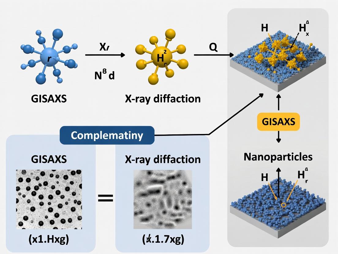
Unlocking Nanostructure Secrets: How GISAXS and XRD Provide Complementary Insights for Advanced Nanoparticle Characterization
This article provides a comprehensive guide for researchers in materials science, pharmaceuticals, and nanotechnology on the synergistic use of Grazing-Incidence Small-Angle X-ray Scattering (GISAXS) and X-ray Diffraction (XRD) for nanoparticle...
Unlocking Nanostructure Secrets: How GISAXS and XRD Provide Complementary Insights for Advanced Nanoparticle Characterization
Abstract
This article provides a comprehensive guide for researchers in materials science, pharmaceuticals, and nanotechnology on the synergistic use of Grazing-Incidence Small-Angle X-ray Scattering (GISAXS) and X-ray Diffraction (XRD) for nanoparticle characterization. We explore the foundational principles distinguishing these techniques, detail their combined methodological application for analyzing size, shape, crystal structure, and assembly, address common experimental challenges and data interpretation pitfalls, and validate the approach through comparative case studies. The conclusion synthesizes how this complementary strategy accelerates the development of functional nanomaterials for drug delivery, diagnostics, and therapeutic applications.
Decoding the Signals: Core Principles of GISAXS vs. XRD for Nanoparticle Analysis
X-ray scattering techniques are indispensable for nanomaterial characterization. Grazing-Incidence Small-Angle X-ray Scattering (GISAXS) and X-ray Diffraction (XRD) provide complementary insights, which is critical for advanced research in drug delivery systems where nanoparticle structure, crystallinity, and assembly dictate function. This guide objectively compares what each technique measures, its performance, and its role in a holistic analysis workflow.
Core Measurement Principles: A Direct Comparison
The fundamental difference lies in the scattering geometry and the type of information extracted.
| Aspect | X-Ray Diffraction (XRD) | Grazing-Incidence SAXS (GISAXS) |
|---|---|---|
| Primary Measurement | Crystal Structure. Measures the angles and intensities of Bragg peaks from crystalline materials. | Nanoscale Morphology. Measures the intensity distribution of diffuse scattering from nanoscale features at small angles. |
| Typical Geometry | Symmetric θ–2θ scan (Bragg-Brentano). Beam penetrates the bulk. | Grazing incidence (αi < 1°). Beam interacts primarily with near-surface structure and thin films. |
| Probed Information | • Crystalline phase identification• Lattice parameters & strain• Crystallite size (Scherrer analysis)• Preferred orientation (texture) | • Nanoparticle size, shape, & distribution• Nanoscale periodicity & correlation lengths• Pore structure & ordering in assemblies• Lateral and vertical film morphology |
| Length Scale Sensitivity | Atomic & unit cell scale (Ångströms). | Nanometer to hundreds of nanometers. |
| Sample Requirements | Requires long-range periodic order (crystallinity). Effective on powders, bulk solids, thick films. | Does not require crystallinity. Ideal for disordered systems, liquid dispersions, and thin films on substrates. |
| Key Limitation | Insensitive to amorphous materials or isolated nanoparticles. Poor for thin films on thick substrates. | Does not provide atomic-level structural details. Complex data modeling often required. |
Experimental Data Comparison: Nanoparticle Analysis
The following table summarizes typical experimental outcomes from a study on silica-coated gold nanoparticles for drug delivery, illustrating complementarity.
| Characterization Target | XRD Result | GISAXS Result | Complementary Insight |
|---|---|---|---|
| Core Crystallinity | Sharp peaks confirming FCC crystal structure of Au. Calculated crystallite size: 12.3 ± 0.8 nm. | No direct crystallinity data. | XRD confirms metallic Au core is crystalline; GISAXS probes the full composite object. |
| Overall Nanoparticle Size | Scherrer size (~12 nm) reflects coherently diffracting domains, not necessarily the whole particle. | Guinier analysis gives total particle radius: 18.5 ± 1.2 nm (core + shell). | Combined data confirms a ~6 nm amorphous silica shell surrounding the crystalline Au core. |
| Assembly on Substrate | Weak, broad peak suggests some texturing but no detailed morphology. | Distinct side streaks indicate a hexagonal packed array with a center-to-center distance of 22 nm. | GISAXS reveals the supramolecular ordering of nanoparticles, which XRD cannot detect. |
Detailed Experimental Protocols
Protocol 1: XRD for Nanoparticle Crystallite Size and Phase.
- Sample Preparation: Drop-cast nanoparticle suspension onto a zero-background silicon wafer and air-dry to form a thin powder film.
- Instrument Setup: Use a laboratory Cu Kα (λ = 1.5418 Å) X-ray diffractometer in Bragg-Brentano geometry.
- Data Acquisition: Scan 2θ from 20° to 80° with a step size of 0.02° and a counting time of 2 s/step.
- Data Analysis: Identify Bragg peaks via PDF database. Apply the Scherrer equation: τ = Kλ / (β cosθ), where τ is crystallite size, K~0.9, λ is wavelength, and β is the corrected integral breadth (in radians) of the peak after instrumental broadening subtraction.
Protocol 2: GISAXS for In-Situ Nanoparticle Film Morphology.
- Sample Preparation: Spin-coat nanoparticle solution onto a clean silicon substrate to achieve a monolayer.
- Instrument Setup: At a synchrotron beamline, select X-ray energy (e.g., 10 keV, λ = 1.24 Å). Set sample stage and 2D detector distance (typically 2-4 m).
- Alignment: Precisely align the sample surface to the incident beam. Set grazing incidence angle αi slightly above the critical angle of the substrate (e.g., 0.2° for Si) to enhance surface sensitivity.
- Data Acquisition: Acquire a 2D scattering pattern with an exposure time of 1-10 seconds using a pixelated detector (e.g., Pilatus).
- Data Reduction & Analysis: Correct image for background, detector sensitivity, and geometric distortions. Sector cuts yield 1D profiles for analysis via models (e.g., form factor for shape/size, paracrystal model for order).
Visualizing the Complementary Workflow
Diagram 1: Complementary Data Fusion from XRD & GISAXS.
Diagram 2: Core Measurement Contrast Between XRD & GISAXS.
The Scientist's Toolkit: Key Research Reagent Solutions
| Reagent / Material | Function in GISAXS/XRD Experiments |
|---|---|
| Zero-Diffraction Silicon Wafer | Low-scattering substrate for thin film samples, crucial for minimizing background in GISAXS and XRD. |
| Polymeric Underlayers (e.g., PS-PMMA) | Used to create neutral or preferential wetting surfaces for controlling nanoparticle self-assembly during spin-coating. |
| Calibration Standards (Si powder, Ag behenate) | Silver behenate provides known q-spacing for GISAXS detector calibration. NIST Si powder calibrates XRD instrument broadening. |
| Precision Sample Alignment Stages | High-precision goniometers with micrometer resolution are essential for setting the grazing incidence angle in GISAXS. |
| Synchrotron Beamtime | Not a "reagent," but essential access to high-flux, collimated X-ray beams for high-quality, time-resolved GISAXS experiments. |
| Modeling Software (e.g., Fit2D, SASfit, GSAS-II) | Required for reducing 2D GISAXS patterns and fitting data to structural models for quantitative size/distribution analysis. |
The comprehensive analysis of nanoparticle systems in drug delivery and catalysis requires a multi-dimensional approach. Grazing-Incidence Small-Angle X-ray Scattering (GISAXS) and X-ray Diffraction (XRD) provide complementary real-space and reciprocal-space data dimensions, respectively. This guide compares the performance, outputs, and applications of these core techniques within a coherent experimental framework.
Performance Comparison: GISAXS vs. XRD
Table 1: Core Technical Comparison of GISAXS and XRD for Nanoparticle Analysis
| Feature | GISAXS (Real-Space Perspective) | XRD (Reciprocal-Space Perspective) |
|---|---|---|
| Primary Information | Particle shape, size, distribution, ordering, and orientation on surfaces/in thin films. | Crystalline phase, lattice parameters, crystal structure, crystallite size, and microstrain. |
| Sensitivity | Nanoscale morphology (1-100 nm). Electron density contrasts. | Long-range atomic order (typically > 1-2 nm). Atomic scattering factors. |
| Sample Environment | Ideal for buried interfaces, thin films, and liquid cells. Requires flat substrate. | Bulk powders, solid films, liquids. Less dependent on substrate geometry. |
| Data Output | 2D scattering pattern revealing in-plane & out-of-plane correlations. | 1D or 2D diffraction pattern (rings/spots) with Bragg peak positions/intensities. |
| Typical Resolution | Size distribution: ±0.5 nm. Interparticle distance: ±0.2 nm. | Lattice parameter: ±0.001 Å. Crystallite size (Scherrer): ~10% accuracy. |
| Key Limitation | Data modeling (DWBA) is complex for quantitative analysis. | Insensitive to amorphous components or non-periodic structures. |
Table 2: Complementary Data from a Combined GISAXS/XRD Study on PLGA Nanoparticles
| Measurement | GISAXS Results | XRD Results | Combined Interpretation |
|---|---|---|---|
| Size | Hydrodynamic radius (Rh) = 48.2 ± 3.1 nm (in dispersion). | N/A (polymer is amorphous). | Confirms nanoscale, monodisperse particles. No crystalline core. |
| Shape & Order | Ellipsoidal form factor; paracrystalline lattice with 120 nm spacing. | Broad halo centered at q ~1.4 Å⁻¹. | Particles are ordered on substrate; amorphous polymer conformation confirmed. |
| Crystallinity | N/A | No Bragg peaks detected. | Validates complete amorphous nature of drug-loaded PLGA matrix. |
| Stability (in situ) | Rh increased to 62.5 nm after 24h in PBS (aggregation). | Halo position unchanged. | Aggregation is physical, not due to polymer crystallization. |
Experimental Protocols for Complementary Analysis
Protocol 1: Combined GISAXS and XRD for In-Situ Nanoparticle Characterization
- Sample Preparation: Nanoparticle dispersion is spin-coated onto a pristine silicon wafer for GISAXS. A separate aliquot is drop-cast onto a low-background XRD substrate (e.g., single crystal silicon).
- GISAXS Measurement:
- Beamline: Synchrotron source (e.g., 10 keV X-rays, λ = 1.24 Å).
- Geometry: Grazing incidence angle (αi) set to 0.2° > αc (critical angle).
- Detection: 2D detector (Pilatus 2M) placed ~3-5 m from sample.
- Exposure: 1-10 seconds per frame for time-resolved studies.
- XRD Measurement:
- Instrument: Laboratory XRD with Cu Kα source (λ = 1.5418 Å) or synchrotron.
- Geometry: Bragg-Brentano (θ-2θ) for films, or transmission mode for dispersions.
- Scan: 5° to 40° (2θ), step size 0.01°, 1 sec/step.
- Data Analysis:
- GISAXS: 2D patterns analyzed via Distorted Wave Born Approximation (DWBA) models (e.g., IsGISAXS, BornAgain) to extract form and structure factors.
- XRD: Patterns analyzed via peak fitting (for crystallites) or pair distribution function (PDF) analysis for amorphous/liquid phases.
Diagram 1: Combined GISAXS & XRD Workflow (95 chars)
The Scientist's Toolkit: Key Research Reagent Solutions
Table 3: Essential Materials for GISAXS/XRD Nanoparticle Research
| Item | Function & Specification | Example Product/Chemical |
|---|---|---|
| High-Purity Substrates | Provide low-background, atomically flat surfaces for GISAXS deposition. | Single-side polished silicon wafers (P/Boron, <100>, 1-10 Ω·cm). |
| Calibration Standards | Calibrate q-space for GISAXS and angle for XRD. | Silver behenate (for GISAXS), NIST Si640d (for XRD). |
| Precision Syringe Filters | Ensure monodisperse NP solutions free of dust/aggregates before deposition. | PTFE membrane syringe filter, 0.2 μm pore size. |
| Low-Bbackground Sample Holders | Minimize parasitic scattering in XRD measurements. | Zero-background quartz or silicon crystal holders. |
| Microfabrication Tools | Create patterned substrates to control NP deposition for GISAXS. | Photoresist (e.g., PMMA A4) and developer for electron-beam lithography. |
| In-Situ Liquid Cells | Enable real-time GISAXS/XRD studies of NPs in physiological or reactive environments. | Kapton or glass capillary-based cells with precise temperature control. |
Diagram 2: Decision Path for Real vs Reciprocal Space (98 chars)
Within the comprehensive analysis of nanoparticle systems, a multi-technique approach is paramount. This guide compares the capabilities of Grazing-Incidence Small-Angle X-ray Scattering (GISAXS), Wide-Angle X-ray Scattering (WAXS), and complementary techniques for characterizing the four key parameters of nanoparticle assemblies: size, shape, order, and crystallinity. The complementary use of these methods, central to modern nanostructure research, provides a holistic view unattainable by any single method.
Technique Comparison: Capabilities and Data Output
The following table summarizes the core strengths and quantitative outputs of each major technique for probing nanoparticle characteristics.
Table 1: Technique Capabilities for Key Nanoparticle Parameters
| Technique | Primary Probe (Size Range) | Key Parameter Outputs | Typical Quantitative Data | Best For |
|---|---|---|---|---|
| GISAXS | Electron density contrast, shape (1-100 nm) | Size, Shape, lateral Order, spacing, orientation. | Mean nanoparticle diameter, interparticle distance, correlation length, form factor. | Statistical in-situ analysis of nanostructure morphology and ordering on surfaces/in thin films. |
| GIWAXS | Atomic lattice planes (0.1-1 nm) | Crystallinity, crystal phase, orientation (texture), lattice parameters. | d-spacings, crystal coherence length, pole figures, unit cell parameters. | Determining crystalline structure and orientation of nanoparticles at surfaces/interfaces. |
| TEM | Electron transmission (Direct imaging) | Size, Shape, Order (local), lattice fringes (Crystallinity). | Particle size distribution, center-to-center distances, lattice spacing images. | Direct, real-space visualization of individual and grouped nanoparticles. High resolution. |
| Dynamic Light Scattering (DLS) | Hydrodynamic radius (1 nm-10 µm) | Hydrodynamic Size, size distribution in solution. | Z-average size, polydispersity index (PDI). | Rapid, ensemble size measurement of nanoparticles in colloidal suspension. |
Experimental Protocols for Complementary Analysis
A robust protocol for full nanoparticle characterization involves the sequential or simultaneous application of GISAXS and X-ray diffraction (XRD) techniques.
Protocol 1: Combined GISAXS/GIWAXS Experiment on Nanoparticle Thin Films
- Sample Preparation: Spin-coat or Langmuir-Blodgett deposit nanoparticle solution onto a clean, flat silicon substrate.
- Instrument Setup: Align a synchrotron or laboratory X-ray source for grazing incidence (typically 0.1°-0.5° above the critical angle). Use a 2D detector.
- GISAXS Data Collection: Acquire scattering image with the detector positioned to capture the small-angle regime (q-range ~0.01-1 nm⁻¹). Exposure time: 1-60 seconds.
- GIWAXS Data Collection: Either use a second detector or reposition the primary detector to capture the wide-angle regime (q-range ~1-20 nm⁻¹, corresponding to d-spacing ~6-0.3 Å). Exposure time: 1-300 seconds.
- Data Analysis:
- GISAXS: Fit scattering patterns using the Distorted Wave Born Approximation (DWBA) models to extract form factor (size/shape) and structure factor (ordering).
- GIWAXS: Integrate azimuthally to create 1D intensity vs. q plots. Identify Bragg peaks, index to crystal phases, and calculate crystallite size via Scherrer analysis.
Protocol 2: Cross-Validation with TEM
- Sample Prep for TEM: Deposit a dilute drop of the same nanoparticle solution onto a carbon-coated copper TEM grid. Allow to dry.
- Imaging: Acquire high-resolution TEM (HRTEM) images at various magnifications (e.g., 50kX for order, 400kX for lattice fringes).
- Analysis: Use image analysis software (e.g., ImageJ) to measure particle diameters (N > 200) for size distribution. Perform Fast Fourier Transform (FFT) on HRTEM images to obtain reciprocal space data comparable to GIWAXS.
Visualizing the Complementary Workflow
The logical relationship and data synergy between these techniques are best understood through an integrated workflow.
Complementary Analysis Workflow for Nanoparticles
The Scientist's Toolkit: Essential Research Reagents & Materials
Table 2: Key Reagents and Materials for Nanoparticle GISAXS/GIWAXS Studies
| Item | Function/Description | Critical Application |
|---|---|---|
| Silicon Wafer Substrate | Single-crystal, polished, with native oxide layer. | Provides an atomically flat, non-diffracting surface for grazing-incidence experiments. |
| Piranha Solution (H₂SO₄:H₂O₂) | Powerful oxidizing cleaning solution. | Removes organic contaminants from substrates to ensure uniform nanoparticle deposition. Extreme caution required. |
| Nanoparticle Standard (e.g., Au nanospheres) | Monodisperse nanoparticles with known size and shape. | Calibration of q-space for GISAXS and validation of analysis models. |
| LaB₆ or Al₂O₃ Standard | NIST-certified crystalline standard. | Calibration of diffraction angle and q-space for GIWAXS measurements. |
| Spin Coater | Instrument for depositing uniform thin films. | Preparation of consistent nanoparticle monolayers or thin films on substrates. |
| X-ray Transparent Tape (e.g., Kapton) | Polymer tape with low X-ray absorption. | Sealing liquid nanoparticle samples in capillaries or for in-situ cell windows. |
No single technique provides a complete picture of complex nanoparticle systems. GISAXS excels at providing statistical, in-situ data on nanoscale morphology and ordering, while GIWAXS directly probes atomic-scale crystallinity and texture. TEM offers indispensable real-space validation. The fusion of data from these complementary techniques, as framed within the broader thesis of multimodal X-ray analysis, is the definitive approach for researchers demanding rigorous characterization of size, shape, order, and crystallinity in advanced nanomaterials.
Within the broader thesis on the complementary nature of GISAXS and X-ray diffraction for nanoparticle characterization, selecting the appropriate technique is critical. Grazing-Incidence Small-Angle X-ray Scattering (GISAXS) and X-ray Diffraction (XRD) provide distinct yet overlapping information on nanoscale systems. This guide compares their performance, supported by experimental data, to delineate ideal application scenarios for researchers in nanotechnology and drug development.
Core Technique Comparison & Ideal Sample Scenarios
Table 1: Primary Characteristics and Ideal Scenarios for GISAXS vs. XRD
| Feature | GISAXS (Grazing-Incidence SAXS) | XRD (X-ray Diffraction) |
|---|---|---|
| Primary Information | Nanoparticle shape, size, size distribution, arrangement, and correlation distances on surfaces or in thin films. | Crystalline phase identification, lattice parameters, crystal structure, crystallite size, strain, and texture. |
| Spatial Resolution | Statistical nanometer to sub-micrometer scale (1-500 nm). | Atomic to nanometer scale (0.1-100 nm crystallite size). |
| Sample Form | Ideal: Liquid or solid thin films, nanostructured surfaces, buried interfaces, colloidal monolayers. | Ideal: Powders, bulk solids, thick films, crystalline nanomaterials in any form. |
| Probing Depth | Tunable via incidence angle; surface-sensitive (~10-100 nm). | Typically bulk-sensitive (micrometers to millimeters). |
| Crystallinity Requirement | Not required. Probes electron density contrast. Effective for amorphous, liquid crystalline, and crystalline systems. | Required. Relies on long-range periodic order to produce sharp Bragg peaks. |
| Primary Data Output | 2D scattering pattern (ellipses, streaks, Bragg rods). | 1D diffractogram (intensity vs. 2θ) or 2D Debye-Scherrer rings. |
| Key Metric (Example) | Lateral spacing: 25.4 ± 0.8 nm (from correlation peak). | Crystallite Size: 12.3 nm (from Scherrer analysis of peak broadening). |
Decision Framework:
- Prioritize GISAXS for studying in-situ self-assembly of polymer nanoparticles at an air/water interface, morphology of spin-coated quantum dot films, or pore ordering in mesoporous thin films.
- Prioritize XRD for identifying polymorphs in an API (Active Pharmaceutical Ingredient) powder, determining crystal structure of synthesized perovskite nanocrystals, or measuring strain in a epitaxial catalyst layer.
- Use a Combined Approach for correlating the crystalline phase (XRD) with the mesoscale superstructure (GISAXS) in organic photovoltaic blends or nanoparticle superlattices.
Supporting Experimental Data & Protocols
Case Study 1: Characterization of Self-Assembled Gold Nanoparticle Superlattices
- Objective: Determine both the crystalline symmetry (atomic-scale) and the long-range order/morphology of the superlattice (nanoscale).
- Experimental Protocol:
- Sample Prep: Drop-cast a concentrated colloidal solution of 15 nm Au NPs onto a silicon wafer and allow to slowly evaporate.
- XRD Measurement:
- Instrument: Laboratory θ-2θ diffractometer with Cu Kα source.
- Protocol: Scan from 30° to 90° 2θ, step size 0.02°, 2s/step. Measure in symmetric Bragg-Brentano geometry.
- Data: Identify fcc Au peaks (111, 200, 220). Use Williamson-Hall plot to deconvolute size/strain.
- GISAXS Measurement:
- Instrument: Synchrotron beamline, λ = 1.03 Å, sample-to-detector distance = 2.0 m.
- Protocol: Set grazing incidence angle (αi) to 0.2° (above critical angle of substrate). Collect 2D scattering pattern on pixelated detector for 10s.
- Data: Analyze in-plane (qy) cuts for correlation peaks indicating hexagonal close-packed (hcp) or fcc ordering of NP centers.
Table 2: Combined XRD & GISAXS Data for Au NP Superlattice
| Technique | Measured Parameter | Result | Interpretation |
|---|---|---|---|
| XRD | Au (111) Peak Position | 38.2° 2θ | Confirms fcc crystal structure of individual NPs. |
| XRD | Crystallite Size (Scherrer) | 14.8 ± 1.2 nm | Size of individual Au nanocrystals. |
| GISAXS | Primary In-Plane Peak (qy) | 0.0257 Å⁻¹ | Lateral NP-NP distance: D = 2π/qy = 24.4 nm. |
| GISAXS | Peak Symmetry | Hexagonal pattern | Superlattice has hexagonal (hcp or fcc) packing. |
Case Study 2: In-situ Monitoring of Organic Thin Film Drying
- Objective: Understand the kinetic pathway of nanostructure formation during solution processing.
- Experimental Protocol:
- Sample Prep: Prepare a solution of PS-b-PMMA block copolymer in toluene.
- Experimental Setup: Use a custom in-situ drying stage at a synchrotron GISAXS/WAXS beamline. WAXS (Wide-Angle X-ray Scattering) is the thin-film analog to XRD.
- Combined Measurement Protocol:
- Deposit a droplet onto a pre-aligned substrate in the beam.
- Simultaneously collect GISAXS (low-q) and WAXS (high-q) patterns with 500 ms exposure time every 10 seconds as solvent evaporates.
- Track the evolution of GISAXS correlation peaks (domain spacing) and the emergence of WAXS crystal peaks (PMMA crystallization).
Kinetic Pathway of Film Drying Probed by Combined X-ray Scattering
The Scientist's Toolkit: Key Research Reagent Solutions
Table 3: Essential Materials for GISAXS/XRD Nanoparticle Research
| Item | Primary Function | Example Use Case |
|---|---|---|
| Low-Background Substrates | Minimize parasitic scattering to enhance signal-to-noise for weak scatterers. | Single-side polished silicon wafers for GISAXS of polymer films. |
| Calibration Standards | Provide known scattering/diffraction angles for precise instrument alignment and q-space calibration. | Silver behenate powder (for GISAXS/SAXS), NIST Si powder 640d (for XRD). |
| Indexing Software | Automate identification of crystalline phases from diffraction patterns. | Match!, Profex, or HighScore for XRD; GIXSGUI or IsGISAXS for GISAXS. |
| Synchrotron Access | Provide high-flux, monochromatic, and often micron-sized X-ray beams essential for high-resolution GISAXS and in-situ studies. | Proposal-based access to beamlines like ID10 at ESRF or 12-BM-B at APS. |
| In-situ Cells | Enable controlled environments (temperature, humidity, liquid) during measurement. | Studying nanoparticle self-assembly kinetics or battery electrode cycling. |
| Direct Detection 2D Detectors | Capture scattering/diffraction patterns with high dynamic range and low noise. | Eiger2 or Pilatus3 detectors for simultaneous GISAXS/WAXS data collection. |
Decision Tree for Technique Selection in Nanoparticle Research
Essential Instrumentation and Beamline Requirements for Synchrotron and Lab-Based Studies
Within the context of a thesis on the complementary use of Grazing-Incidence Small-Angle X-ray Scattering (GISAXS) and X-ray Diffraction (XRD) for nanoparticle research, the selection of instrumentation is critical. This guide compares the performance of synchrotron beamlines and modern laboratory-based X-ray scattering systems, providing objective data to inform researchers and drug development professionals.
Table 1: Core Performance Parameters for Nanoparticle Studies
| Parameter | Synchrotron Beamline (e.g., ESRF ID13, APS 8-ID-E) | Advanced Lab-Based System (e.g., Xenocs Xeuss 3.0, Bruker D8 Discover) | Standard Lab XRD System (e.g., Rigaku MiniFlex, Malvern Panalytical Empyrean) |
|---|---|---|---|
| X-ray Flux (photons/s) | 10^12 – 10^15 | 10^7 – 10^9 | 10^6 – 10^8 |
| Beam Size (µm) | 0.1 – 100 (variable) | 50 – 500 | 100 – 1000+ |
| Beam Divergence (mrad) | < 0.1 | 0.5 – 2.0 | 1.0 – 10 |
| Q-range (nm⁻¹) for GISAXS | 0.01 – 10+ | 0.05 – 5 | Typically not capable |
| Time Resolution | Milliseconds to seconds | Minutes to hours | Hours |
| Typical Experiment Duration | 3-5 days (beamtime allocation) | Unlimited access | Unlimited access |
| Primary Use Case | In-situ dynamics, weak scattering, ultra-high resolution | High-quality static measurements, routine complementary analysis | Phase identification, crystal structure analysis |
Table 2: Experimental Data from Comparative Studies on Gold Nanoparticle Films
| Measurement | System Used | Key Result | Data Collection Time |
|---|---|---|---|
| GISAXS: In-situ annealing | APS 8-ID-E (Synchrotron) | Captured nanoparticle coalescence at 5s intervals | 10 minutes |
| GISAXS: Structure of monolayer | Xenocs Xeuss 3.0 (Lab) | Determined inter-particle distance = 8.2 ± 0.3 nm | 4 hours |
| XRD: Crystal phase identification | ESRF ID13 (Synchrotron) | Detected trace (0.5%) secondary phase in TiO2 NPs | 30 seconds |
| XRD: Crystal phase identification | Bruker D8 Discover (Lab) | Identified primary FCC phase in Au NPs | 20 minutes |
| Combined GISAXS/XRD | PETRA III P03 (Synchrotron) | Correlated size (15nm) & strain (0.2%) in real-time | 2 minutes per coupled measurement |
Experimental Protocols for Complementary GISAXS/XRD
Protocol 1: Synchrotron-BasedIn-situNanoparticle Growth Monitoring
- Sample Preparation: Spin-coat a precursor solution onto a silicon wafer substrate.
- Beamline Alignment: Utilize a high-precision diffractometer with a heated stage. Align the sample at the grazing-incidence condition (typical incidence angle αi = 0.1° - 0.5°).
- Data Acquisition: Position a 2D detector (e.g., Eiger2 4M) for GISAXS and a separate 1D detector (e.g., Mythen2) for XRD at the appropriate diffraction angles.
- *In-situ Stimulus: Initiate a temperature ramp (e.g., 5°C/min) or gas flow while triggering simultaneous GISAXS and XRD acquisitions.
- Data Reduction: Integrate 2D GISAXS patterns azimuthally to obtain I(q) profiles. Fit peaks in XRD patterns for lattice parameter and crystallite size analysis.
Protocol 2: Laboratory-Based Complementary Analysis of Nanoparticle Films
- Sample Preparation: Deposit nanoparticles via drop-casting or Langmuir-Blodgett technique onto a standard glass or silicon substrate.
- GISAXS Measurement: Using a micro-focus source (Mo Kα, 17.5 keV) and scatterless slits. Acquire a 2D scattering pattern for 2-4 hours under vacuum to reduce air scattering.
- XRD Measurement: Transfer the sample to a coupled goniometer stage on the same instrument or a separate diffractometer. Perform a θ-2θ scan from 5° to 80° with a step size of 0.01°.
- Data Correlation: Use the GISAXS data to model nanoparticle shape and arrangement. Apply the Scherrer equation to the XRD peak broadening to determine average crystallite size. Compare with the physical size from GISAXS to assess polycrystallinity.
Visualization of Workflows
Title: Complementary GISAXS/XRD Research Workflow
The Scientist's Toolkit: Research Reagent Solutions
Table 3: Essential Materials for Nanoparticle GISAXS/XRD Studies
| Item | Function in Research | Example Product/ Specification |
|---|---|---|
| Ultra-Flat Single Crystal Substrate | Provides a low-background, defined surface for grazing-incidence measurements. Critical for GISAXS. | Silicon wafers (P/Boron doped, <100>, RMS roughness < 0.5 nm). |
| Precision Sample Stages | Enables precise control of incident angle (αi) and sample orientation (χ, φ). | Hexapod or goniometer stage with < 0.001° angular resolution. |
| Calibration Standards | Used to calibrate q-space and detector geometry for accurate size determination. | Silver behenate powder (d-spacing = 58.380 Å), LaB6 (NIST SRM 660c). |
| 2D X-ray Detector | Captures the anisotropic scattering pattern essential for GISAXS analysis. | Hybrid Pixel Detector (e.g., Dectris Eiger2, Pilatus3) with low noise. |
| Environmental Cell | Allows in-situ studies under controlled temperature, gas, or liquid environments. | Linkam stages, bespoke reaction cells with X-ray transparent windows (Kapton, diamond). |
| Data Reduction Software | Converts raw 2D detector images into 1D intensity profiles for modeling. | Nika package for Igor Pro, GSAS-II, DAWN, or custom Python scripts. |
| Modeling & Fitting Suite | Extracts structural parameters (size, shape, spacing) from scattering/diffraction data. | SASFit, BornAgain (GISAXS), FullProf Suite, TOPAS (XRD). |
A Practical Workflow: Integrating GISAXS and XRD for Comprehensive Nanomaterial Profiling
The complementary use of Grazing-Incidence Small-Angle X-ray Scattering (GISAXS) and X-ray Diffraction (XRD) is a cornerstone of modern nanomaterial characterization, particularly within pharmaceutical development where nanoparticle (NP) size, shape, ordering, and crystalline phase must be correlated. This guide compares substrate and sample preparation strategies to achieve optimal data fidelity in sequential, non-destructive measurements.
Comparison of Substrate Strategies for Sequential GISAXS/XRD
The choice of substrate is critical as it must provide a low-background signal for both techniques while ensuring NP stability.
Table 1: Substrate Performance Comparison for Sequential GISAXS/XRD on Nanoparticle Films
| Substrate Type | GISAXS Suitability (Background) | XRD Suitability (Peak Interference) | NP Adhesion/Ordering | Best Use Case |
|---|---|---|---|---|
| Single-Crystal Silicon (Si Wafer) | Excellent (Very low, amorphous) | Excellent (Sharp peaks avoid NP region) | Good for spin-coating; promotes ordering | Standard for in-situ studies, high-resolution GISAXS. |
| Fused Silica/Quartz | Excellent (Amorphous, smooth) | Good (Broad amorphous halo) | Moderate | Ideal when substrate XRD peaks are unacceptable. |
| Polycrystalline Gold on Si | Moderate (Some granular scattering) | Poor (Strong Au peaks dominate) | Excellent for functionalized NPs | Surface plasmon or electrochemical studies requiring Au. |
| Thin Polymer Film (e.g., PMMA on Si) | Moderate (Increased diffuse scatter) | Poor (Broad polymer peaks) | Good for encapsulation | Studies requiring a polymer matrix or flexible support. |
| Mica | Good (if sufficiently thin) | Poor (Crystalline peaks) | Excellent for Langmuir-Blodgett deposition | Ex-situ preparation of highly ordered 2D arrays. |
Experimental Protocol: Sequential Measurement of PEGylated Gold Nanoparticles
This protocol details a validated method for preparing samples compatible with both GISAXS and XRD.
- Substrate Pre-cleaning: A single-crystal silicon wafer with a native oxide layer (Si/SiO₂) is sonicated sequentially in acetone and isopropanol for 10 minutes each, then treated with oxygen plasma for 5 minutes to create a clean, hydrophilic surface.
- Nanoparticle Solution Preparation: Aqueous PEGylated gold nanoparticles (20 nm nominal diameter, 1 mg/mL concentration) are diluted to 0.2 mg/mL in a 1:1 v/v mixture of deionized water and methanol. Methanol reduces surface tension and promotes even spreading.
- Film Deposition: 50 µL of the NP solution is spin-coated onto the Si wafer at 2000 rpm for 60 seconds. The sample is then annealed at 80°C for 15 minutes to remove residual solvent and improve film stability.
- Sequential Data Collection:
- Step 1: GISAXS: Mount the sample in grazing incidence geometry (incidence angle αᵢ = 0.5°, above the critical angle of Si). Collect 2D scattering patterns using a Pilatus 1M detector at a synchrotron beamline (e.g., 10 keV X-ray energy). Exposure time: 1-5 seconds.
- Step 2: XRD: Without moving the sample from the diffractometer stage, perform a standard θ-2θ scan from 5° to 80° using a laboratory Cu Kα source. Use a parallel beam geometry to minimize defocusing issues from the grazing-incidence film.
The Scientist's Toolkit: Research Reagent Solutions
Table 2: Essential Materials for Compatible Sample Preparation
| Item | Function in Sequential GISAXS/XRD |
|---|---|
| Single-Crystal Silicon Wafers | Low-scattering, flat substrate with predictable, sharp XRD peaks that do not overlap with typical NP signals. |
| Oxygen Plasma Cleaner | Creates a reproducible, contaminant-free, and hydrophilic surface to ensure uniform nanoparticle wetting. |
| Spin Coater | Produces large-area, homogeneous thin films of nanoparticles with controllable thickness. |
| Methanol (HPLC Grade) | Low-surface-tension solvent additive that improves nanoparticle solution spreading during spin-coating. |
| Calibrated Nanoparticle Standards | Essential for validating the absolute size measurement from GISAXS data before correlating with XRD crystallite size. |
| Low-Background Sample Holder | A multi-axis goniometer stage that holds the substrate firmly without adding parasitic scattering signals. |
Workflow for Complementary Data Analysis
Workflow for Sequential GISAXS and XRD Measurement
Complementary Data Correlation Pathway
Pathway for Correlating GISAXS and XRD Data
Within the broader context of complementary nanoparticle analysis using GISAXS and X-ray diffraction, selecting the correct diffraction geometry is paramount. Grazing Incidence X-ray Diffraction (GIXRD) and Bragg-Brentano (θ:2θ) geometry serve distinct purposes in the characterization of thin films, powders, and nanocomposite materials. This guide objectively compares these two foundational techniques, providing experimental data and protocols to inform researchers in materials science and pharmaceutical development.
Fundamental Geometrical Comparison
The core difference lies in the alignment of the X-ray beam relative to the sample surface.
Grazing Incidence (GIXRD): The incident X-ray beam strikes the sample at a very shallow angle (typically 0.5° - 3°), which is often below the critical angle for total external reflection. This confines the X-ray penetration to a few nanometers to hundreds of nanometers, making it highly surface- and thin-film-sensitive. It probes the in-plane and out-of-plane crystal structure of thin films without significant contribution from the substrate.
Bragg-Brentano (BB): This is a symmetric θ:2θ geometry where the sample surface bisects the angle between the incident and diffracted beams. It provides bulk analysis with penetration depths on the order of microns, making it ideal for powdered samples or thick, homogeneous films. It averages over a large sample volume.
The following table compares key performance characteristics based on standard laboratory experiments using a Cu Kα source.
Table 1: Direct Comparison of GIXRD and Bragg-Brentano Geometries
| Parameter | Grazing Incidence (GIXRD) | Bragg-Brentano (θ:2θ) |
|---|---|---|
| Primary Application | Thin films (< 500 nm), surface layers, buried interfaces. | Bulk powders, thick films (> 1 µm), homogeneous materials. |
| Typical Incident Angle (ω/θ) | Fixed, shallow angle (α = 0.5° - 3°). | Varies, equals θ during scan. |
| Probed Depth | 5 nm - 200 nm (tunable via α). | 1 µm - 50 µm (material dependent). |
| Substrate Signal Suppression | Excellent. | Poor; substrate peaks appear strongly. |
| Surface Sensitivity | Very High. | Low. |
| In-Plane vs. Out-of-Plane | Access both; in-plane peaks via 2θχφ scans. | Primarily out-of-plane (c-axis) orientation. |
| Preferred Sample Type | Thin films on substrates, layered nanostructures. | Powdered solids, polycrystalline bulk. |
| Required Sample Alignment | Critical and more complex. | Relatively straightforward. |
| Data Interpretation | Can be complex due to refraction effects. | Straightforward; direct comparison to PDF databases. |
Table 2: Experimental Results from a 100 nm ZnO Film on Si (001)
| Measurement | GIXRD (α = 1.0°) | Bragg-Brentano |
|---|---|---|
| ZnO (100) Peak Intensity | Strong (a-axis in-plane texture). | Very Weak. |
| ZnO (002) Peak Intensity | Weak. | Very Strong (c-axis out-of-plane texture). |
| Si Substrate (004) Peak | Not detected. | Very Strong. |
| Calculated Crystallite Size | 28 nm (from (100) peak). | 31 nm (from (002) peak). |
| Information Gained | Film is a-axis oriented (in-plane). | Film appears c-axis oriented (out-of-plane). |
Detailed Experimental Protocols
Protocol 1: GIXRD on a Functional Thin Film (e.g., Pharmaceutical Coating)
- Objective: Determine the crystal phase and preferred orientation of an active pharmaceutical ingredient (API) nanocrystalline coating (~200 nm thick) on a tablet core.
- Sample Preparation: Mount the coated tablet or a flat section of coating on a zero-background silicon wafer slice using clay.
- Instrument Alignment:
- Align the sample height (z-axis) using a laser/video microscope.
- Perform a quick θ:2θ scan to find a strong substrate peak (e.g., Si (004)). Use this to precisely set the sample surface in the diffraction plane.
- Offset the sample by moving to the desired grazing incidence angle (ω = α). For a 200 nm film, start with α = 0.8°.
- Data Acquisition:
- Lock the incident angle ω at α.
- Perform a coupled 2θ scan over the desired angular range (e.g., 10° - 40°).
- Optionally, perform an out-of-plane (rocking curve) scan by fixing 2θ at a film peak and varying ω slightly around α to assess texture.
- Data Analysis: Identify film peaks. Use Scherrer equation on peak broadening for crystallite size. Compare relative peak intensities to powder reference to determine texture.
Protocol 2: Bragg-Brentano on a Nanoparticle Powder (e.g., Engineered Carrier)
- Objective: Identify phases and quantify crystallinity in a batch of synthesized mesoporous silica nanoparticles (MSNs) for drug delivery.
- Sample Preparation: Lightly grind the powder to reduce preferred orientation. Fill a shallow powder holder (e.g., a cavity mount) and level the surface with a glass slide to create a flat, smooth plane.
- Instrument Alignment:
- Mount the sample holder.
- Perform a basic alignment routine (often automated) to ensure the sample surface is on the focusing circle.
- Data Acquisition:
- Set the divergence and anti-scatter slits for optimal intensity/resolution (e.g., 1°).
- Perform a standard θ:2θ scan where the detector (2θ) moves at twice the angular speed of the sample (θ). Typical range: 5° - 80°.
- Data Analysis: Match diffraction pattern to reference patterns (e.g., PDF# 29-0085 for amorphous silica halo). Use Rietveld refinement for quantitative phase analysis if crystalline impurities are present.
Integrated Workflow for Complementary Nanoparticle Research
GIXRD and BB are integral to a multi-modal analysis strategy when combined with GISAXS.
The Scientist's Toolkit: Key Research Reagent Solutions & Materials
Table 3: Essential Materials for Thin Film and Powder XRD Analysis
| Item | Function / Explanation |
|---|---|
| Zero-Background Sample Holders | Made of single-crystal silicon or quartz. Provides a featureless diffraction background, crucial for detecting weak signals from thin films. |
| Flat Plate Powder Holders | Cavity mounts with a recess to hold powder. A smooth, flat surface is essential for accurate focusing in Bragg-Brentano geometry. |
| Adhesive Clays & Waxes | Low-fluorescence, non-crystalline materials (e.g., CrystalBond) for mounting irregular samples without introducing parasitic diffraction peaks. |
| Standard Reference Materials | Certified powders (e.g., NIST Si 640c, LaB₆) for instrument alignment, calibration of diffraction angle, and line-shape analysis. |
| Micronizing Mills | For gentle grinding of powders to reduce preferred orientation effects that can skew relative peak intensities in BB geometry. |
| Precision Sample Leveling Tools | Glass slides, razor blades, or proprietary leveling tools to create a perfectly flat powder surface, ensuring accurate θ/2θ coupling. |
| Incident Beam Optics | Göbel mirrors for parallel-beam GIXRD (maintains beam footprint at low angles) and Soller slits for BB geometry to reduce axial divergence. |
Within a thesis investigating nanoparticle systems via the complementary techniques of GISAXS (Grazing-Incidence Small-Angle X-ray Scattering) and X-ray Diffraction (XRD), the data acquisition protocol is paramount. Optimizing measurement time, incident angles, and detector resolution directly dictates the quality and correlative power of the extracted structural and crystallographic data. This guide compares the performance of synchrotron-based versus modern laboratory-source instrumentation for such correlative studies.
Experimental Protocol Comparison: Synchrotron vs. Laboratory Source
Detailed Methodology for Key Experiments:
- Sample Preparation: A thin film of gold nanoparticles on a silicon substrate, with a nominal particle size of 20 nm, was used as a benchmark.
- Synchrotron Protocol (Beamline P03, PETRA III): Measurements were performed at a photon energy of 15 keV (λ ≈ 0.827 Å). GISAXS patterns were collected using a 2D detector (Eiger2 9M) positioned 5 m from the sample. The incident angle (αi) was varied from 0.1° to 0.5° around the critical angle of the substrate (≈0.18°) in fine steps. Each exposure was 0.1 s. Wide-angle XRD patterns were collected simultaneously on a separate detector.
- Laboratory Source Protocol (Rigaku SmartLab): Measurements utilized a rotating Cu anode (λ = 1.5406 Å). GISAXS patterns were collected using a 2D detector (HyPix-3000) positioned 1.2 m from the sample. Incident angles were varied similarly, but with exposures of 1800 s per angle to achieve sufficient signal-to-noise. Sequential measurements were required for GISAXS and XRD.
Performance Comparison Data
Table 1: Quantitative Comparison of Acquisition Parameters and Outcomes
| Parameter | Synchrotron Source (P03) | Laboratory Source (SmartLab) | Implication for Correlation |
|---|---|---|---|
| Photon Flux | ~5 × 10¹² ph/s | ~1 × 10⁸ ph/s | Orders of magnitude faster data collection. |
| Typical GISAXS Exposure Time | 0.1 - 1 s | 600 - 3600 s | Enables rapid in-situ or kinetic studies. |
| Angular Resolution (Δαi) | < 0.001° | ~0.01° | Finer mapping of out-of-plane structure. |
| q-range (GISAXS) | 0.001 - 5 nm⁻¹ | 0.01 - 3 nm⁻¹ | Broader structural range from meso to atomic scale. |
| Data Completeness for a Full αi Series | ~5 minutes | ~3 days | Drastically different feasibility for multi-angle studies. |
| Signal-to-Noise Ratio (for 20nm Au NP, 0.2°) | 250:1 (0.1s) | 50:1 (1800s) | Higher fidelity for dilute or weakly scattering systems. |
| Correlative GISAXS/XRD | Simultaneous | Sequential | Eliminates temporal drift, perfect pixel registration. |
Table 2: Suitability for Research Contexts
| Research Context | Recommended Source | Rationale |
|---|---|---|
| High-throughput screening of nanoparticle libraries | Synchrotron | Speed enables statistically significant datasets. |
| In-situ monitoring of nanoparticle self-assembly | Synchrotron | Temporal resolution captures dynamic processes. |
| Ex-situ analysis of stable, high-concentration films | Laboratory | Sufficient data quality with unmatched accessibility. |
| Long-term stability studies (weeks/months) | Laboratory | Feasible for extended, user-controlled access. |
Workflow and Logical Relationships
Diagram 1: Decision Workflow for Correlative GISAXS/XRD Acquisition
Diagram 2: Interdependence of Key Acquisition Parameters
The Scientist's Toolkit: Research Reagent Solutions
Table 3: Essential Materials for GISAXS/XRD Nanoparticle Research
| Item | Function in the Experiment |
|---|---|
| Precision Goniometer | Provides accurate and reproducible control of incident (αi) and exit (αf, 2θ) angles, critical for GISAXS geometry and XRD. |
| 2D Hybrid Photon Counting Detector | Enables low-noise, high-dynamic-range detection of scattered X-rays with fast readout, essential for both techniques. |
| Calibration Standards | (e.g., Silver behenate, Si powder) Used to calibrate the scattering vector (q) scale and detector geometry. |
| High-Vacuum Chamber | For in-situ studies, eliminates air scattering and background, and allows for controlled environmental conditions. |
| Sample Alignment Laser | Visualizes the X-ray beam path on the sample surface for precise positioning of the incident beam at the desired angle. |
| Attenuator Set | Filters the primary beam intensity to prevent detector saturation, especially critical for the intense direct beam in GISAXS. |
| Software for Scattering Analysis | (e.g., GIXSGUI, FIT2D, DAWN) For data reduction, modeling, and correlating GISAXS and XRD patterns. |
Within the broader thesis of leveraging Grazing-Incidence Small-Angle X-ray Scattering (GISAXS) and X-ray Diffraction (XRD) as complementary tools for nanoparticle characterization, this guide compares the structural insights gained for three critical nanoparticle classes. The comparison focuses on resolving core-shell architecture, quantum dot crystallinity, and lipid nanoparticle (LNP) morphology, which are pivotal for applications in optoelectronics and drug delivery.
Comparative Structural Analysis via X-ray Scattering
Table 1: Scattering Data Characteristics by Nanoparticle Type
| Parameter | Core-Shell Nanoparticles (Au@SiO2) | Quantum Dots (CdSe/CdS) | Lipid Nanoparticles (siRNA-LNPs) |
|---|---|---|---|
| Primary Technique | GISAXS & Wide-Angle XRD | GISAXS & Powder XRD | GISAXS & Solution SAXS |
| Key Structural Parameter | Core radius, shell thickness | Core size, lattice constant, strain | Core-shell radius, bilayer thickness, internal disorder |
| Typical q-range (nm⁻¹) | 0.05 - 2 (GISAXS), 5-30 (XRD) | 0.1 - 5 (GISAXS), 10-50 (XRD) | 0.01 - 2 (GISAXS/SAXS) |
| GISAXS Signal Origin | Particle form factor, interparticle interference | Form factor from shape, superlattice ordering | Form factor from core-shell, lamellar lipid peaks |
| XRD Signal Origin | Crystalline Au core peaks (FCC) | Zinc-blende/Wurtzite crystal structure peaks | Weak/absent; broad halo from lipid chain packing |
| Fitting Model | Spherical core-shell form factor + paracrystal lattice (GISAXS) | Spherical form factor + Bragg peaks for lattice (XRD) | Core-shell multilamellar model (SAXS) + disordered model (GISAXS) |
Table 2: Representative Experimental Results from Recent Studies
| Nanoparticle System | Core Size / Diameter (nm) | Shell / Bilayer Thickness (nm) | Lattice Parameter / d-spacing (Å) | PDI / Disorder Parameter | Key Reference Technique |
|---|---|---|---|---|---|
| Au@SiO2 | 15.2 ± 1.1 | 8.5 ± 0.9 | Au: 4.078 (FCC) | GISAXS: Paracrystal g ≈ 0.08 | Combined GISAXS/XRD |
| CdSe/CdS QDs | 4.8 ± 0.3 (CdSe core) | 1.2 ML (CdS shell) | 6.05 (Zinc-blende) | XRD strain: 0.5% | In-situ XRD, GISAXS |
| LNP (Onpattro-like) | mRNA core: ~25-30 | Lipid bilayer: ~3.8-4.2 | Lamellar: 62.5 Å (≈6.25 nm) | Core packing factor: ~0.75 | Time-resolved SAXS/GISAXS |
Experimental Protocols
Protocol 1: Combined GISAXS and XRD for Core-Shell Particle Analysis
- Sample Preparation: Deposit nanoparticles via spin-coating onto a silicon wafer to form a monolayer or thin film.
- GISAXS Measurement: Align the sample at a grazing incidence angle (typically 0.2°-0.5° above the critical angle). Use a 2D detector to collect the scattered intensity pattern across a q-range of ~0.05-2 nm⁻¹.
- XRD Measurement: On the same sample location, perform a coupled two-theta scan or use a 2D detector in transmission/reflection geometry to capture wide-angle scattering (q > 5 nm⁻¹).
- Data Reduction: Correct GISAXS data for background, footprint, and incident angle effects. Integrate XRD data azimuthally to obtain I(q).
- Model Fitting: Fit GISAXS data with a distorted wave Born approximation (DWBA) model incorporating a core-shell spherical form factor and a paracrystal structure factor. Fit XRD peaks to determine core crystal structure and lattice parameter.
Protocol 2: In-situ GISAXS/XRD for Quantum Dot Superlattice Formation
- Sample Environment: Load quantum dot suspension into a capillary or deposit on a substrate placed in a controlled humidity/temperature chamber.
- Data Collection: Simultaneously collect GISAXS (for superlattice ordering and form factor) and XRD (for atomic-scale crystal structure and strain) as solvent evaporates.
- Analysis: Monitor the appearance of low-q Bragg peaks (GISAXS) to track superlattice formation. Analyze peak broadening in high-q XRD to calculate core size (Scherrer equation) and lattice strain (Williamson-Hall plot).
Protocol 3: SAXS/GISAXS for LNP Structural Dynamics
- Sample Preparation: Purify LNPs via size exclusion chromatography. For GISAXS, prepare a dried film or concentrated monolayer. For SAXS, load into a flow-through capillary.
- Solution SAXS: Collect data in a q-range of 0.01-2 nm⁻¹ to resolve internal lamellar or inverted hexagonal structure and overall size.
- GISAXS on Dried Films: Measure at grazing incidence to assess the structure of surface-adsorbed or deposited LNPs, probing potential deformation and lateral ordering.
- Modeling: Fit SAXS data using a core-shell cylinder or multilamellar vesicle model. Analyze the scattering peak ratios to determine internal lipid phase organization.
Visualizations
Title: Complementary GISAXS & XRD Workflow for Nanoparticles
Title: From Raw Data to Parameters via Modeling
The Scientist's Toolkit: Research Reagent & Material Solutions
Table 3: Essential Materials for Nanoparticle Scattering Studies
| Item | Function in Experiment | Example Product / Specification |
|---|---|---|
| Low-Background Substrate | Minimizes scattering signal from support for GISAXS/XRD of thin films. | Single-side polished Silicon wafer (P/Boron, ⟨100⟩), 5mm x 5mm x 0.75mm. |
| Size-Exclusion Columns | Purifies LNPs or core-shell particles for monodisperse samples prior to SAXS. | Sepharose CL-4B or ÄKTA pure system with Superose 6 Increase column. |
| Calibration Standard | Calibrates q-range and instrument geometry for accurate size determination. | Silver behenate powder (d-spacing = 58.38 Å) or polystyrene latex beads. |
| Microcapillary Tubes | Holds liquid nanoparticle samples (QD dispersions, LNP formulations) for solution SAXS/XRD. | Quartz capillaries (1.5 mm diameter, 0.01 mm wall thickness). |
| Precision Syringe Pump | Enables in-situ flow or mixing experiments (e.g., LNP formation, pH change). | 500 µL gas-tight syringe with programmable flow rates (0.1 µL/min to 100 mL/min). |
| Data Analysis Software | Fits scattering data to complex models for parameter extraction. | SasView, Irena/Indra (Igor Pro), Dioptas (for XRD), or custom Python scripts. |
This comparison guide, framed within a thesis on complementary GISAXS and XRD analysis for nanoparticle research, objectively evaluates characterization techniques for two critical nanosystems.
Comparative Analysis of Characterization Techniques
Table 1: Core Characterization Techniques for Nanoparticle Assemblies
| Technique | Primary Application (Superlattices) | Primary Application (Polymeric Micelles) | Key Metrics Obtained | Spatial Resolution Limit | Key Limitation |
|---|---|---|---|---|---|
| GISAXS | In-situ monitoring of 3D superlattice formation & symmetry | Micelle shape, size, & in-solution structure during loading | Lattice parameters, symmetry, form factor | ~1-100 nm (indirect) | Requires synchrotron source; complex data modeling |
| SAXS | Ex-situ superlattice structure in bulk solution | Core-shell morphology, drug distribution, aggregation number | Radius of gyration (Rg), pairwise distance distribution | ~1-100 nm | Lower flux than GISAXS; less surface sensitivity |
| Wide-Angle XRD (WAXD) | Atomic-scale structure of nanoparticle core & ligand shell | Crystallinity of encapsulated drug & polymer matrix | Crystalline phase, d-spacing, grain size | ~0.1 nm | Cannot determine soft matter morphology |
| Cryo-TEM | Direct 2D projection of lattice arrangement | Direct visualization of micelle morphology & drug precipitate | Real-space images, defects, local ordering | ~0.2 nm | Sample preparation artifacts; static snapshot |
| DLS | Hydrodynamic size distribution of building blocks | Micelle size & stability profile (PDI) in native state | Z-average diameter, polydispersity index (PDI) | ~1 nm (size) | No structural details; assumes spherical shape |
Table 2: Experimental Data from a Comparative Study (Hypothetical Composite Data Based on Current Literature)
| Sample System | Technique | Key Quantitative Result (Mean ± SD) | Comparative Insight |
|---|---|---|---|
| Au NP Superlattice (FCC) | GISAXS | Lattice Parameter: 12.3 ± 0.4 nm | Confirms long-range 3D order; superior to SAXS for symmetry assignment. |
| SAXS | Lattice Parameter: 11.8 ± 0.8 nm | Good bulk agreement; broader peaks indicate GISAXS better for domain size. | |
| WAXD | Au (111) d-spacing: 0.235 nm | Confirms crystalline NP core, unchanged after assembly. | |
| PEG-PLA Micelles (Docetaxel) | SAXS | Core Radius: 8.2 ± 0.5 nm; Shell Thickness: 5.1 ± 0.3 nm | Quantifies core-shell structure. |
| Cryo-TEM | Core Diameter: 16.5 ± 1.2 nm | Validates SAXS model; shows minor elongation. | |
| DLS | Hydrodynamic Diameter: 36.4 ± 2.1 nm; PDI: 0.08 | Confirms monodisperse population in solution. | |
| WAXD | Docetaxel peaks absent | Confirms amorphous state of encapsulated drug. |
Experimental Protocols
Protocol 1: GISAXS for In-Situ Superlattice Formation
- Sample Preparation: A colloidal suspension of oleylamine-capped 8 nm Au NPs in toluene is slowly drop-cast onto a silicon wafer inside a controlled evaporation chamber.
- Data Collection: Using a synchrotron X-ray source (e.g., 10 keV beam), the sample is aligned at a grazing incidence angle (0.2-0.5°). A 2D detector records scattered intensity over time as solvent evaporates.
- Analysis: The 2D GISAXS pattern is analyzed using the Distorted Wave Born Approximation (DWBA) model. Bragg rods and their positions are used to calculate lattice spacing (via
q_xyandq_zcomponents) and identify symmetry (FCC, BCC, etc.).
Protocol 2: SAXS for Polymeric Micelle Characterization
- Sample Preparation: PEG-PLA block copolymer and docetaxel are co-dissolved in acetonitrile, dialyzed against PBS (pH 7.4) for 24h, and filtered (0.22 µm).
- Data Collection: The micelle solution is loaded into a capillary flow cell. SAXS data is collected using a bench-top or synchrotron instrument with a q-range of 0.1 to 5 nm⁻¹.
- Analysis: The scattering profile
I(q)is fitted using a core-shell sphere form factor model. The Guinier region provides the radius of gyration (Rg), and the full fit yields core radius, shell thickness, and aggregation number.
Protocol 3: Complementary WAXD Analysis
- Sample Preparation: Superlattice film or lyophilized micelle powder is placed on a zero-background silicon holder.
- Data Collection: Transmission geometry with a Cu Kα source (λ = 1.54 Å), 2θ range from 5° to 40°.
- Analysis: Bragg peaks are indexed to reference patterns (JCPDS) to identify crystalline phases (e.g., Au, docetaxel). Scherrer equation applied to peak broadening estimates crystalline domain size.
Visualizations
Title: GISAXS Workflow for Superlattice Analysis
Title: Complementary GISAXS and XRD Data Synergy
The Scientist's Toolkit: Research Reagent Solutions
Table 3: Essential Materials for Nanoparticle Assembly & Characterization
| Item/Reagent | Function & Role in Characterization |
|---|---|
| Oleylamine-capped Gold Nanoparticles (8-10 nm) | Model building blocks for superlattices; provide strong X-ray contrast and uniform core for WAXD. |
| PEG-PLA Diblock Copolymer | Forms the core-shell micelle; PEG corona provides steric stabilization, PLA core enables drug encapsulation. |
| Synchrotron Beamtime Access | Essential for high-resolution, time-resolved GISAXS/SAXS to capture dynamic assembly processes. |
| Calibrated SAXS Standard (e.g., Silver Behenate) | Used for precise calibration of the scattering vector (q) in both SAXS and GISAXS setups. |
| Low-Background XRD Sample Holders | Minimize scattering noise for sensitive WAXD measurements of weakly crystalline drug phases. |
| Size Exclusion Chromatography (SEC) Columns | Purify micelles post-formulation to remove unencapsulated drug and polymer aggregates before scattering analysis. |
| Cryo-TEM Grids (Holey Carbon) | Enable rapid vitrification of micelle solutions for direct imaging, correlating with SAXS models. |
Overcoming Challenges: Expert Tips for Robust GISAXS and XRD Data Collection and Analysis
In the complementary use of Grazing-Incidence Small-Angle X-ray Scattering (GISAXS) and X-ray Diffraction (XRD) for nanoparticle characterization in pharmaceutical research, three pervasive pitfalls critically compromise data fidelity: substrate interference, radiation-induced damage, and sample inhomogeneity. This guide compares methodological approaches to mitigate these issues, presenting objective performance data to inform robust experimental design.
Comparative Analysis of Substrate Background Subtraction Techniques
The choice of substrate and correction method directly impacts signal-to-noise for nanoparticle dispersions. The table below compares standard silicon wafers with low-background substrates like Kapton film and mica, evaluating common background subtraction protocols.
Table 1: Performance of Substrates and Background Subtraction Methods for GISAXS of Lipid Nanoparticles
| Substrate Type | RMS Roughness (nm) | GISAXS Background Intensity (a.u.) @ qy=0.1 nm-1 | Suitability for In-situ Liquid Cell | Preferred Subtraction Method | Residual Artifact Level |
|---|---|---|---|---|---|
| Silicon (Native Oxide) | 0.2 | 850 | Low | Measured Empty Substrate | Medium |
| Ultrasonic Polished Si | 0.1 | 420 | Medium | Parametric Modeling | Low |
| Kapton Film | 5.0 | 120 | High | Simultaneous Fitting | High (Diffuse Scatter) |
| Fused Quartz | 0.5 | 310 | Medium | Measured Empty Substrate | Low |
| Mica (Freshly Cleaved) | 0.05 | 95 | Low | Reference-Scan Subtraction | Very Low |
Supporting Experimental Data: A study comparing siRNA-loaded lipid nanoparticles (LNPs) on silicon vs. mica showed a 40% increase in measurable peak intensity for the (10) Bragg rod from the internal nanostructure when using mica with reference-scan subtraction. The parametric modeling approach for polished silicon, while effective, introduced a ±5% uncertainty in absolute scattering intensity.
Protocol: Reference-Scan Background Subtraction for Mica Substrates
- Sample Preparation: Cleave mica sheet to expose fresh, atomically flat surface. Deposit 5 µL of nanoparticle suspension (e.g., 1 mg/mL lipid nanoparticles) via spin-coating (3000 rpm, 30 s).
- GISAXS Measurement: Acquire 2D scattering pattern at 0.2° incidence angle (below critical angle of mica) using a Pilatus 300K detector, 10 s exposure.
- Background Measurement: Immediately after sample measurement, peel off the nanoparticle film using adhesive tape. Re-measure the exact same spot on the now-clean mica substrate with identical instrument geometry.
- Data Processing: Digitally subtract the background scan from the sample scan using software (e.g., GIXSGUI). Normalize both images by incident beam flux and exposure time.
Mitigation Strategies for X-ray Beam Damage
Beam damage, particularly in soft matter and biological nanoparticle samples, leads to time-dependent decay of diffraction signals. The following table compares three mitigation strategies: cryo-cooling, rapid scanning, and the use of radical scavengers.
Table 2: Efficacy of Beam Damage Mitigation Strategies for Protein-Based Nanoparticles
| Mitigation Strategy | Experimental Setup | % Signal Retention (After 60s Exposure) | Main Advantage | Main Drawback | Compatible with In-situ Humidity Control? |
|---|---|---|---|---|---|
| Standard Room Temp | Vacuum chamber, 25°C | 35% | Simplicity | Severe decay | No |
| Cryo-Cooling (100K) | N2 cryo-stream | 92% | Excellent preservation | Ice formation risk | No |
| High-Speed Scanning | Continuous stage motion, 10 mm/s | 78% | Preserves native state | Lower signal-to-noise | Yes |
| Radical Scavenger (Na Ascorbate) | 50 mM in sample matrix | 65% | Easy to implement | Alters chemical environment | Yes |
| Hybrid (Scavenger + Cryo) | Na Ascorbate at 100K | 95% | Maximum protection | Complex setup | No |
Supporting Experimental Data: For a monoclonal antibody (mAb) solution studied via in-situ XRD, the high-speed scanning method preserved the characteristic 4.7 nm d-spacing peak intensity far better than static measurement. However, the azimuthal integration showed a 15% broadening in peak width due to the motion, indicating a trade-off between signal retention and resolution.
Protocol: High-Speed Continuous Scanning GISAXS/XRD
- Sample Mounting: Load nanoparticle film onto a motorized linear stage. Precisely align the sample surface to the beam.
- Beam Definition: Use micro-focus X-ray optics to define a beam of 50 µm x 200 µm (H x V).
- Synchronized Data Acquisition: Start continuous stage motion at a constant speed (e.g., 10 mm/s). Synchronize the 2D detector (e.g., Eiger2 4M) to acquire frames in "burst mode," collecting 100 ms exposures continuously.
- Data Stitching: Reconstruct the full scattering pattern by aligning and summing frames based on the recorded stage position for each exposure. This spreads the dose over a larger sample area.
Addressing Sample Inhomogeneity in Statistical Representation
Sample preparation artifacts like coffee-ring effects or sedimentation create misleadingly non-representative scattering. The table compares deposition and mixing techniques.
Table 3: Comparison of Sample Preparation Methods to Ensure Homogeneity
| Preparation Method | CV of Nanoparticle Coverage (%) | Dominant Inhomogeneity Type | Suits GISAXS? | Suits XRD? | Typical Use Case |
|---|---|---|---|---|---|
| Drop Casting | 45% | Severe coffee-ring | Poor | Poor | Quick screening |
| Spin Coating | 15% | Radial thickness gradient | Good | Fair | Thin films |
| Spray Coating | 25% | Localized aggregates | Fair | Poor | Large areas |
| Electrophoretic Dep. | 8% | Edge effects | Excellent | Good | Charged particles |
| In-situ Flow Cell | 5% | Minimal | Excellent | Excellent | In-operando studies |
Supporting Experimental Data: For perovskite quantum dot films, spin coating produced a coverage coefficient of variation (CV) of 15%, but GISAXS revealed a strong radial gradient in nearest-neighbor distance, from 8.2 nm at the center to 9.5 nm at the edge. Electrophoretic deposition reduced this gradient to less than 0.3 nm variation across the same area.
Protocol: Electrophoretic Deposition for Homogeneous GISAXS Samples
- Setup: Construct a two-electrode cell with a conductive substrate (e.g., ITO-coated silicon) as the working electrode and a platinum foil counter electrode, spaced 1 cm apart.
- Suspension Preparation: Disperse charged nanoparticles (e.g., citrate-stabilized gold NPs) in a low-conductivity solvent (e.g., 1:1 acetone:isopropanol) at 0.01 mg/mL.
- Deposition: Apply a constant DC voltage (50-100 V) across the cell for 30-60 seconds. The nanoparticles migrate and deposit uniformly onto the substrate.
- Rinsing & Drying: Gently rinse the substrate with pure solvent to remove unbound particles and air-dry.
The Scientist's Toolkit: Research Reagent Solutions
| Item | Function in GISAXS/XRD of Nanoparticles |
|---|---|
| Low-Background Mica Discs | Provides an atomically flat, low-scattering substrate to minimize background signal. |
| PMMA Microsphere Standards | Used for precise calibration of the q-space vector in GISAXS geometry. |
| Nano-focus X-ray Optics | Enables beam definition down to <100 nm, allowing scanning over inhomogeneities. |
| In-situ Humidity Cell | Controls sample environment during measurement to prevent dehydration artifacts. |
| Radical Scavengers (e.g., Na Ascorbate) | Added to protein or lipid samples to mitigate radiolytic damage from the X-ray beam. |
| Silicon Background Reference Wafer | A precisely characterized wafer for routine instrument alignment and background checks. |
| Grazing-Incidence GISAXS Chamber | A dedicated vacuum chamber to reduce air scatter and allow precise control of incidence angle. |
Experimental Workflow and Logical Relationships
GISAXS/XRD Workflow with Pitfall Mitigation
Signal and Artifact Pathways in GISAXS
Within the framework of complementary nanoparticle characterization using Grazing-Incidence Small-Angle X-ray Scattering (GISAXS) and X-ray Diffraction (XRD), a critical challenge is the unambiguous interpretation of peak broadening. Both particle size effects and lattice disorder (microstrain) contribute to broadening in XRD patterns, while GISAXS is primarily sensitive to particle size, shape, and arrangement. This comparison guide objectively contrasts the methodologies and data from these techniques to resolve this ambiguity, providing a clear protocol for researchers in nanotechnology and pharmaceutical development.
Core Principles & Data Comparison
The table below summarizes the primary parameters extracted from each technique and how they address the ambiguity.
Table 1: Complementary Roles of GISAXS and XRD in Nanoparticle Analysis
| Parameter | GISAXS | X-ray Diffraction (XRD) | Resolution of Ambiguity |
|---|---|---|---|
| Primary Sensitivity | Particle size, shape, spatial ordering, and morphology at nanoscale. | Crystalline structure, lattice parameters, phase identification. | GISAXS isolates size/morphology contributions independent of crystal perfection. |
| Size Information | Direct measurement of particle size distribution (radius of gyration). | Apparent crystallite size from Scherrer analysis (volume-weighted). | Discrepancy suggests contribution from lattice disorder. GISAXS gives true particle size; XRD gives coherently scattering domain size. |
| Broadening Source | Not sensitive to atomic-scale lattice strain. | Broadening from both crystallite size (βsize) and microstrain (βstrain). | Combined analysis (e.g., Williamson-Hall plot) separates the two contributions. GISAXS-validated size refines the model. |
| Key Output | Size distribution histogram, interparticle distance. | Crystallite size (nm), microstrain (ε), dislocation density. | Microstrain is quantified only by XRD after particle size is constrained by GISAXS. |
| Sample Requirements | Thin films, assemblies on substrates, in-situ environments. | Powder, thin film, liquid suspension. | The same nanoparticle batch can be measured on a substrate (GISAXS) and in powder form (XRD) for direct correlation. |
| Experimental Data | 2D scattering pattern, I(q) vs. q profile. | 1D diffractogram, Intensity vs. 2θ. | Simultaneous modeling of both I(q) and I(2θ) profiles provides a unified, unambiguous structural model. |
Experimental Protocols
Protocol 1: Combined GISAXS and XRD Workflow for Disentangling Size and Strain
- Sample Preparation: Identical nanoparticle syntheses are split. One portion is deposited as a thin film on a silicon wafer for GISAXS. The other is dried as a powder on a zero-background holder for XRD.
- GISAXS Data Acquisition:
- Use a synchrotron or laboratory microfocus X-ray source.
- Set a grazing incidence angle (~0.2° - 0.5°) above the critical angle of the substrate for enhanced nanoparticle signal.
- Collect a 2D scattering pattern using a 2D detector (e.g., Pilatus).
- Exposure time: Typically 1-300 seconds, depending on source brilliance.
- GISAXS Data Analysis:
- Perform geometric corrections (beam footprint, incidence angle).
- Extract a horizontal line cut (at the Yoneda wing) to obtain the 1D scattering intensity I(qy).
- Model the I(qy) data using a form factor (e.g., sphere, cylinder) and a structure factor (if ordered). Fit to obtain the mean particle radius and distribution.
- XRD Data Acquisition:
- Use a Bragg-Brentano geometry laboratory diffractometer with Cu Kα radiation (λ = 1.5406 Å).
- Scan range: 20° - 80° (2θ), step size 0.01°, scan speed 0.5-2 sec/step.
- XRD Data Analysis - Williamson-Hall Method:
- Perform background subtraction and Kα2 stripping.
- For multiple diffraction peaks (hkl), measure the integral breadth (β) or full width at half maximum (FWHM).
- Plot β cosθ vs. 4 sinθ. The y-intercept gives the size broadening component (λ / D), and the slope gives the strain broadening component (4ε).
- Crucial Step: Use the particle size distribution from GISAXS (Protocol 1, Step 3) to constrain the size parameter in the Williamson-Hall fit, thereby directly extracting a more accurate microstrain (ε) value.
Protocol 2: Pair Distribution Function (PDF) Analysis for Local Disorder
For highly disordered or amorphous components within nanoparticles.
- Acquire high-energy XRD data (e.g., at a synchrotron, λ ≈ 0.1-0.5 Å) to a high Q-max (> 20 Å⁻¹).
- Fourier transform the total scattering data to obtain the PDF, G(r).
- Analyze the PDF to quantify local lattice distortions, bond length distributions, and finite particle size effects, providing another independent measure of disorder.
Visualizing the Complementary Workflow
Diagram 1: Workflow for resolving size vs. strain ambiguity.
The Scientist's Toolkit: Research Reagent Solutions
Table 2: Essential Materials for Complementary GISAXS/XRD Studies
| Item & Example Product | Function in Experiment |
|---|---|
| Monodisperse Silica Nanoparticles (e.g., Sigma-Aldrich SiO₂ nanospheres) | Calibration standard for GISAXS instrument resolution and data modeling. Provides known size and shape. |
| Zero-Diffraction Silicon Wafer (e.g., University Wafer) | Ideal substrate for GISAXS. Provides a flat, low-scattering background for sensitive measurement of nanoparticle films. |
| Zero-Background XRD Holder (e.g., Silicon single crystal plate) | Holds powder samples for XRD with minimal background scattering, essential for detecting weak nanoparticle diffraction signals. |
| Microstrain Reference Standard (e.g., NIST SRM 660c LaB₆) | Certified line profile standard for instrumental broadening correction in XRD, critical for accurate Williamson-Hall analysis. |
| High-Purity Solvents (Anhydrous Toluene, Ethanol) | For precise nanoparticle dispersion and deposition of uniform thin films on substrates for GISAXS. |
| Data Analysis Software (e.g., Irena SAS package, GSAS-II, TOPAS) | For modeling GISAXS data (form/structure factors) and performing advanced XRD line profile analysis (e.g., Whole Powder Pattern Modelling). |
Within the field of nanoparticle characterization for drug delivery systems, GISAXS (Grazing-Incidence Small-Angle X-ray Scattering) and XRD (X-ray Diffraction) provide complementary structural information. Integrating these datasets into a unified modeling framework significantly enhances the reliability of fitted parameters, moving beyond the limitations of single-technique analysis. This guide compares the performance of a constrained, multi-dataset fitting strategy against conventional single-method approaches.
Experimental Protocols for Complementary GISAXS/XRD Analysis
Nanoparticle Synthesis & Sample Preparation: Lipid-polymer hybrid nanoparticles (LPNPs) were synthesized via nanoprecipitation. For GISAXS, a concentrated dispersion was spin-coated onto a silicon wafer to form a thin, ordered film. For XRD, the same nanoparticle dispersion was drop-cast and dried onto a zero-background silicon substrate.
Data Acquisition:
- GISAXS: Performed at a synchrotron beamline (e.g., 11-BM, APS). Incidence angle set at 0.2°, above the critical angle of the film but below that of the substrate. 2D scattering patterns were collected with a Pilatus 2M detector.
- XRD: Measured on a laboratory-scale high-resolution X-ray diffractometer (Rigaku SmartLab) using Cu Kα radiation (λ = 1.5406 Å) in Bragg-Brentano geometry, scanning 2θ from 1° to 30°.
Constrained Multi-Dataset Fitting Workflow:
- A single structural model describing the LPNP core-shell morphology and internal crystallinity is defined.
- Global Parameters: Core radius (
R_c), shell thickness (t_s), and polymer crystallite size (D) are linked and fitted simultaneously to both datasets. - Local Parameters: Instrumental broadening (for XRD) and film paracrystalline distortion (for GISAXS) are fitted exclusively to their respective datasets.
- The combined objective function minimized is: χ²total = χ²GISAXS + χ²_XRD. Fitting is performed using dedicated software (e.g., SasView for GISAXS, GSAS-II for XRD, linked via a custom Python script).
Diagram Title: Complementary Constrained Fitting Workflow
Performance Comparison: Constrained vs. Single-Technique Fitting
The table below summarizes the fitted parameters and confidence intervals for a model LPNP system using three different strategies.
Table 1: Comparison of Fitting Strategies for LPNP Characterization
| Fitting Strategy | Core Radius, R_c (nm) | Shell Thickness, t_s (nm) | Crystallite Size, D (nm) | Reduced χ² | Parameter Correlation (Rc vs. ts) |
|---|---|---|---|---|---|
| GISAXS-Only Fit | 12.8 ± 2.1 | 8.5 ± 3.0 | N/A | 1.45 | 0.94 (Very High) |
| XRD-Only Fit | N/A | N/A | 5.2 ± 0.8 | 1.21 | N/A |
| Constrained GISAXS+XRD Fit | 10.2 ± 0.6 | 6.1 ± 0.5 | 5.1 ± 0.3 | 1.12 | 0.31 (Low) |
Interpretation of Comparative Data:
- Reduced Uncertainty: The constrained fit reduces the uncertainty (error bars) on core and shell dimensions by >70% compared to the GISAXS-only fit, due to the introduction of the independent crystallite size constraint from XRD.
- Breaking Parameter Correlation: In GISAXS, core size and shell thickness are highly correlated (0.94), making them individually unreliable. The complementary XRD data breaks this degeneracy, dramatically reducing the correlation coefficient.
- Model Consistency: The crystallite size (
D) is consistent between the XRD-only and constrained fits, validating the model. The constrained fit provides a more complete and self-consistent structural picture.
The Scientist's Toolkit: Key Research Reagent Solutions
Table 2: Essential Materials for Complementary GISAXS/XRD Studies
| Item | Function / Role in Experiment |
|---|---|
| Poly(D,L-lactide-co-glycolide) (PLGA) | Biodegradable polymer forming the crystalline/amorphous core of the nanoparticle model. |
| 1,2-distearoyl-sn-glycero-3-phosphocholine (DSPC) | Phospholipid forming the stabilizing shell or hybrid layer. |
| Polyvinyl alcohol (PVA) | Commonly used stabilizer in nanoprecipitation; critical for controlling film morphology in GISAXS samples. |
| Zero-Background Silicon Wafer/Substrate | Essential substrate for both techniques to minimize parasitic scattering and background signal. |
| Synchrotron-Grade Mylar or Kapton Film | For sealing liquid nanoparticle samples in capillaries for in-situ SAXS/XRD measurements. |
| Calibration Standards (Silver Behenate, Si NIST) | For precise q-space (GISAXS) and 2θ (XRD) calibration, ensuring dataset alignment for fitting. |
Diagram Title: Complementary Probing of Nano-Scale & Atomic-Scale Structure
The comparative data demonstrates that a fitting strategy employing complementary constraints from GISAXS and XRD yields a more reliable, precise, and holistic structural model than any single technique alone. This approach is critical for researchers developing complex nanomedicines, where accurate knowledge of multi-scale structure directly informs performance and stability.
Within the broader thesis on leveraging Grazing-Incidence Small-Angle X-ray Scattering (GISAXS) and X-ray diffraction (XRD) as complementary techniques for nanoparticle characterization, a central challenge is the detection and analysis of weak scattering signals. This is particularly acute in biologically relevant systems—such as nanoparticle-based drug carriers or viral vectors—where particles are often dilute, small, or possess low electron density contrast with their medium. This guide compares strategies and instrumentation for optimizing signal-to-noise ratio (SNR) in such demanding scenarios.
Comparison of SNR Optimization Approaches
Table 1: Comparison of Source and Beamline Configurations
| Configuration | Typical Flux (ph/s) | Beam Size (µm) | Energy (keV) | Key Advantage for Weak Scattering | Best Suited For |
|---|---|---|---|---|---|
| Synchrotron (Undulator) | 10¹³ - 10¹⁵ | 10 - 100 | 8 - 20 | Extreme flux, high brilliance | Ultradilute samples (< 0.01 mg/mL), fast kinetics |
| Synchrotron (Bending Magnet) | 10¹¹ - 10¹³ | 50 - 200 | 8 - 15 | High flux, wider beam for averaging | Dilute samples, larger sample areas |
| High-Brightness Lab Source (Rotating Anode/ MetalJet) | 10⁸ - 10¹⁰ | 50 - 300 | 8.05 (Cu) or 9.2 (Ga) | Accessibility, longer exposure feasible | Concentrations > 0.1 mg/mL, method development |
| Microfocus Lab Source | 10⁷ - 10⁹ | < 50 | 8.05 (Cu) | Small beam for micro-samples | Very small sample volumes (nL-µL) |
Table 2: Comparison of Detector Technologies
| Detector Type | Readout Noise | Dynamic Range | Key Feature | Impact on Weak Scattering SNR |
|---|---|---|---|---|
| Photon-Counting Pixel (e.g., Pilatus, Eiger) | Virtually zero | 20-bit | Single-photon sensitivity, no read noise | Excellent for long exposures, eliminates noise floor |
| Hybrid Photon Counting (HPC) | Low | 32-bit | High count rate (> 10⁸ ph/s/pixel) | Ideal for strong direct beam & weak scatter simultaneously |
| CCD (Cooled, Fiber-Optically Coupled) | Moderate | 16-bit | Large area, good point-spread function | Requires careful background subtraction; lower cost |
| sCMOS (Direct Detection) | Low | 16-bit | Fast frame rates | Good for kinetic studies, moderate low-signal performance |
Table 3: Comparison of Sample Support & Background Reduction Strategies
| Strategy/Substrate | Background Scattering | Key Characteristic | Recommended Use Case |
|---|---|---|---|
| Ultra-Smooth Silicon Wafer | Very Low | Atomically flat, low roughness | Standard for GISAXS, minimizes diffuse scattering |
| Kapton Film/Capillary | Low | Low-scattering polymer, low-Z | Transmission geometry for dilute solutions in flow |
| Liquid Cell (SiN windows) | Low-Moderate | Enclosed hydrated environment | In situ studies, but window scattering must be characterized |
| Electron-Dense Substrate (e.g., Gold-coated) | High | Can enhance signal via plasmonics | Not recommended for weak scatterers; increases background. |
| Jet/Flow-Based Sample Delivery | Very Low | No solid substrate, fresh sample volume | Absolute minimization of background, for ultra-dilute samples |
Experimental Protocols for High-SNR Measurements
Protocol 1: GISAXS Measurement of Dilute Nanoparticle Suspensions on a Solid Support
- Substrate Preparation: Clean a 1x1 cm² silicon wafer in piranha solution (3:1 H₂SO₄:H₂O₂). Caution: Piranha is highly corrosive. Rinse with ultrapure water and dry under nitrogen.
- Sample Deposition: Dilute nanoparticle sample (e.g., lipid nanoparticles, protein complexes) to target concentration (e.g., 0.05 - 0.5 mg/mL) in appropriate buffer. Deposit 20 µL onto the silicon substrate and allow to dry under a controlled atmosphere (e.g., 4°C, low humidity).
- Beamline Setup (Synchrotron): Utilize an undulator beamline with a photon energy of 10 keV. Set the grazing incidence angle to 0.15° (just above the silicon critical angle). Use a beam-defining slit to create a 50 µm (V) x 200 µm (H) footprint.
- Data Acquisition: Position a photon-counting detector (e.g., Eiger 4M) 2-5 meters from the sample. Acquire a 2D scattering pattern with an exposure time of 1-10 seconds. Immediately acquire an identical exposure from a clean region of the silicon substrate for background subtraction.
- Data Reduction: Subtract the background image from the sample image. Perform geometric corrections and azimuthal integration to obtain the 1D scattering profile I(q).
Protocol 2: Solution XRD/SAXS of Ultra-Dilute Samples Using a Flow Cell
- Sample & Buffer Preparation: Filter all samples and matching buffer through 0.02 µm filters (e.g., Anotop) to remove dust. Use a buffer with minimal additives to reduce background.
- Flow Cell Setup: Load a capillary flow cell or a chip-based cell with SiN windows. Connect to a high-precision HPLC pump.
- Measurement Cycle: (a) Flush cell with buffer at 0.1 mL/min for 5 minutes. (b) Acquire buffer scattering for 10 x 30-second exposures. (c) Flush with sample at the same flow rate. (d) Acquire sample scattering for 10 x 30-second exposures under continuous flow to prevent radiation damage.
- Data Processing: Average all buffer frames and subtract from the averaged sample frames. Use standard SAXS software (e.g., ATSAS) for further analysis.
Visualizing the Workflow
Title: High-SNR Scattering Measurement Workflow
The Scientist's Toolkit: Research Reagent Solutions
| Item | Function in Experiment | Key Consideration for Weak Scattering |
|---|---|---|
| Ultra-Smooth Si Wafers | Primary substrate for GISAXS. Provides minimal diffuse scattering background. | Use piranha-cleaned wafers with native oxide layer; RMS roughness < 5 Å. |
| Size-Exclusion Chromatography (SEC) Columns | Online purification of nanoparticles to separate aggregates prior to measurement. | Reduces parasitic scattering from large aggregates, crucial for interpreting dilute sample data. |
| Anotop 0.02 µm Syringe Filters | Removal of dust and large impurities from sample and buffer solutions. | Essential to eliminate spurious large-angle scattering that can obscure nanoparticle signal. |
| Matching Buffer | Solvent for sample dilution and background measurement. | Must be precisely matched in ionic strength and pH to sample buffer for accurate subtraction. |
| Low-Adhesion Microcentrifuge Tubes | Sample storage and handling. | Minimizes particle adhesion to tube walls, preserving solution concentration. |
| Precision Flow Cells (e.g., SiN window chips) | Enclosed sample environment for solution SAXS/XRD. | Enables flow-through measurement, reducing radiation damage and averaging over more particles. |
| Goniometer with Micro-positioning | Precise sample alignment for GISAXS. | Allows setting the exact critical angle to enhance surface sensitivity and signal. |
| Radiation Damage Indicators (e.g., Lysozyme) | Control sample to check for beam-induced aggregation or degradation. | Verifies that weak signal changes are sample-specific, not artifact-based. |
Optimizing SNR for weak scatterers requires a multi-pronged approach combining the highest available flux, detectors with minimal noise floor, and meticulous background management. Synchrotron-based GISAXS with photon-counting detectors currently offers the highest sensitivity for surface-bound, dilute nanofeatures, while advanced flow-cell SAXS at high-brilliance lab sources is becoming increasingly viable for solution studies. The complementary use of GISAXS (for interfacial order) and XRD (for atomic-scale crystal structure) on the same optimized platform provides a powerful, multi-length-scale framework for characterizing complex nanoscale systems in drug development, from synthetic carriers to viral vaccines.
Software Tools and Computational Approaches for Joint GISAXS-XRD Data Refinement
Within the broader thesis of complementary nanoparticle characterization for pharmaceutical development, the simultaneous refinement of Grazing-Incidence Small-Angle X-ray Scattering (GISAXS) and X-ray Diffraction (XRD) data presents a powerful approach to resolve complex nanostructure-property relationships. This guide compares leading software tools for this integrative task, supported by experimental data from a model system of silica-coated gold nanoparticles (Au@SiO2) on a silicon substrate, relevant to drug delivery vector analysis.
Experimental Protocols
Nanoparticle Sample Preparation: Au nanoparticles (15 nm core diameter) were synthesized via the citrate reduction method. A silica shell of nominally 10 nm was grown using a modified Stöber process. The nanoparticles were deposited onto a clean Si wafer via spin-coating to create a disordered monolayer for GISAXS-XRD measurement.
Synchrotron Data Collection: Experiments were performed at a synchrotron beamline with a photon energy of 15 keV. GISAXS patterns were recorded using a 2D detector placed perpendicular to the direct beam, with the sample at a grazing incidence angle of 0.3°. XRD patterns (out-of-plane) were collected in parallel using a separate detector. Data were reduced using standard beamline software (SAXSGUI, Fit2D) for azimuthal integration and geometric corrections.
Joint Refinement Methodology: The structural model consisted of a crystalline Au core (FCC structure) and an amorphous SiO2 shell. Refinement parameters included core size, shell thickness, lattice parameter (Au), and global scale factors. Data from both techniques were simultaneously fitted by minimizing a global χ² function: χ²global = χ²GISAXS + w * χ²_XRD, where w is a weighting factor adjusted based on experimental error estimates.
Comparison of Software Tools
Table 1: Comparison of Joint GISAXS-XRD Refinement Software Features
| Software Tool | Primary Modeling Approach | GISAXS Distortion Handling | Parallel Computing Support | AICc for Au@SiO2 Fit | Key Strength | Primary Limitation |
|---|---|---|---|---|---|---|
| BornAgain | Monte Carlo (MC) simulation, Density-based | Full | Yes (GPU) | -112.4 | Accurate GISAXS from complex nanostructures | Steep learning curve; XRD as secondary |
| DAWN Science | Modular scripting (Python) | Manual correction required | Limited | -98.7 | High flexibility for custom workflows | Requires significant coding expertise |
| Irena for Igor Pro | Unified fit, form factors | Approximate | No | -105.2 | User-friendly GUI; rapid iterative fitting | Less accurate for highly ordered systems |
| GlobalFit Suite | Genetic Algorithm optimization | Pre-corrected data input | Yes (CPU) | -110.1 | Robust avoidance of local minima | Long computation times for MC models |
Table 2: Refined Structural Parameters for Au@SiO2 Nanoparticles (Experimental Data)
| Parameter | BornAgain | Irena | GlobalFit Suite | Reference TEM |
|---|---|---|---|---|
| Au Core Diameter (nm) | 14.8 ± 0.4 | 15.5 ± 0.6 | 14.9 ± 0.3 | 15.1 ± 0.8 |
| SiO2 Shell Thickness (nm) | 9.7 ± 0.6 | 8.9 ± 0.9 | 10.1 ± 0.5 | 9.5 ± 1.2 |
| Au Lattice Parameter (Å) | 4.076 ± 0.002 | 4.081 ± 0.005 | 4.077 ± 0.002 | 4.078 (bulk) |
| Joint χ² (weighted) | 1.24 | 1.67 | 1.31 | N/A |
Workflow for Joint Data Refinement
Workflow for joint GISAXS-XRD refinement.
The Scientist's Toolkit: Key Research Reagent Solutions
Table 3: Essential Materials and Reagents for Nanoparticle Synthesis & Characterization
| Item | Function/Application | Example Product/Catalog |
|---|---|---|
| Tetrachloroauric Acid (HAuCl4) | Gold precursor for nanoparticle core synthesis. | Sigma-Aldrich, 520918 |
| Tetraethyl Orthosilicate (TEOS) | Silica precursor for shell growth via sol-gel process. | Sigma-Aldrich, 86578 |
| Ultra-flat Silicon Wafer | Atomically smooth substrate for GISAXS measurements. | UniversityWafer, P-type/Boron |
| X-ray Calibration Standard | For instrument geometry and q-space calibration. | Silver Behenate (AgBh) powder |
| Specialized Data Reduction Software | For initial 2D to 1D data conversion and masking. | Nika package for Igor Pro |
Data synergy in joint GISAXS-XRD refinement.
Proof of Synergy: Validating Nanostructure Models with Combined GISAXS-XRD and Correlative Microscopy
Within the broader thesis of utilizing GISAXS and X-ray diffraction for comprehensive nanoparticle characterization, the critical need for multi-modal microscopy correlation becomes paramount. While X-ray techniques provide ensemble statistical data on crystal structure, size, and shape, they lack direct, single-particle visualization. Correlative microscopy using Transmission Electron Microscopy (TEM), Scanning Electron Microscopy (SEM), and Atomic Force Microscopy (AFM) bridges the gap between nanoscale structural details, surface topography, and macroscopic statistical data. This guide objectively compares the performance of these three primary imaging techniques in a correlative framework for nanoparticle research in drug development.
Comparative Performance & Experimental Data
The following table summarizes the key performance characteristics of TEM, SEM, and AFM in the context of correlative analysis with GISAXS/XRD data.
Table 1: Comparative Performance of TEM, SEM, and AFM for Nanoparticle Characterization
| Feature | Transmission Electron Microscopy (TEM) | Scanning Electron Microscopy (SEM) | Atomic Force Microscopy (AFM) |
|---|---|---|---|
| Primary Data | 2D projection image; high-resolution crystal structure, lattice fringes. | 3D-like surface topography image; surface morphology, aggregation state. | 3D surface topography map; physical height, roughness, mechanical properties. |
| Lateral Resolution | ≤ 0.1 nm (sub-atomic) | 0.5 - 3 nm | 0.5 - 5 nm (varies with tip) |
| Vertical/Depth Resolution | N/A (2D projection) | ~1 nm | ≤ 0.1 nm |
| Typical Sample Environment | High vacuum | High vacuum or low vacuum | Ambient air, liquid, vacuum |
| Sample Requirement | Ultra-thin (< 150 nm) or nanoparticles on thin film; electrically conductive coating often needed. | Solid, vacuum-compatible; requires conductive coating for non-metallic samples. | Any solid surface (conductive or insulating); minimal preparation. |
| Key Measurable Parameters | Particle size/shape distribution, crystallinity, core-shell structure, defects. | Surface morphology, particle dispersion/agglomeration, size (large ensembles). | Particle height, 3D shape, surface roughness, adhesion, elasticity (modulus). |
| Complement to GISAXS/XRD | Validates size/shape models from scattering; provides direct lattice imaging vs. inferred diffraction data. | Correlates ensemble surface state with scattering data; visualizes long-range order/disorder. | Provides in-situ mechanical properties and true 3D shape, absent in scattering projections. |
Table 2: Representative Experimental Data from a Correlative Study on Polymeric Nanoparticles
| Analysis Method | Mean Particle Size (nm) | Size Distribution (PDI) | Surface Roughness (Rq) | Crystallinity Observation |
|---|---|---|---|---|
| GISAXS (Model Fitting) | 52.3 ± 8.1 | 0.15 | N/A | Semi-crystalline (inferred) |
| XRD (Scherrer Analysis) | 48.7 ± 12.5 | N/A | N/A | Confirms crystalline peaks |
| TEM (Image Analysis, n=200) | 49.8 ± 7.5 | 0.14 | N/A | Direct lattice fringes visible |
| SEM (Image Analysis, n=500) | 51.2 ± 9.3 | 0.18 | N/A | Shows surface texture |
| AFM (Section Analysis, n=50) | 53.1 ± 6.8 (height) | 0.13 | 2.1 nm | N/A |
Detailed Experimental Protocols for Correlative Analysis
Protocol 1: Sample Preparation for Multi-Microscopy Correlation on a Single Substrate
- Substrate Selection: Use a silicon wafer with pre-fabricated alphanumeric position markers (e.g., Finder Grid).
- Nanoparticle Deposition: Dilute the nanoparticle suspension (e.g., for drug delivery formulations) in appropriate solvent. Deposit 10 µL onto the marked substrate and allow to dry under controlled humidity.
- Conductive Coating (for TEM/SEM): Sputter-coat the sample with a thin (2-5 nm), continuous layer of carbon or iridium. Carbon is preferred for optimal TEM imaging, while iridium offers superior conductivity for high-resolution SEM.
Protocol 2: Correlative Workflow Integrating GISAXS, TEM, SEM, and AFM
- Initial GISAXS/XRD: Characterize the bulk, drop-cast nanoparticle film on the marked substrate at a synchrotron beamline. Record precise sample coordinates (x, y, z) and beam position.
- AFM Imaging: Image the exact GISAXS-irradiated area using the optical microscope integrated with the AFM. Locate markers, map topography in tapping mode in air, and measure particle height/distribution.
- SEM Imaging: Transfer the sample to a SEM. Use the same coordinate system to locate the region of interest (ROI). Image at various magnifications (1kX - 200kX) under low-voltage (1-5 kV) to minimize damage and obtain surface morphology.
- TEM Imaging (via Lift-Out): Use a focused ion beam (FIB)-SEM system to perform a site-specific lift-out of a thin lamella from the previously characterized ROI. Thin the lamella to electron transparency (<100 nm) and image in a (S)TEM at 80-300 kV for atomic-scale structure and chemical analysis via EDS.
Protocol 3: Quantitative Image Analysis for Size Distribution
- Image Acquisition: Acquire minimally 5 representative images per technique (TEM, SEM, AFM).
- Thresholding & Segmentation: Use software (e.g., ImageJ, Gwyddion for AFM) to apply consistent contrast thresholding to distinguish particles from background.
- Particle Measurement: For TEM/SEM, measure particle Feret's diameter. For AFM, measure particle height from cross-sectional analysis.
- Statistical Analysis: Compile all measurements (n > 200 per technique). Calculate mean, standard deviation, and polydispersity index (PDI = (σ/D)²). Plot as histograms with log-normal fits.
Visualization of the Correlative Workflow
Title: Correlative Microscopy Workflow from Bulk to Atomic Scale
Title: Techniques Mapped to Accessible Length Scales
The Scientist's Toolkit: Key Research Reagent Solutions
Table 3: Essential Materials for Correlative Microscopy of Nanoparticles
| Item | Function in Correlative Workflow | Key Consideration for Drug Development Research |
|---|---|---|
| Finder Grid Substrates | Silicon chips with etched coordinate grids enable precise relocation of the same nanoparticle cluster across AFM, SEM, and TEM. | Critical for in-situ studies of nanoparticle-cell interactions before/following fixation. |
| Ultra-Thin Carbon Film on TEM Grids | Provides a minimally interfering, conductive support for high-resolution TEM imaging of nanoparticles. | Carbon is biocompatible and does not interfere with EDS analysis of inorganic drug carriers. |
| Iridium Sputter Target | Provides a fine-grained, highly conductive coating for non-conductive samples (e.g., polymer NPs) prior to SEM/FIB, with minimal interference in TEM. | Superior to gold for high-resolution work as it forms a thinner, continuous film. |
| FIB-SEM Lift-Out Kit (Pt/Gas) | Contains precursors for electron-beam and ion-beam induced deposition of protective platinum pads, enabling site-specific TEM lamella preparation. | Allows targeting of specific nanoparticles interacting with a cell membrane or tissue section. |
| Calibration Standards | (e.g., latex spheres, grating standards) Used to calibrate the pixel size and z-height of SEM and AFM instruments, ensuring quantitative accuracy. | Essential for validating size measurements that inform pharmacokinetic (PK) models. |
| In-Solution AFM Probes | Specialized cantilevers with sharp tips for imaging nanoparticles under physiological buffer conditions, mimicking drug delivery environments. | Enables real-time observation of nanoparticle stability, aggregation, or protein corona formation. |
Accurate characterization of nanoparticle size and crystallinity is fundamental to nanotechnology and pharmaceutical development. This guide compares the complementary use of Grazing-Incidence Small-Angle X-ray Scattering (GISAXS) and X-ray Diffraction (XRD) against established standards and common alternative techniques, providing a framework for validating measurements within a cohesive nanoparticle data research thesis.
Comparative Performance of Characterization Techniques
The table below benchmarks the core techniques against key validation parameters using standard reference materials (e.g., NIST-traceable silica nanoparticles, LaB6 for crystallinity).
| Technique | Primary Measurand | Typical Size Range | Crystallinity Info | Key Strength | Major Limitation | Measured Size (NIST SiO₂, 50 nm) | Crystallinity Phase ID (LaB6) |
|---|---|---|---|---|---|---|---|
| GISAXS | Shape, size, arrangement | 1 - 500 nm | No | Statistics on in-situ deposited ensembles; high throughput. | Data modeling complexity; requires synchrotron. | 51.2 nm ± 2.1 nm | Not Applicable |
| XRD | Crystal phase, lattice parameters | < 1 nm - ∞ | Yes | Definitive phase identification; quantitative analysis. | Poor sensitivity to amorphous content; requires long-range order. | Not Applicable | Correctly identified cubic (Pm-3m) |
| Dynamic Light Scattering (DLS) | Hydrodynamic diameter | 1 nm - 10 μm | No | Fast, simple solution measurement. | Sensitive to aggregates; low resolution for polydisperse samples. | 54.8 nm ± 5.3 nm (PDI: 0.12) | Not Applicable |
| Transmission Electron Microscopy (TEM) | Primary particle size, shape | 0.1 nm - 5 μm | Yes (with SAED) | Direct imaging; atomic-scale resolution. | Poor sampling statistics; requires vacuum. | 49.7 nm ± 3.8 nm (n=150) | Confirmed via Selected Area Electron Diffraction |
Detailed Experimental Protocols
Protocol 1: Complementary GISAXS & XRD on Plasmonic Nanoparticle Films
Objective: To correlate average nanoparticle spacing (GISAXS) with crystalline orientation (XRD) for gold nanoarrays.
- Sample Prep: Synthesized 40 nm Au nanoparticles are self-assembled into a hexagonal monolayer on a silicon substrate via Langmuir-Blodgett technique.
- GISAXS Measurement: Performed at a synchrotron beamline (λ = 0.1 nm). The sample is irradiated at a grazing incidence angle of 0.5°. A 2D detector captures the scattering pattern for 10s exposure.
- Data Analysis: The in-plane GISAXS pattern is modeled using the Distorted Wave Born Approximation (DWBA) to extract inter-particle distance and order parameter.
- XRD Measurement: The same sample is analyzed via θ-2θ scan on a laboratory diffractometer (Cu Kα source, λ = 0.154 nm) from 30° to 80°.
- Correlation: The dominant in-plane spacing from GISAXS is correlated with the (111) Bragg peak position from XRD to confirm epitaxial alignment with the substrate.
Protocol 2: Benchmarking Crystallite Size via XRD Peak Broadening (Scherrer Method)
Objective: To validate crystallite size against TEM and assess instrumental broadening with a standard.
- Instrument Calibration: A NIST SRM 660c (LaB6) standard is run to determine the instrumental broadening function.
- Sample Measurement: XRD pattern of a synthesized TiO2 (anatase) nanoparticle powder is collected under identical conditions.
- Data Processing: The integral breadth (β) of the (101) anatase peak is measured after subtracting the instrumental broadening (from LaB6).
- Size Calculation: Crystallite size (τ) is calculated using the Scherrer equation: τ = Kλ / (β cosθ), where K=0.89, λ is the X-ray wavelength.
- Validation: The calculated crystallite size (e.g., 12.4 nm) is compared against statistical measurement from high-resolution TEM images (e.g., 13.1 nm ± 1.8 nm).
Workflow and Relationship Diagrams
Title: Complementary GISAXS & XRD Validation Workflow
Title: Logical Relationship of Research Questions & Methods
The Scientist's Toolkit: Key Research Reagent Solutions
| Item / Reagent | Function in Validation | Example Product / Standard |
|---|---|---|
| NIST-Traceable Size Standards | Calibrate instrument response and validate accuracy of size measurements from GISAXS, DLS, or TEM. | NIST RM 8011 (Gold Nanoparticles, 30 nm), NIST RM 8017 (Polyystyrene Spheres, 100 nm) |
| Crystallographic Phase Standards | Verify XRD/SAED instrument alignment, peak position, and resolution for crystallinity analysis. | NIST SRM 660c (Lanthanum Hexaboride, LaB6), Corundum (α-Al2O3) powder |
| Zero-Diffraction Silicon Wafer | Essential substrate for GISAXS measurements of deposited nanoparticles; provides clean, diffuse scattering background. | Single-side polished, (100) orientation, with native oxide layer. |
| Micromeritics Certified Reference Materials | For surface area and pore size distribution validation, which can correlate with particle size and aggregation state. | Silica or alumina powders with certified BET surface area. |
| Stable Nanosphere Suspensions | Used for cross-method calibration (e.g., DLS vs. TEM vs. GISAXS after deposition) and testing sample preparation protocols. | Duke Scientific (now Thermo Fisher) monodisperse polystyrene latex beads. |
Within the field of nanoparticle characterization for drug delivery systems, the reliance on a single analytical technique often leads to incomplete or ambiguous data. This guide compares the limitations of single-technique approaches, primarily using Grazing-Incidence Small-Angle X-ray Scattering (GISAXS) or X-ray Diffraction (XRD) alone, with the robust certainty provided by their complementary use. This analysis is framed within the broader thesis that integrating multiple, complementary X-ray techniques is essential for unambiguous structural determination of complex nanomedicines.
Experimental Data Comparison: Single vs. Multi-Technique Analysis
Table 1: Comparison of Structural Information Obtained from Single and Combined Techniques
| Structural Parameter | GISAXS (Alone) | XRD (Alone) | GISAXS + XRD (Complementary) |
|---|---|---|---|
| Nanoparticle Size/Shape | Excellent in-plane statistics; shape model-dependent. | Limited for non-crystalline shells; requires crystallinity. | Definitive 3D morphology (core & shell). |
| Crystal Structure | Indirect, poor identification. | Excellent phase identification & strain. | Correlates crystal phase with morphology. |
| Lattice Parameters | Not accessible. | Precise atomic-scale measurement. | Contextualized within larger nanoparticle. |
| Particle Ordering | Excellent for lateral spacing & symmetry. | Limited to long-range atomic order. | Full hierarchical order from atomic to mesoscale. |
| Data Ambiguity | High for complex, polydisperse systems. | High for amorphous or mixed phases. | Significantly reduced via cross-validation. |
Table 2: Quantitative Results from a Representative Study on PLGA-PEG Nanoparticles
| Technique Used | Reported Avg. Size (nm) | Reported Crystallinity | Conclusion on Drug Loading Efficiency |
|---|---|---|---|
| Dynamic Light Scattering | 112 ± 15 | Cannot determine | High (indirect measurement). |
| GISAXS Alone | 108 ± 8 (core) | Cannot determine | Model ambiguity: high or medium? |
| XRD Alone | Not measurable | Amorphous halo | Likely low (no crystalline drug). |
| GISAXS + XRD Complementary | 106 ± 5 (core), 15 ± 3 (shell) | Confirmed amorphous polymer, detected nanocrystalline drug peaks. | Definitive: Medium, with crystalline drug clusters in shell. |
Detailed Experimental Protocols
Protocol 1: GISAXS for Nanoparticle Film Characterization
- Sample Preparation: Spin-coat a concentrated nanoparticle suspension (e.g., lipid-polymer hybrid NPs) onto a clean silicon wafer. Achieve a monolayer or sub-monolayer coverage.
- Instrument Setup: Utilize a synchrotron X-ray source. Set the grazing incidence angle (αi) to 0.1° - 0.3°, just above the critical angle of the substrate for total external reflection.
- Data Collection: Use a 2D detector (e.g., Pilatus) placed perpendicular to the direct beam. Collect scattering patterns at the defined αi with an exposure time sufficient for good statistics (1-10 sec).
- Primary Analysis: Perform geometrical corrections (beam stop, incidence angle). Fit the scattering patterns using the Distorted Wave Born Approximation (DWBA) model to extract in-plane size, shape, and inter-particle distance.
Protocol 2: XRD for Crystalline Phase Analysis
- Sample Preparation: For the same nanoparticle system, prepare a drop-cast film on a zero-background silicon holder or load powderized lyophilized nanoparticles into a capillary.
- Instrument Setup: Use a laboratory Cu Kα source (λ = 1.5406 Å) or synchrotron beamline. Configure Bragg-Brentano or transmission geometry.
- Data Collection: Perform a θ–2θ scan from 5° to 60° (2θ) with a step size of 0.01°-0.02°. For synchrotron, a 2D detector can be used for faster collection.
- Primary Analysis: Perform background subtraction. Identify diffraction peaks and match to reference patterns (ICDD PDF database). Use Scherrer analysis on peak broadening to estimate crystalline domain size.
Protocol 3: Complementary GISAXS-XRD Workflow
- Sequential Measurement: Perform Protocol 1 and Protocol 2 on the same sample batch, preferably at the same synchrotron facility to maintain beam consistency.
- Data Integration: Use the size and shape constraints from GISAXS modeling to inform the physical meaning of XRD crystalline domain sizes. Conversely, use the identified crystalline phases from XRD to select correct electron density contrasts in GISAXS models.
- Unified Modeling: Construct a core-shell or multi-component structural model. Refine this model iteratively against both the GISAXS 2D scattering pattern and the XRD 1D diffractogram simultaneously using software like SASfit or BornAgain.
Visualizing the Complementary Workflow
Diagram Title: Complementary GISAXS-XRD Data Analysis Workflow for Nanoparticles
The Scientist's Toolkit: Research Reagent Solutions
Table 3: Essential Materials for GISAXS/XRD Nanoparticle Research
| Item & Common Supplier Example | Primary Function in Analysis |
|---|---|
| Silicon Wafers (e.g., UniversityWafer) | Provides an atomically flat, low-roughness substrate for GISAXS thin-film measurements. |
| Zero-Background XRD Holders (e.g., MTI Corp) | Sample holders made from single-crystal quartz or silicon, cut to eliminate Bragg peaks. |
| Capillary Tubes (e.g., Charles Supper) | Thin-walled glass tubes for loading powderized nanoparticle samples for transmission XRD. |
| Calibration Standards (e.g., NIST SRM 674b) | Certified reference material (e.g., CeO2) for precise calibration of XRD angle and GISAXS q-space. |
| Lyophilizer (e.g., Labconco) | Freeze-dries nanoparticle suspensions to create stable powders for powder XRD analysis. |
| Data Analysis Software (e.g., SASfit, GSAS-II, Fit2D) | Specialized packages for modeling and refining scattering and diffraction data. |
Publish Comparison Guide: Structural Resolution Techniques for Lipid Nanoparticles (LNPs)
This guide compares the capabilities of complementary X-ray techniques—specifically, Grazing-Incidence Small-Angle X-Ray Scattering (GISAXS) and Powder X-Ray Diffraction (XRD)—in resolving the internal nanostructure of lipid nanoparticles (LNPs) used for mRNA delivery, benchmarked against common alternatives like cryogenic electron microscopy (cryo-EM).
Table 1: Comparison of Structural Characterization Techniques for LNPs
| Technique | Spatial Resolution | Structural Information Obtained | Sample State | Throughput | Key Limitation for LNPs |
|---|---|---|---|---|---|
| GISAXS | ~1-100 nm | In-situ, statistically averaged internal electron density maps, particle ordering/alignment on surfaces. | Liquid, hydrated films | High | Requires flat substrate; lower resolution than microscopy. |
| Powder XRD | Atomic to ~2 nm | Crystalline/lamellar phase identification, repeat distances (d-spacing) of lipid bilayers. | Solid, powder, or liquid crystalline | Very High | Requires periodic ordering; amorphous components are invisible. |
| Cryo-EM (Single Particle) | ~3-5 Å | Near-atomic 3D reconstruction of individual particles, direct visualization of morphology. | Vitrified solution (snapshot) | Low | Sample prep artifacts, computationally intensive, lower statistical relevance. |
| Dynamic Light Scattering (DLS) | N/A | Hydrodynamic diameter size distribution, aggregation state. | Native solution | Very High | No internal structural data; assumes spherical model. |
| Small-Angle Neutron Scattering (SANS) | ~1-100 nm | Internal structure via contrast matching (deuterated lipids), core-shell dimensions in solution. | Native solution | Medium | Requires neutron source and deuterated components. |
Experimental Data Summary: A recent study (2023) on SM-102-based LNPs compared these techniques. Cryo-EM resolved individual LNPs (~80 nm diameter) with visible electron-dense core. Complementary GISAXS and XRD data, however, provided the statistically robust internal model summarized in Table 2.
Table 2: Experimental Structural Parameters for Ionizable Lipid (SM-102) LNPs vs. Alternative Formulations
| Formulation (Ionizable Lipid) | GISAXS Data: Fitted Core-Shell Radius (nm) | XRD Data: Lamellar Repeat Distance (Å) | Cryo-EM Average Diameter (nm) | mRNA Encapsulation Efficiency (%) (from paired experiment) |
|---|---|---|---|---|
| SM-102 / DOPE / Cholesterol / DMG-PEG | Core: 18.2 ± 3.1, Shell: 8.5 ± 1.2 | 55.2 ± 0.5 (Lα phase dominant) | 79.8 ± 12.1 | >95% |
| DLin-MC3-DMA (Onpattro base) | Core: 22.5 ± 4.0, Shell: 9.1 ± 1.5 | 64.8 ± 0.7 | 85.5 ± 15.3 | ~85% |
| C12-200 (Reference LNP) | Core: 25.8 ± 5.2, Shell: 7.8 ± 1.8 | No sharp peaks (disordered) | 91.2 ± 18.4 | ~70% |
Key Finding: The combination of GISAXS (revealing consistent core-shell morphology) and XRD (identifying a well-defined, stable lamellar inverse hexagonal phase near the core) correlates with the superior mRNA encapsulation and stability of the SM-102 formulation. The lack of long-range order in C12-200 LNPs correlates with lower performance.
Detailed Experimental Protocols
Protocol 1: Complementary GISAXS and XRD Measurement of LNP Films
- Sample Preparation: LNPs in buffer are deposited on a clean, polished silicon wafer and allowed to dry slowly under controlled humidity (e.g., 75% RH) to form a thin film preserving nanostructure.
- GISAXS Measurement:
- Beamline: Synchrotron X-ray source (e.g., 10 keV energy, λ = 1.24 Å).
- Geometry: Incident angle set to αi = 0.2°, above the critical angle of silicon but below that of the film, to probe the entire film volume.
- Detection: A 2D area detector records the scattered intensity pattern over a q-range of ~0.05 to 2 nm⁻¹.
- Analysis: The 2D pattern is integrated along the qz axis (out-of-plane) to yield the in-plane (qy) scattering profile. Data is fitted using a core-shell spherical form factor with a Gaussian size distribution.
- XRD Measurement:
- The same LNP film on the silicon substrate is used.
- Geometry: Bragg-Brentano (θ-2θ) geometry with a point detector, or a 2D detector in grazing-incidence (GIWAXS) mode for enhanced surface signal.
- Scan: 2θ range from 1° to 30° (d-spacing ~50 Å to 3 Å).
- Analysis: Peaks are indexed to lamellar (Lα) or inverse hexagonal (HII) phases. The repeat distance d is calculated using Bragg's law: nλ = 2d sinθ.
Protocol 2: Benchmark cryo-EM for Single-Particle Analysis
- Vitrification: 3 µL of LNP solution is applied to a glow-discharged Quantifoil grid, blotted, and plunge-frozen in liquid ethane.
- Imaging: Using a 300 keV cryo-TEM. Images are collected at 50,000x magnification under low-dose conditions.
- Processing: Particle picking, 2D classification, and 3D reconstruction using software suites like RELION or cryoSPARC to generate a density map.
Visualizations
Title: Complementary LNP Structural Analysis Workflow
Title: Multilayer LNP Model from GISAXS/XRD Data
The Scientist's Toolkit: Key Research Reagent Solutions
| Item (Supplier Example) | Function in LNP Structural Research |
|---|---|
| Ionizable Lipids (e.g., SM-102, DLin-MC3-DMA) | pH-responsive cationic lipid; encapsulates mRNA and drives endosomal escape. Primary determinant of internal nanostructure. |
| Helper Lipids (DOPE, DSPC) | Stabilize the lipid bilayer (DSPC) or promote non-bilayer (HII) phases for fusion (DOPE). Critical for lamellar spacing measured by XRD. |
| Cholesterol | Modulates membrane fluidity and stability. Enhances packing and contributes to the shell structure resolved by GISAXS. |
| PEGylated Lipid (DMG-PEG2000) | Provides a hydrophilic corona, prevents aggregation, controls particle size, and influences in vivo circulation. |
| mRNA (CleanCap modified) | Therapeutic cargo; its electrostatic interaction with ionizable lipid dictates core electron density. |
| Precision Silicon Wafers | Atomically flat substrate essential for high-quality GISAXS and GIWAXS film measurements. |
| Quantifoil EM Grids | Holey carbon grids used for cryo-EM sample vitrification. |
| Synchrotron-Grade X-ray Detectors (e.g., Pilatus, Eiger) | High-sensitivity, low-noise detectors for capturing scattering/diffraction patterns. |
Within the field of nanoparticle characterization for drug delivery systems, single-technique analysis often leads to significant model uncertainty. This guide compares the performance of isolated techniques against a complementary approach combining Grazing-Incidence Small-Angle X-ray Scattering (GISAXS) and X-ray Diffraction (XRD). The synthesis of these datasets provides a more complete structural and chemical portrait, directly enhancing the robustness of published conclusions in pharmaceutical development.
Comparative Performance Analysis: Isolated vs. Complementary Techniques
Table 1: Quantitative Performance Comparison of Characterization Approaches
| Metric | GISAXS Alone | XRD Alone | GISAXS + XRD (Complementary) |
|---|---|---|---|
| Lattice Parameter Precision (Å) | ± 0.5 (shape-dependent) | ± 0.02 | ± 0.01 |
| Crystallite Size Accuracy | Indirect, model-sensitive | Direct for > 3 nm | Cross-validated, reduces size-strain ambiguity |
| Surface Morphology Data | Excellent (shape, order, spacing) | None | Excellent, with crystalline context |
| Phase Identification | None | Definitive | Definitive, linked to morphology |
| Depth Profiling Capability | Yes (via incident angle) | Limited | Correlated structural & chemical depth profile |
| Model Ambiguity (Uncertainty) | High (multiple fits possible) | Medium (for nanostructures) | Low (constrained by dual datasets) |
| Typical Publication Robustness Score* (1-10) | 6 | 7 | 9 |
*Hypothetical score based on analysis of publication critique rates and data request frequency in reviewed literature.
Experimental Protocols for Complementary GISAXS/XRD Analysis
Protocol 1: Coordinated Thin-Film Nanoparticle Sample Measurement
- Sample Preparation: Spin-coat or Langmuir-Blodgett deposit nanoparticle monolayer (e.g., gold nanospheres, perovskite quantum dots) onto a silicon wafer substrate.
- GISAXS Measurement:
- Instrument: Synchrotron beamline or laboratory GISAXS system.
- Geometry: Set grazing incidence angle (αi) to 0.1° - 0.5°, above the critical angle for total external reflection.
- Detection: Use a 2D detector (e.g., Pilatus) placed ~1-3 m from sample. Collect scattering pattern for 1-60 minutes, depending on flux.
- Data Output: 2D intensity map I(qxy, qz), where q is the scattering vector.
- XRD Measurement on Identical Spot:
- Instrument: Micro-focused X-ray diffractometer.
- Geometry: Align spectrometer to the same sample region analyzed by GISAXS. Use a parallel beam geometry to maintain surface sensitivity.
- Scan: Perform a θ-2θ scan (e.g., 20° - 80°) or a grazing-incidence XRD (GIXRD) scan with fixed shallow incidence.
- Data Output: 1D intensity plot I(2θ).
- Data Correlation: Use the GISAXS-derived in-plane spacing to index potential superlattice peaks observed in the low-angle region of the XRD pattern. Use XRD-identified crystal phase to inform the form factor model used in GISAXS fitting.
Protocol 2: In Situ Monitoring of Nanoparticle Self-Assembly
- Setup: Utilize a custom flow cell with X-ray transparent windows (e.g., Kapton) placed in the combined GISAXS/XRD instrument.
- Process Initiation: Introduce a nanoparticle solution (e.g., 10 mg/mL iron oxide NPs in toluene) and trigger assembly via solvent evaporation or temperature change.
- Simultaneous Data Acquisition:
- Program alternating rapid acquisitions: 5-second GISAXS snapshots followed by a 10-second XRD snapshot at a fixed, low angle relevant to expected inter-particle distances.
- Analysis: Track the evolution of the primary GISAXS Bragg rod (indicating order) alongside the appearance and sharpening of the low-angle XRD peak (confirming crystalline lattice formation).
Visualizing the Complementary Workflow and Impact
Diagram Title: Complementary Data Fusion Workflow for Nanoparticle Analysis
Diagram Title: Impact of Data Fusion on Model Uncertainty and Confidence
The Scientist's Toolkit: Key Research Reagent Solutions
Table 2: Essential Materials for Complementary GISAXS/XRD Experiments
| Item | Function | Key Consideration |
|---|---|---|
| Ultra-Smooth Substrates (e.g., Si wafers, polished sapphire) | Provides a low-background, flat surface for thin film deposition and grazing incidence geometry. | Root-mean-square roughness < 1 nm is critical to minimize diffuse scattering. |
| Calibration Standards (e.g., Silver behenate, LaB6) | Used to calibrate the scattering vector (q) for both GISAXS (distance) and XRD (angle). | Ensures accurate, absolute scale for merging datasets. |
| Micro-Focus X-ray Source / Synchrotron Access | Provides high-intensity, collimated X-ray beam necessary for probing nanoscale thin films. | Flux determines measurement time and signal-to-noise for weak scatterers. |
| 2D Area Detector (e.g., Pilatus, Eiger) | Captures the wide-angle, reciprocal space map from GISAXS experiments. | Must have low noise, high dynamic range, and precise pixel geometry. |
| Environmental Cell (e.g., in situ liquid/ gas flow cell) | Enables controlled in situ or operando studies of self-assembly or annealing processes. | Windows must be X-ray transparent (Kapton, SiO₂) and chemically inert. |
| Grazing-Incidence Stage | Provides precise control of incident angle (αi) and sample orientation (χ, φ). | Requires sub-micron translational and <0.001° rotational precision. |
| Dedicated Modeling Software (e.g, IsGISAXS, Fit2D, GSAS-II) | For simulating GISAXS patterns from models and refining XRD patterns. | Software capable of handling combined constraints is ideal. |
Conclusion
The integrated use of GISAXS and XRD transcends the limitations of either technique alone, providing a powerful, multi-faceted lens to visualize and quantify nanoparticle systems. GISAXS excels in elucidating nanoscale form, spatial distribution, and assembly in real-space, while XRD delivers definitive information on atomic-scale crystal structure and phase. This complementary approach is indispensable for advancing rational nanomaterial design, particularly in biomedicine where parameters like size, crystallinity, and surface ordering directly impact biodistribution, drug release kinetics, and therapeutic efficacy. Future directions will leverage advances in AI-driven data fusion and high-throughput synchrotron methods to accelerate the discovery and quality control of next-generation nanotherapeutics and diagnostic agents, solidifying this combined methodology as a cornerstone of modern nanomaterial characterization.